Antibodies
1/32
There's no tags or description
Looks like no tags are added yet.
Name | Mastery | Learn | Test | Matching | Spaced | Call with Kai |
|---|
No analytics yet
Send a link to your students to track their progress
33 Terms
Anatomy of an antibody
Y shape
they cab be divided into two
Antibodies
Made by plasma cells, they are immunoglobulins (ig) that help the immune system recognize and fihgt foreign substances
Structure of antibodies
4 polypeptide chains
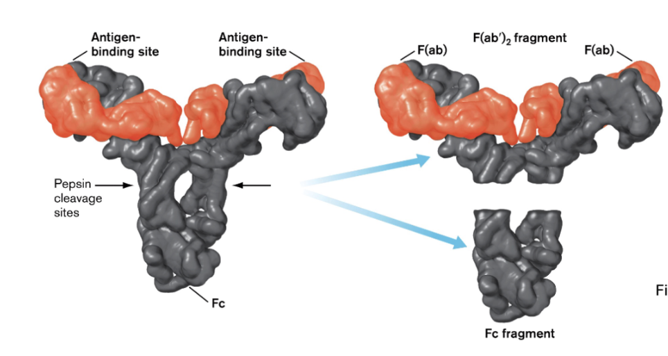
2 large heavy chains and and 2 smaller light chains bound together by disulfide bonds.
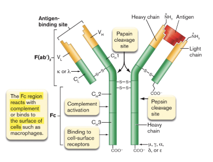
the bottom part of the Y binds to immune cells (like B cells)
Antibodies have constant and variable regions
Constant Region: The part of an antibody that has a conserved amino acid sequence.
Varivale region :The part of an antibody that differs between antibodies and allows for specific antigen binding.
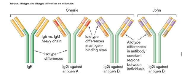
Isotype
A class of antibody common to a species, such as human IgE being the same in all humans but different from that in monkeys.
What determines the class (isotype) of an antibody?
The type of heavy chain it has.
5 kinds (α (IgA), μ (IgM), γ (IgG), δ (IgD), ε (IgE)
Light chain types
κ (kappa) and λ (lambda)
Acronym for remembering the antibodies
Antibodies protect your body from DAMaGE
IgD, IgA, IgM, IgG, IgE – the five major antibody classes
Allotype vs idiotype
Allotype: differences in the constant region shared by some but not all members of a species
Idiotype: differences in the hypervariable region within an individual.
IgG
The most abundant, smallest, and simplest antibody in blood and tissue fluids; always a monomer.
within it has 4 classes IG1-4 that vary in amino acid sequence
Opsanization
The process where antibodies coat a microbe to make it easier for phagocytes to recognize and engulf it.
What are the main functions of IgG?
Opsonizes microbes to enhance phagocytosis
Neutralizes viruses
Activates the classical complement pathway
Do antibodies kill pathogens directly?
No, antibodies act as signaling molecules that guide and activate parts of the immune system to respond.
they can neutralize viruses by blocking attachment to host cell receptors, in igG this activates complement pathway.
IgA
The major antibody found on mucosal surfaces, often secreted as a dimer linked by disulfie bonds to the J-chain protein in purple. also known as secretory IgA.
during secretion, they heavy chains bind to the secretory piece (orange)
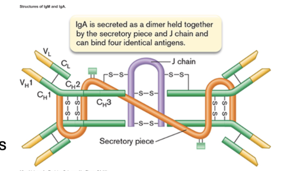
secretory igA (slgA) found in secreted things like tears, breast milk, and mucosal surfaces.
if you look at the antigen binding site, we can see that it can bing 4 antigens.
IgM
The first antibody isotype produced during an infection; found as a pentamer in circulation (held together by j-chains) and as a monomer on B-cell surfaces.
5 y shaped molecules joined together. in this example 10 antigens can bind.
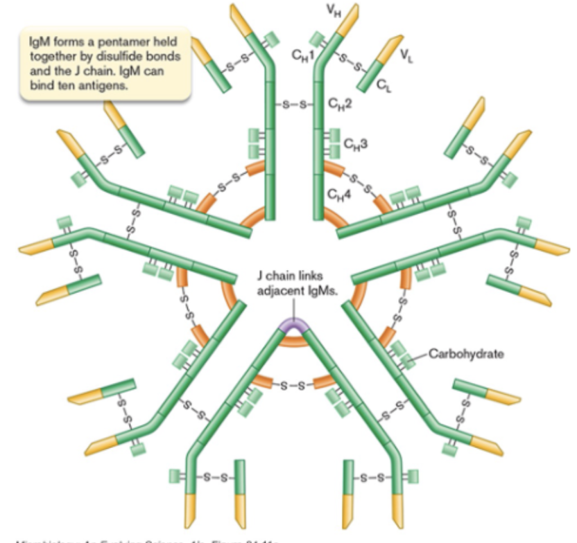
first responder, grabbing many antigens quickly to jumpstart the response.
relativelt weak binding, but compensates by binding to many things at once.
igD
An antibody present in trace amounts in the blood and found in monomer form on the surface of B-cells.
does NOT activate complement system
main function: plays a role in B-cell activation, but function not really understood.
recent work suggests that it may enhance mucosal homeostasis and immune surveillance.
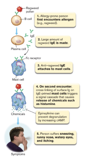
igE
Present in trace amounts in blood, found primarily on mast cells and basophils, where it triggers rapid immune amplification.
when 2 igE molecules on a mast cell are cross-linked by an antigen, the cell degranulates, releasing inflammatory mediators to amplify the immune response.
this response needs memory.
Function: amplify bodies immune response to invaders.
Degranulation
cells release their contents from granules
Allergy
An inappropriate immune response where harmless antigens (like pollen or dust) are treated as threats by the immune system.
anti-allergen igE made for the antigen and triggers degranulation of chemicals like histamine from mast cells.
How does early microbiome exposure affect allergies?
It helps teach the immune system tolerance—without this, people may develop hypersensitivities like allergies.
Anaphylaxis
A life-threatening allergic reaction caused by massive histamine release, leading to smooth muscle contraction and blood vessel leakage.
this can lead to trouble breathing, fluid loss into tissues, and widespread inflammation.
How do we combat anaphylaxis?
Using an epipen
which delivers epinephrin
function:
Raises cyclic AMP levels in cells
Stops mast cell degranulation
Reduces histamine release
Relaxes airway muscles
Does an epipen cure anaphylaxis?
No—it slows the reaction temporarily. Call 911 immediately after using it.
Which antibody has the longest serum half-life?
IgG1 and IgG4 – both last 21 days.
Which antibody has the most antigen-binding sites?
IgM – 2–10 binding sites (as a pentamer).
Which anitbodies bind complement?
IgG (not iG4) and IgM.
Which complement pathway do antibodies activate?
The classical pathway
requires a few extra proteins but c3 is the major player still
The classical pathway
also converges onto C3 (all three complement pathways do)
the other pathway to know is the lectin-mediated pathway which is similar to the classical pathway.
After C3 convertase is formed, the rest of the pathway follows the same steps as the classical pathway.
Why do we need 3 complement pathways?
to provide multiple ways to recognize and eliminate pathogens, especially those that can evade the immune system.
Phase variation
the ability of pathogens to regularly change their antigens to avoid detection by the immune system.
The innate immune system can respond quickly to pathogens even when the adaptive immune system is less effective due to phase variation.
the gut immune system (how to fight off invaders of the gut but not harm the microbiome)
Epithelial layer has T cells (Intraepithelial lymphocytes)
Dendritic cells reach between epithelial cells to sample antigens from microbiota
Specialized M cells (in Peyer’s patches) also sample antigens
sampled antigens are presented to B and T cells under epitheral cells.
slgA
sIgA is secreted into the lumen of the gut, where it coats microbiota components considered to be threats by doing this it prevents bound microbes from penetrating the gut barrier.
may promote the colonization of certain important beneficial microbes
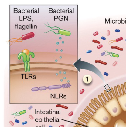
TLR positioning in the gut
TLRs on the epithelial cell side facing the gut lumen. (may see many antigens)
also some TLRs on basal side (bottom) which are much more reactive then the ones on the lumen side.
as well, many beneficial micrbobes have evolved to dampen TLR signalling.