Brain Imaging
1/15
Earn XP
Name | Mastery | Learn | Test | Matching | Spaced | Call with Kai |
|---|
No analytics yet
Send a link to your students to track their progress
16 Terms
What is brain imaging?
It refers to techniques used to visualize the structure and function of the brain. These methods allow researchers to study brain activity, identify abnormalities, and understand how different brain areas contribute to behaviour and cognition.
What types of brain imaging are used?
MRI.
fMRI.
What is MRI?
MRI or Magnetic Resonance Imaging is a structural brain imaging technique that produces high-resolution, three-dimensional images of the brain.
It does not use ionizing radiation, making it safer for repeated use.
It is primarily used for:
Brain tumours,
Strokes,
Brain injuries or
Neurodegenerative diseases (Alzheimer’s, Parkinson’s)
What are the advantages of MRI?
High spatial resolution → produces highly detailed images, allowing doctors to detect even small abnormalities.
Non-invasive and safe → does not use radiation, making it suitable for repeated use.
Can scan soft tissues → it can also provide detailed images of brain matter, differentiating grey and white matter.
Useful for clinical diagnosis → helps detect tumour, and neurodegenerative diseases.
What are the disadvantages of MRI?
Expensive → MRI machines are costly to purchase and maintain, limiting availability in some hospitals.
Time-consuming → scans can take up to 40 minutes, requiring the patient to remain completely still.
Claustrophobia concerns → the enclosed space of the MRI machines can be distressing for some patients.
Not suitable for people with metal implants → the strong magnetic field can interfere with pacemakers, metal plates, or screws in the body.
What is an fMRI?
fMRI or functional Magnetic Resonance Imaging is a functional brain imaging technique that measures brain activity in real time.
Unlike MRI, which inly shows structure, fMRI allows researchers to see which brain areas are active during specific tasks.
Used in:
Cognitive neuroscience (memory, language, problem-solving).
Clinical diagnosis (brain disorders, schizophrenia, depression).
Research on emotions and behaviour.
What are some advantages of fMRI?
High spatial resolution → provides precise localisation of brain function.
Non-invasive → no radiations or injections are required.
Can study cognitive processes in real time → allows researchers to examine memory, emotions, and problem-solving.
Can be used in clinical diagnosis → helps identify abnormalities in brain function related to disorders like schizophrenia and depression.
What are some disadvantages of fMRI?
Poor temporal resolution → fMRI detects blood flow changes, which lag behind actual neural activity, making it less accurate for tracking fast brain processes.
Expensive and not widely available → fMRI scanners require specialised equipment and trained professionals.
Motion sensitivity → small movements can create artefacts in the scan, reducing accuracy.
Complex data interpretation → requires advanced statistical analysis to differentiate real brain activity from noise.
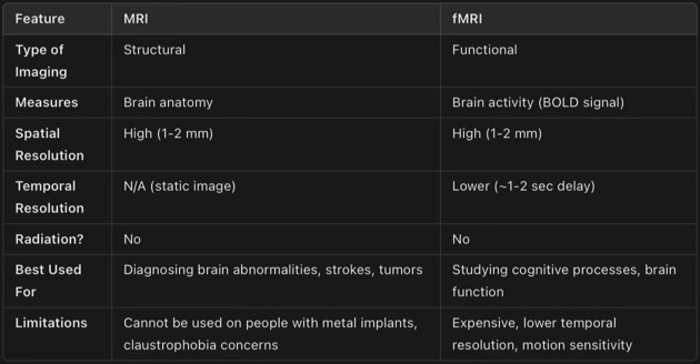
Compare them.
What studies help support this theory?
Maguire.
Draganski.
Tell me about the study of Maguire.
When: 2000
Aim: to see whether the brains of London taxi drivers would somehow be different as a result of their knowledge of te city and the many hours that they spend behind the wheel.
Sample: 16 male taxi drivers
Condition: take a “”Knowledge” test and to have their license for more than 1,5 years.
Procedure: they did an MRI scan and the researchers compared it to the MRI of 50 male non taxi drivers, which was taken out of an MRI data base. The data from the MRI was measured using two methods.
VBM (Voxel-based Morphometry) → measure the density of grey matter.
Pixel counting → to calculate the area of the hippocampus.
Results:
Pixel counting revealed that the posterior hippocampi of taxi drivers were significantly larger relative to the control subjects, and the right posterior hippocampi were significantly smaller.
VBM showed that the size of the right posterior hippocampi correlated with the years being a taxi driver.
Conclusion: this demonstrates that the hippocampus may change in response to environmental stimuli.
How do you link this study to the theory?
This study utilizes brain imaging techniques, such as MRI, to reveal structural changes in the hippocampus of London taxi drivers, demonstrating the brain’s ability to adapt to extensive navigation experience.
Evaluate the study.
Maguire
Quasi experiment → no cause and effect relationship → unable to control IV
Single blind study (researcher did not know whether she was looking at the scan of a taxi driver or a control) → avoid researcher bias
Low ecological validity → MRI scans
Sampling bias → as the sample was only male taxi drivers → difficult to generalize
Sample size → only 16 participants → hard to generalize
Ethically sound → MRI does not cause harm and they all gave consent and had the right to withdraw.
Tell me about the study of Draganski.
Date: 2004
Aim: whether learning a new skill -in this case juggling- would affect the brains of participants.
Sample: 24 non-juggling volunteers (21 females and 3 males)
Conditions: allocated into one of two conditions
Groups 1 → juggling condition
Group 2 → non-juggling condition
Procedure:
They had an MRI before the start of the experiment.
Group 1 had to learn a three-ball cascade routine. They were asked to practice this routine and to notify the researchers when they had mastered it.
Then they had a second MRI.
Then they were told to notify practice anymore.
The last MRI took place three months later.
Group 2 —> control condition for the duration of study.
To analyze the MRI they used Voxel-Based Morphometry (VBM) → determine if there were significant differences in the density of their grey matter.
Results:
1st MRI → no significant differences between the two conditions.
2nd MRI → jugglers showed a significantly larger amount of grey matter in the mid-temporal area in both hemispheres (are associated with visual memory).
3rd MRI → the amount of grey matter in these areas had decreased (after they had forgotten completely the routine).
No change in grey matter for the non-jugglers throughout the experiment.
Conclusion: it appears that juggling relies more in visual memory -that is, the perception and spatial anticipation of moving objects- that on “procedural memory” which would more likely show a change in the cerebellum or basal ganglia.
How do you link this study to the theory?
This study utilizes advanced brain imaging techniques, such as MRI, to observe structural changes in the brains of individuals learning new skills like juggling. Highlighting how these techniques can reveal neuroplastic changes in specific brain regions.
Evaluate the study.
Pre-test/Post-test design → show differences in neural density over time.
Experimental → cause-and-effect relationship.
Control group to alleviate the confounding variables.
Small sample → data may not be neither reliable neither able to be generalised.
Field experiment → IV was manipulated under natural conditions; therefore, the study has potential problems with internal validity as the participants were in their home environments for a good part of the study.