PDHPE - preliminary
1/121
Earn XP
Description and Tags
Name | Mastery | Learn | Test | Matching | Spaced |
|---|
No study sessions yet.
122 Terms
What is the anatomical position?
The anatomical position is a standard reference position in which the body is standing upright, facing forward, arms at the sides, and palms facing forward.
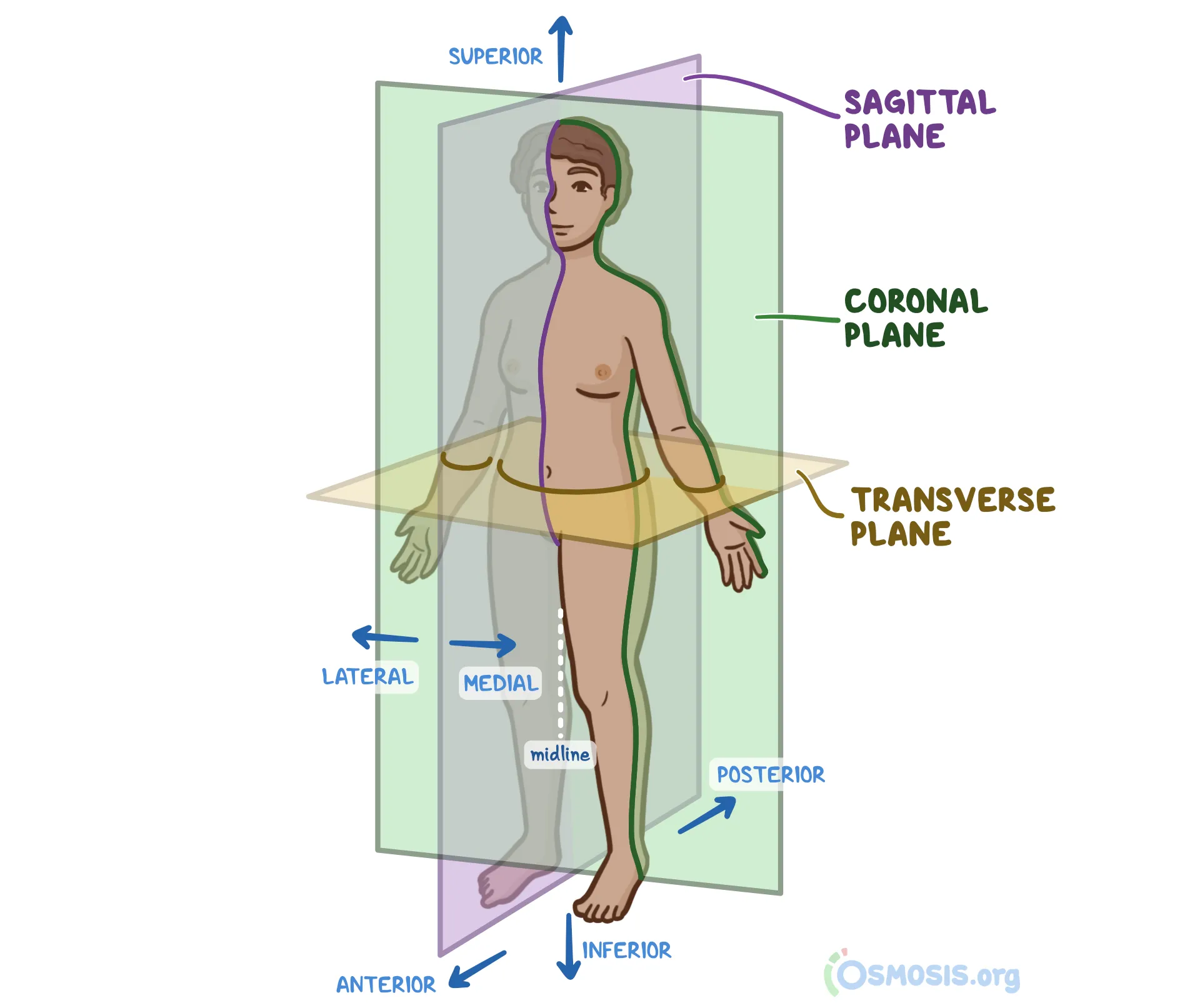
Superior
Towards the head; e.g. the chest is superior to the hips
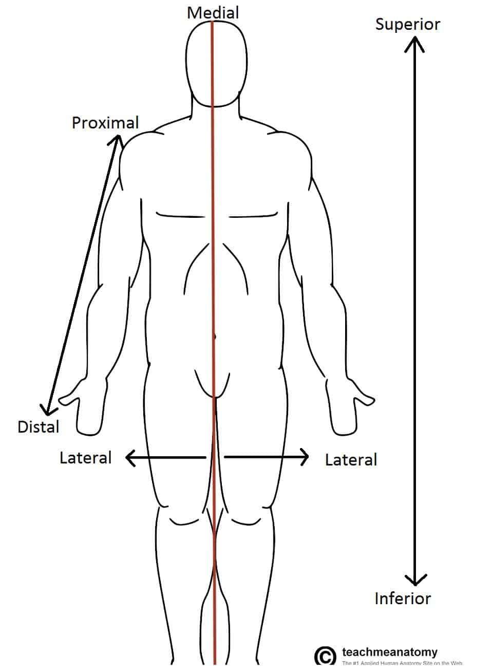
Inferior
Towards the feet; e.g., the foot is inferior to the leg
Anterior
Towards the front; e.g., the breast is on the anterior chest wall
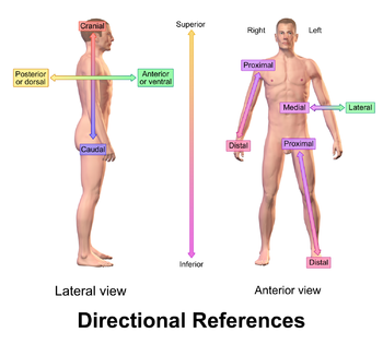
Posterior
Towards the back, e.g., the backbone is posterior to the heart
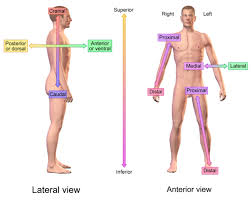
Medial
Towards the midline of the body; e.g the big toe is on the medial side of the foot
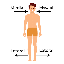
Lateral
towards the side of the body; e.g, the little toe is on the lateral side of the foot
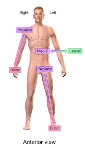
Proximal
towards the body’s mass; e.g, the shoulder is proximal to the elbow
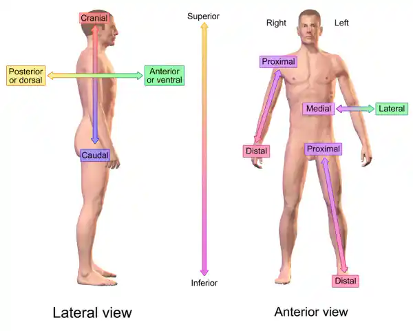
Distal
away from the body’s mass; e.g, the elbow is distal to the shoulder
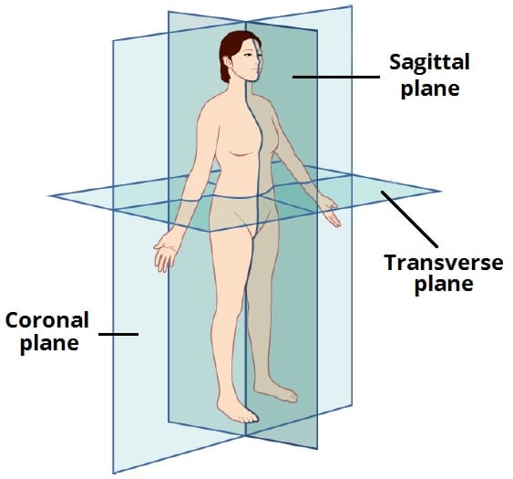
What are the anatomical planes?
Anatomical planes are imaginary lines describing body part orientation:
Sagittal - left/right
Coronal - front/back
Transverse - top/bottom
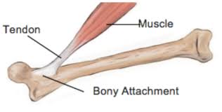
Tendon
Connects muscle to bone
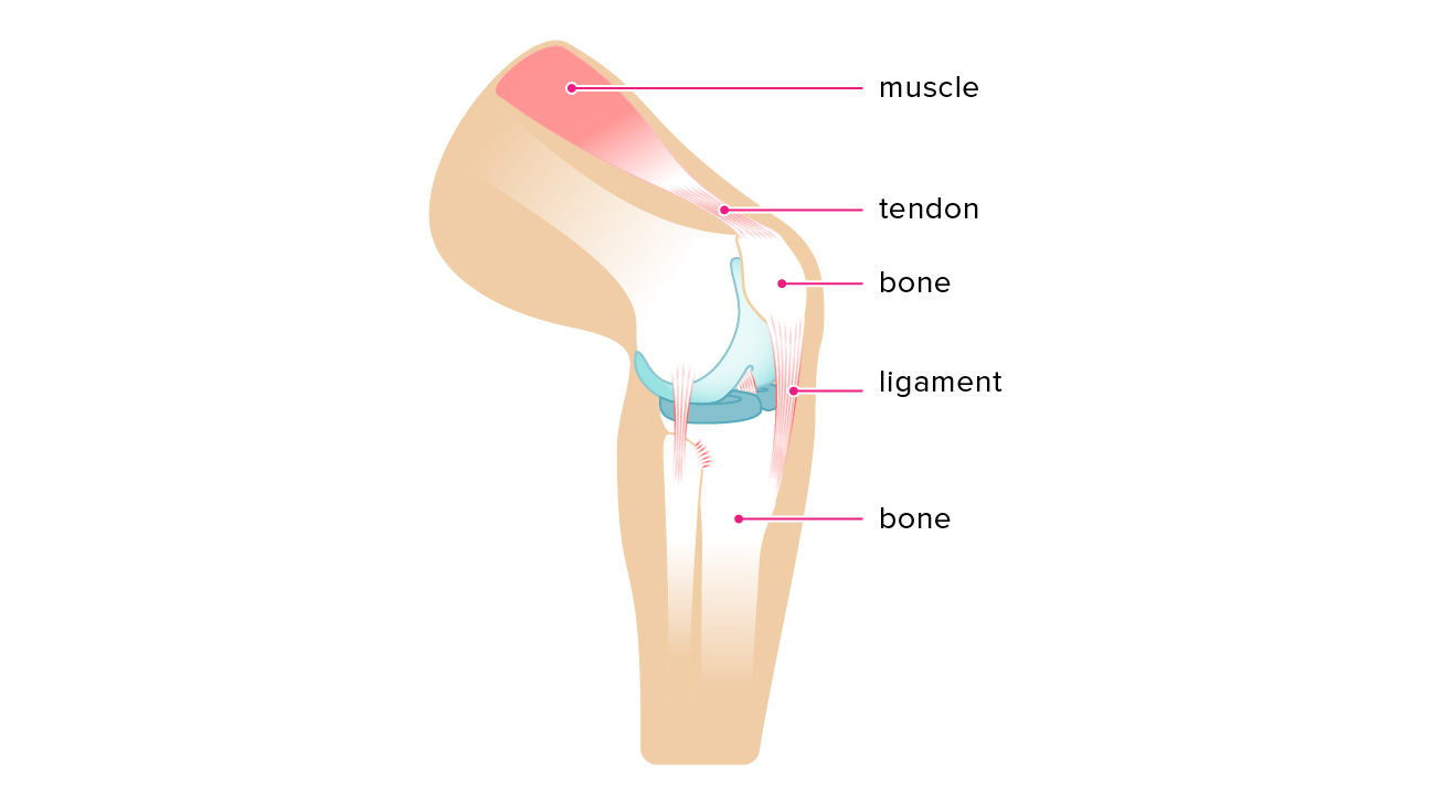
Ligaments
Connects bone to bone
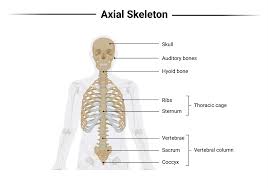
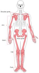
Axial skeleton
includes skull, vertebral column, and rib cage. It provides support and protection for vital organs and structures in the central axis of the body.
Appendicular skeleton
Has more tendons and ligaments and is mostly for movement. Tend to tear, dislocate, etc. more because the tendons and ligaments make it more unstable.
Roles of bones
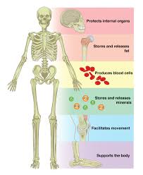
Bones provide structure and support for the body.
They protect vital organs like the brain and heart.
Bones produce red and white blood cells in the bone marrow.
They store minerals such as calcium and phosphorus.
Bones aid in movement by providing attachment points for muscles.
What are bones made of?
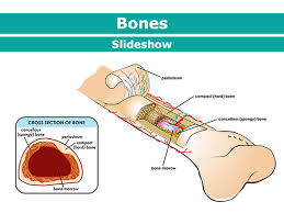
Cartilage: Provides a smooth surface for joint movement, shock absorption and flexibility.
Periosteum: A dense layer of connective tissue covering the bone surface, essential for bone growth and repair.
Bone Marrow: Produces blood cells and stores fat within the bone.
Compact Bone: Dense and strong outer layer of bone, providing support and protection.
Spongy Bone: Porous inner layer of bone, containing bone marrow and contributing to bone strength.
What is the process for formation of bones?
Formation of Bones:
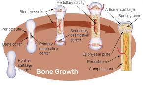
Begin as cartilage in the womb
Ossification process
Requires mineral and vitamin absorption
Vitamin D aids in bone building
Calcium maintains bone strength
Continual replacement throughout adulthood
Malnutrition can severely affect bone health
Bone structure hardens with diet and exercise.
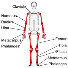
Long bones
Longer than they are wide
Slightly curved to enable shock absorption
Femur, phalanges, arm and leg bones
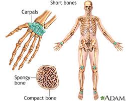
Short bones
Wider than they are long
Strong irregular cubes made of spongy bone covered with compact tissue
Just synovial fluids, not a lot of cartilage
Smaller and often found with many others
Carpals and tarsals
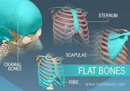
Flat bones
Flat, thin
Two parallel plates of compact bone enclosing a layer of spongy bone
Usually forms a protective surface around organs
Provide some protection
Lots of muscle attached
Skull, ribs, sternum
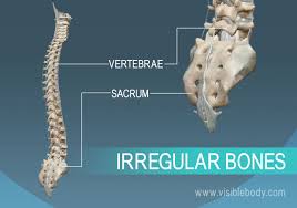
Irregular bones
Sutural - small bones which are located between the joints of certain cranial bones. Their number varies from person to person.
Sesamoid - small bones wrapped in tendons where considerable pressure develops e.g. patella
Vertebral column function
Movement: The joints of the spine mean you can bend and twist
Support: The spine is long and strong to support other body parts, e.g. the head
Protection: The spine is hard and protects the spinal cord (main pathway for information connecting the brain and peripheral nervous system)
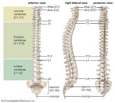
Composition of vertebral column
Made up of 33 vertebrae divided into 5 regions
Cervical Vertebrae (7): supports the head, allowing it to bend and twist
Thoracic vertebrae (12): the ribs are connected to these and there is little movement
Lumbar vertebrae (5): these are big and allow powerful twisting and bending of the back
Sacrum vertebrae (5): these form one solid mass → fused to the pelvis
Coccyx vertebrae (4): the remains of our tail
Disks (cartilage)
Function of joints
To move for flexibility
What are joints?
Point of contact between bones, between cartilage and bone or between teeth and bone
Types of joints
Fixed joint
Ball and socket
Hinge
Gliding /sliding
Condyloid
Saddle
Pivot
Classifications of joints
Fibrous or immovable - joints firmly held together by a layer of strong connective tissue. No movement between bones e.g. between bones of skull
Cartilaginous or slightly moveable - where the bones forming the joints atre attached to each other by means of discs and ligaments which only allow a limited degree of movement e.g. vertebrae in spine
Synovial or freely movable - freely movable e.g. wrist, ankle and shoulder
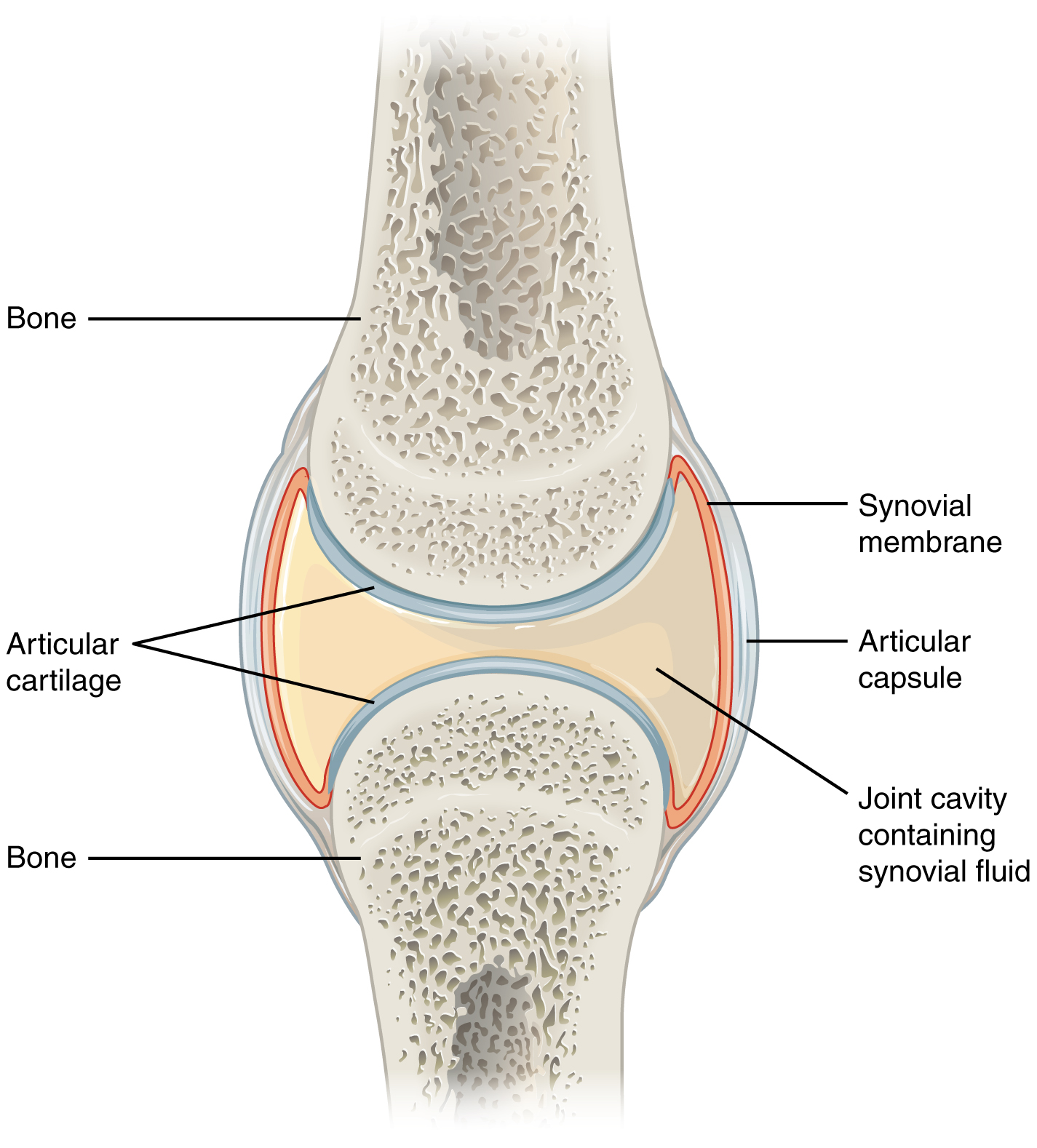
All synovial joints must have:
Cartilage - smooth, slightly elastic connective tissue which acts as a shock absorber and stop bones wearing on each other
Hyaline cartilage: In a synovial joint the ends of bones are covered in a smooth, shiny cartilage
Synovial fluid - fills the synovial cavity of a joint to lubricate it and nourish the cartilage like an oil to stop friction
Ligaments - provide stability, connective tissue joining bone to bone, slightly elastic to allow small movement of bones at joint
Tendons - inelastic and very strong, assist in movement by helping the muscle pull on a bone across a joint, connective tissue which attach the muscles to the bone to allow movement to occur
Bursa - small sac between tendon and bone, reduce friction between bone when movement occurs
Articular capsule - an envelope surrounding a synovial joint, ensures stability of joint and keeps it moving freely
Hinge joints
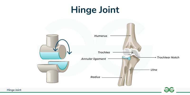
Structure: Convex surface of one bone fits into the concave surface of another.
Movement: Allows movement in one direction (like a door hinge).
Types of Movement: Flexion and extension.
Examples: Knee, elbow.
Ball and Socket joints
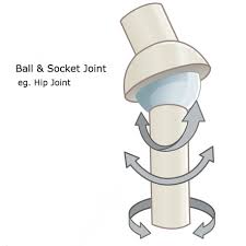
Structure: Ball-shaped surface of one bone fits into the cup-shaped depression of another.
Movement: Allows a high range of movement in multiple directions.
Types of Movement: Circumduction, adduction, abduction, some rotation.
Examples: Hip, shoulder.
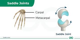
Saddle joints
Structure: One bone surface is shaped like a saddle; the other fits like a rider.
Movement: Allows movement in two directions.
Examples: Thumb.
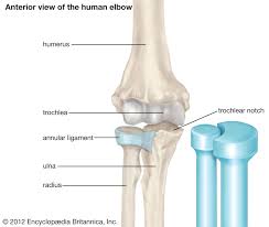
Pivot joints
Structure: Rounded or pointed surface of one bone moves within a ring formed partly by another bone and partly by a ligament.
Movement: Rotation of a bone around its own axis.
Examples: Between the atlas and axis vertebrae in the neck, between the radius and ulna.
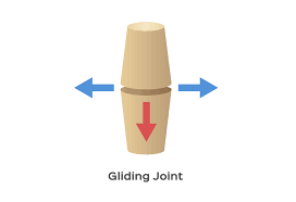
Gliding joints
Structure: Surfaces of the bones are flat or slightly curved, allowing them to glide over each other.
Movement: Allows movement in two directions.
Examples: Between the carpal bones in the hand, between the tarsal bones in the ankle.
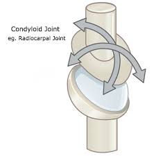
Condyloid (Ellipsoidal) Joints
Structure: Oval-shaped surface of one bone fits into the oval-shaped cavity of another.
Movement: Allows movement in two directions, and can combine these movements for semi-circumduction.
Examples: Between the radius and carpals in the wrist, between the tibia and tarsals in the ankle.
Categories of the muscular system
Skeletal
Cardiac (the heart)
Smooth (cover the outside of organs)
Skeletal muscles
Striated: twisted and bounded muscles that allow for strength
Voluntary: CNS decides when and how to use them
Structure of skeleton muscles
Connects to bone via tendon
Movement of skeletal muscles
They can only pull - Muscltendon must be on both ends to allow movement
When muscle moves, opposite muscle relaxes (basically lengthens)
E.g. contract the biceps brachii, triceps brachii relaxes to allow the elbow to bend
How to name the muscles
Action - e.g. adductor, extensor
Shape - e.g. trapezius
Origin and/or insertion - e.g. biceps femoris
Number in group - e.g. biceps and triceps brachii
Location (e.g. rectus abdominis)
Direction of fibres (e.g. transversus)
Action
Contraction of a muscle to allow movement, categorized as:
Concentric: Shortening
Eccentric: Lengthening
Isometric: Remains the same
Origin
Fixed attachment of a muscle that doesn’t move
Generally proximal
Basis for muscle action on the more stationary bone
Insertion
Moveable attachment of a muscle
Generally distal
Effects movement and is close to the ends of limbs
Antagonist
Muscle that supports contraction
Example: In a bicep curl, the tricep is the antagonist
Agonist
Muscle that is contracting
Example: In a bicep curl, the bicep is the agonist
The relationships between muscles
In muscle movements, the agonist (prime mover) shortens to perform the main action, while the antagonist relaxes or lengthens to allow it. Synergists assist the agonist by providing extra strength and control, and stabilizers support the joint to ensure safe and controlled movement.
Types of muscle contractions
Concentric Contractions:
Muscle shortens.
Example: Biceps brachii during a bicep curl.
Agonist.
Eccentric Contractions:
Muscle lengthens.
Example: Triceps during a bicep curl.
Antagonist.
Isotonic Contractions:
Includes both concentric and eccentric contractions.
Muscle changes length.
Isometric Contractions:
Muscle remains the same length.
Respiration system
The respiration system involves the processes of taking in oxygen (inspiration) and expelling carbon dioxide (expiration) to release energy from food. This crucial function, known as gaseous exchange, ensures that all body cells receive the oxygen they need and remove carbon dioxide.
Process of gaseous exchange
Air Intake: Oxygen enters through the nose/mouth, passing through nasal cavities to be filtered, warmed, and moistened.
Pharynx: Common passage for air and food; directs air to the trachea.
Trachea: Hollow tube that splits into bronchial tubes leading to the lungs.
Bronchial Tubes: Lined with mucus and cilia to trap and remove dirt and germs.
Bronchioles and Alveoli: Bronchioles branch into alveoli, where gas exchange (O₂ in, CO₂ out) occurs.
Lungs: Bag-like organs in the thoracic cavity with folded tissue and air pockets (alveoli).
Lung function
Breathing involves two main processes:
Inspiration:
Air enters the lungs from outside.
Diaphragm contracts and flattens, while external intercostal muscles lift the ribs.
Chest cavity volume increases, decreasing air pressure, causing air to rush into the lungs.
Expiration:
Diaphragm relaxes and moves upwards; internal intercostal muscles and ribs return to resting position.
Chest cavity volume decreases, increasing pressure inside the lungs, forcing air out.
Breathing Rate: Typically 12-18 BPM; increases with activity, excitement, or high body temperature; higher in babies and young children due to smaller lung capacity.
What is the circulatory system?
The circulatory system delivers oxygen and nutrients to tissues and removes waste. Blood flows from the heart to cells and back via the cardiovascular system, which includes the heart, blood vessels, and blood.
What are the functions of the blood?
Transport of oxygen and nutrients to the tissues and removal of carbon dioxide and wastes
Protection of the body using the immune system, and by clotting to prevent blood loss
Regulation of the body’s temperature and fluid content of the body’s tissues
What are the components of blood?
Blood consists of plasma (55%) and solid components (45%), including red blood cells, white blood cells, and platelets. An average person has about 5 liters of blood, varying with height and weight.
What is plasma?
Liquid component of blood, making up 55% of its volume.
90% water and 10% dissolved substances, including proteins, nutrients, hormones, mineral salts, CO2, and O2.
Helps regulate body temperature and is crucial for maintaining blood volume and hydration.
What are red blood cells?
Formed in the bone marrow and are responsible for carrying oxygen and carbon dioxide throughout the body.
They contain hemoglobin, which binds oxygen, and iron.
Flat discs, numerous (about 700:1 compared to white blood cells), and are replaced every second, living for about 4 months
Men typically have a higher oxygen-carrying capacity due to higher hemoglobin levels.
What are white blood cells?
Formed in the bone marrow and lymph nodes
Also called leukocytes
Provide mobile protection against disease
Two main types:
Phagocytes: engulf foreign material and bacteria
Lymphocytes: produce antibodies to fight disease
Diseases like HIV/AIDS can disrupt their normal function
What are platelets?
Tiny structures made from bone marrow, with no nucleus
Help to produce clotting substances to prevent blood loss in an injury
What is the structure of the heart?
Size: About the size of a clenched fist, pear-shaped.
Protected by ribs, sternum, and surrounded by lungs and diaphragm.
Divided into right and left sides by a muscular wall, each with two chambers:
Atria: Upper, thin-walled chambers that receive blood.
Ventricles: Lower, thick-walled chambers that pump blood.
Four one-way valves ensure blood flows in one direction:
Atrioventricular valves (atrium to ventricles)
Arterial valves (ventricles to arteries)
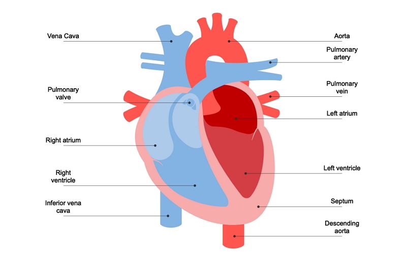
What is the function of the heart?
A muscular pump that contracts rhythmically to circulate blood throughout the body.
It beats involuntarily, about 70 times per minute (1,000,000 beats per day), moving 12,000 liters of blood.
What is a heartbeat?
The heartbeat is the sound made by the heart as it pumps blood, heard as a two-stage sound (dum-dum).
It consists of an initial low-pressure sound (AV valves closing) and a second high-pressure sound (arterial valves closing).
How is a heartbeat formed?
The heart beats involuntarily due to electrical impulses from the pacemaker in the right atrium wall.
Fibrillations help control the rate of breathing.
Each ventricular contraction produces a wave of blood flow.
How does blood supply get to the heart?
Blood supply to the heart is through coronary circulation.
Cardiac blood vessels branch from the aorta and spread over the heart wall (myocardium).
The heart needs a rich blood supply to meet its high oxygen demands, especially during exercise.
It extracts more than 75% of the oxygen delivered to it.
Coronary circulation accounts for about 10% of the total blood volume from the left ventricle during exercise, compared to 3% at rest.
What is the structure of arteries?
Thick, strong, elastic walls
Smooth muscles to handle high pressure
Valves to prevent backflow
Branch into smaller arteries, arterioles, and capillaries
What is the function of arteries?
Carry blood away from the heart
Blood from the left ventricle travels through the aorta to the body
Blood from the right ventricle goes through the pulmonary artery to the lungs for oxygenation
What is the structure of capillaries?
Smallest of all blood vessels
Extremely thin walls, consisting of a single layer of flattened cells
What is the function of capillaries?
Exchange oxygen and nutrients for waste
Link between arterioles and veins
Rejoin to form tiny veins called venules
Dense network in active tissues (e.g., muscle, brain) for extensive material exchange
Large surface area facilitates exchange with interstitial fluid
Blood pressure helps force fluid out; carbon dioxide and wastes return to capillaries
What is capillary exchange?
Oxygen, nutrients, and hormones diffuse from blood into interstitial fluid and cells
Carbon dioxide and cell wastes diffuse from cells into capillaries
What is the structure of veins?
Collect deoxygenated blood from venules and return it to the right atrium
Thinner walls than arteries, allowing for more flexibility
Valves at regular intervals prevent backflow of blood
What is the function of veins?
Carry deoxygenated blood from body tissues back to the heart
Blood flow mainly against gravity, assisted by gravity in veins above the heart
Pressure changes from heart pumping and rhythmic muscle contractions (muscle pump) help draw blood into veins
Adjacent arterial pressure surges push against veins to assist blood flow
What is blood pooling?
Blood pooling can occur if exercise stops abruptly or standing still for long periods, causing possible fainting
Cool-down period after exercise is important to gradually lower heart rate and prevent blood pooling
What is pulmonary circulation?
Carries deoxygenated blood from the right side of the heart to the lungs
Blood picks up oxygen and releases carbon dioxide in the lungs
Oxygenated blood returns to the left side of the heart

What is systemic circulation?
Carries oxygenated blood from the left side of the heart to the rest of the body
Delivers oxygen and nutrients to tissues and collects waste products
Deoxygenated blood returns to the right side of the heart

What is blood pressure?
The force exerted by blood on the walls of blood vessels
What are the phases of blood pressure?
Systolic Pressure:
Pressure in the arteries when the heart pumps (during contraction)
Diastolic Pressure:
Pressure in the arteries when the heart fills with blood (during relaxation)
What are the determining factors for the level of blood pressure?
Cardiac Output:
Increased output leads to higher blood pressure
Volume of Blood in Circulation:
Increased blood volume raises blood pressure (e.g., water retention)
Blood pressure falls during hemorrhage
Resistance to Blood Flow:
Increased viscosity or narrower vessels raise resistance and blood pressure
Elasticity of arterial walls helps maintain blood flow; decreased elasticity (e.g., due to deposits) increases resistance
Venous Return:
Affects cardiac output, which in turn affects blood pressure
What are the measurements and normal ranges of blood pressure?
Recorded by: Sphygmomanometer
Measured in: mm Hg
Expression: Systolic pressure/Diastolic pressure
Normal Ranges:
Systolic Pressure: 100-130 mm Hg
Diastolic Pressure: 60-80 mm Hg
How can blood pressure vary?
Blood pressure can vary with posture, breathing, emotion, exercise, and sleep
Can rise with excitement, stress, or physical exertion but typically normalizes with rest
What is motion used to describe?
Movement and path of a body (e.g. person or object)
Linear Motion
Occurs when the human body, human limb or an object propelled by a human moves in the same direction at the same speed over the same distance
Examples |
|---|
Vertical jump |
Downhill skiing (crouching position) |
Swimming (torso) |
Cycling |
Sprinting (torso) |
Angular Motion
This type of motion occurs when the human body, human limb or an object propelled by a human moves in a circular path about some fixed point at the same time, in the same direction and at the same angle
Examples (rotation on an axis) |
|---|
arm and legs in sprinting |
Somersault (diving/gymnastic) |
Golf swing |
Figure skating (doing a triple quad) |
General Motion
This type of motion is a combination of both linear and angular motion. Most sport movements are generally general motion.
Examples |
|---|
Whole movement of sprinting |
Run up in fast bowling |
Rowing |
Distance & Displacement ez
Distance - The length of the path through which a body travels.
Displacement - measured by the length of a line drawn between the starting and finishing points. (‘As the crow flies’.)
● If you are running a 4km cross country then your distance is 4 km, however your displacement will be different as it will draw a straight line from start to finish (if start and finish are different)
Speed and Velocity
Speed - describes only the magnitude of the speed of the body (i.e. how quickly the body is moving). Speed = distance/time
Velocity - measures the displacement of the body and is divided by the time taken to get from point A to point B. It measures both magnitude and direction. (10m east) Velocity = displacement/time
● Speed and velocity are only the same when the object moves in a straight line
● We tend to use speed and velocity as the same.
Instantaneous speed/velocity
done by calculating the average speed of a body over a short distance
What is the advantage of using instantaneous speed over average speed?
It shows the speed at different times of the race so you are able to see how fast the body is moving in segments during the race. Determine where we are strong and weak and also depicts opponent’s strengths and weaknesses.
Acceleration
Is the rate at which velocity changes over a period of time. Athletes need to be able to increase and decrease their velocity rapidly
● In an 100m sprint the acceleration increases until the end of the race. Acceleration = change in velocity/time
● An increase in velocity is called acceleration
● Zero acceleration tells us that velocity is not changing.
● Negative acceleration is when a body is moving forward with positive velocity but their acceleration is decreasing
Momentum
Is the ‘quantity of motion’. Calculated by: Momentum =Mass x Velocity. The main variable that changes in momentum is velocity.
e.g. 1kg discus x 20m per second= 20 kg/m/s
1.5kg discus x 20 m per second= 30 kg/m/s = therefore the 1.5 kg discus will have more momentum
Angular velocity/momentum
bringing mass towards the point to the axis
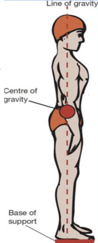
Centre of Gravity
Centre of Gravity (or COG) is the point around which a body’s weight is equally balanced, no matter how the body is positioned. In most spherical objects (e.g. a soccer ball), the COG is found in the centre of the object. In the human body, the position of the COG depends upon how the body parts are arranged (that is, the position of the arms and legs relative to the trunk). Because the human body is flexible and can assume a variety of positions, the location of the centre of gravity can vary.
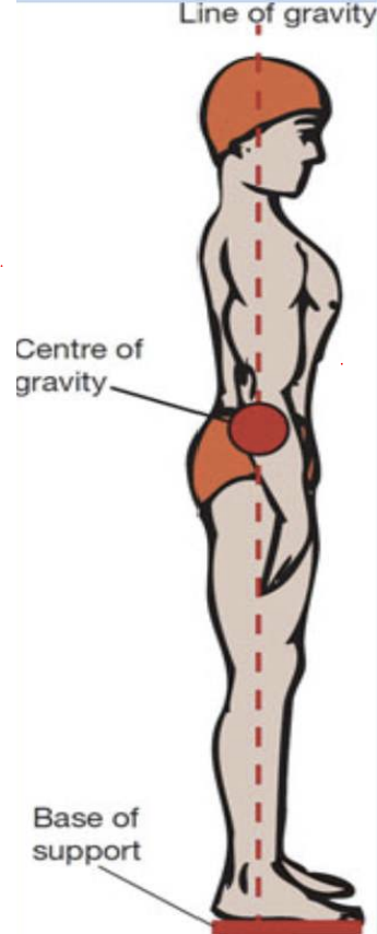
Line of Gravity
The line of gravity is an imaginary vertical line passing through the centre of gravity and extending to the ground. It indicates the direction that gravity is acting on the body. When we are standing upright, the line of gravity dissects the centre of gravity so that we are perfectly balanced over our base of support.
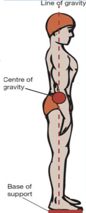
Base of support
Is an imaginary area that surrounds the outside edge of the body when it is in contact with a surface.
● A narrow base of support allows the COG to fall close to the edge of the base of support (therefore less stable)
● A wide base of support is essential for stability because the COG is located well within the boundaries
Our base of support has a limited area. Widening our stance increases the size of the base of support. However, rules of some sports and competitions limit the size of the base of support; for example, the starting blocks in athletics.
How does the body absorb force?
- Absorbed through joints, which bend or flex in response to the impact
- When landing you exert force on the surface and the surface exerts force on
the body
- Absorb force over a longer period of time
- Absorb force over a larger surface area
Applying force to an object
Quality of force applied to the object
The greater the force, the greater the acceleration of the object
Mass of an object is increased; more force is needed to move the object the same distance
Objects of greater mass require more force to move them than objects of smaller mass
How does the body apply internal force?
those that develop with the body; the contraction of a muscle group
causing a joint angle to decrease.
How does the body apply applied forces?
generated by muscles working on joints
How does the body apply external force?
Come from outside the body and act on it in one way or another
Centripetal force
is a force directed towards the centre of a rotating body
Centrifugal force
is a force directed away from the centre of a rotating body.
● Greater speed about the axis, the greater the force produced
How can an athlete use base of support to their advantage (think of examples)
● The dancer performing a pirouette has a very narrow base of support and must work hard to ensure that their centre of gravity remains within the base.
● Wrestlers widen their base of support to prevent their opponents from moving them into a disadvantageous position.
● Tennis players lower the centre of gravity and widen the base of support in preparation to receive a fast serve. This enhances balance and enables the centre of gravity to be moved in the desired direction more readily.
● Golfers spread their feet to at least the width of their shoulders to enhance balance when they rotate their body during the swing
stability and balance
Stability is the resistance of a body to change in its equilibrium; that is, change in its linear or angular acceleration. When an individual can assume a stable position and then control that position, he or she is said to be in a state of balance.
Static balance
the ability to maintain equilibrium while the body is stationary