Anatomy test #4... prayers prayers
1/70
There's no tags or description
Looks like no tags are added yet.
Name | Mastery | Learn | Test | Matching | Spaced | Call with Kai |
|---|
No analytics yet
Send a link to your students to track their progress
71 Terms
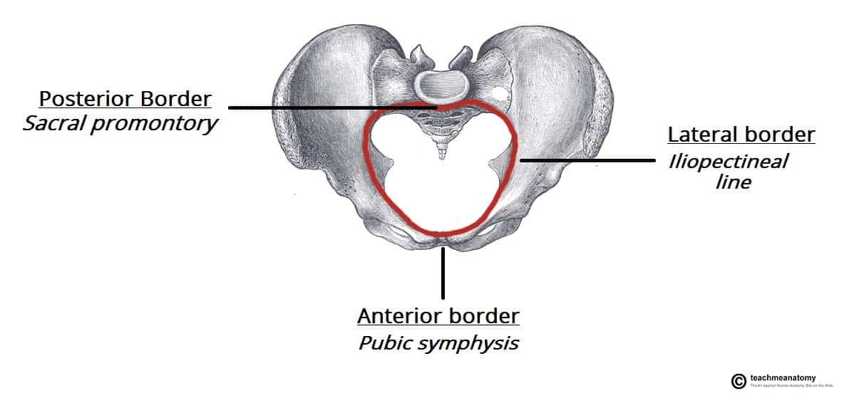
What are the borders of the pelvic region
Supierior = pelvic inlet
inferior = pelvic outlet/ pelvic diaphrgm
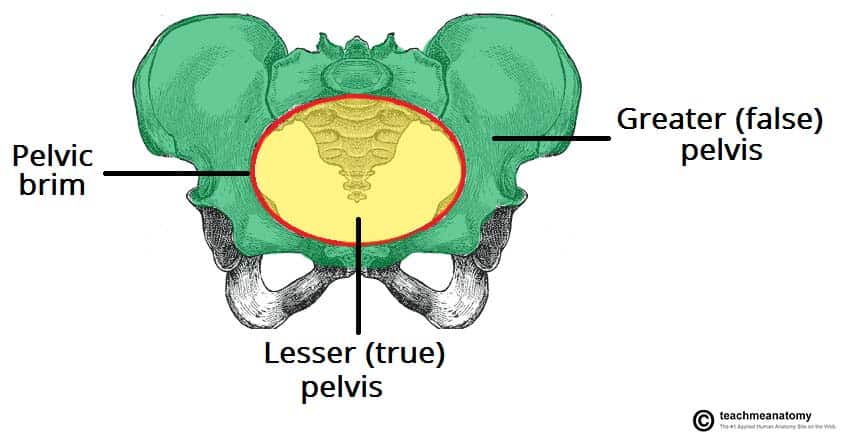
What are the regions in the pelvise bone area (in gen)
pelvic brim
greater false pelvis
inferior lesser pelvis
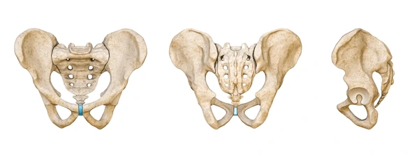
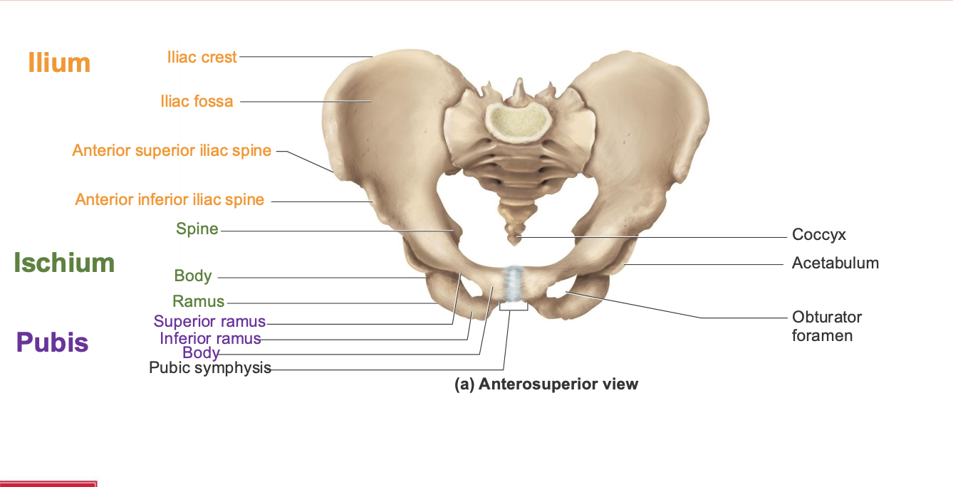
What are the 3 sections of the pelvis and what bones are found in there + what other structures can be found (anteriror side)
Ilium
Iliac crest
Iliac fossa
Anterior superior iliac spine
Anterior inferior iliac spine
Ishium
Spine
Body ramus
Pubis
Superior ramus
Inferior ramus
Body
Pubic symphysis
Coccyx
Acetabulum
Obturator foramen

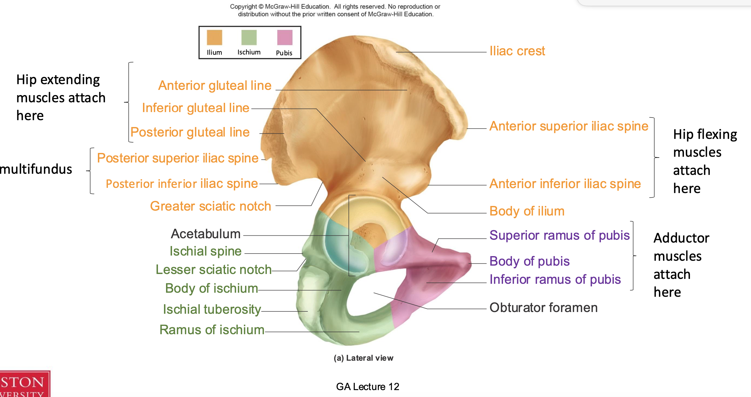
Waht are the components of the lateral pelvis labled:
Anteiror gluteal line
Inferior gluteal line
Posterior gluteal line
Minifundus
Posterior superior iliac spine
Posterior inferior iliac spine
Greater sciatic notch
Hip flexor muscle attachments
Anteiror superior iliac spine
Anterriror inferior iliac spine
Body of ilium
Iliac crest
Ishial spine
Lesser sciatic notch
Body of ishium
Ishial tuberosity
Ramus of ishium
Adductor muscles attach here
Superiror ramus of pubis
Body of pubis
Inferior ramus of pubis
Obturator foramen
Ilium
Hip extending muscle attachments of the ilium
Anteiror gluteal line
Inferior gluteal line
Posterior gluteal line
Minifundus
Posterior superior iliac spine
Posterior inferior iliac spine
Greater sciatic notch
Hip flexor muscle attachments
Anteiror superior iliac spine
Anterriror inferior iliac spine
Body of ilium
Iliac crest
Ishium
Ishial spine
Lesser sciatic notch
Body of ishium
Ishial tuberosity
Ramus of ishium
Pubis
Adductor muscles attach here
Superiror ramus of pubis
Body of pubis
Inferior ramus of pubis
Obturator foramen
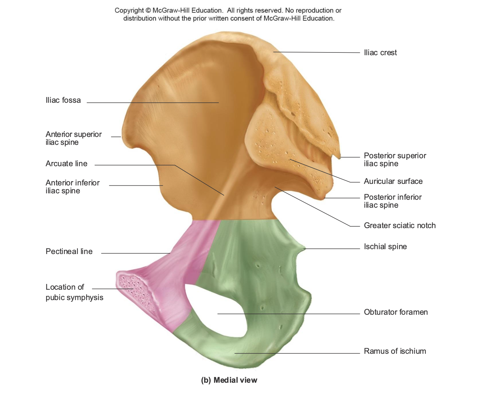
What are the components of the medial spine. label the diagram
Iliac fossa
Iliac crest
Anterior superior iliac spine
Arculate line
Anterior inferior iliac spine
Posterior superior riliac spine
Auriticular surface
Psteriror inferior iliac spine
Greater sciatic notch
Ischilal spine
Ramus of ischum
Pectineal line
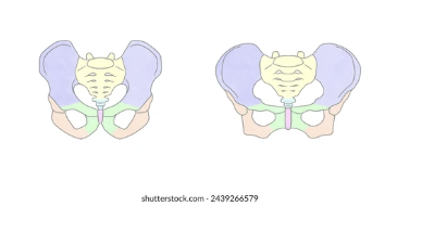
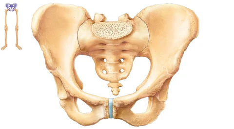
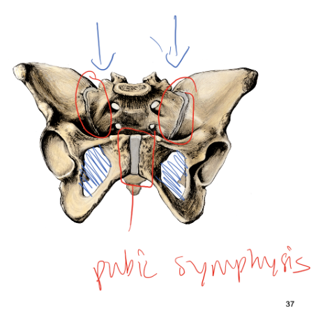
Location of pubic symphasis
Ilium
Iliac fossa
Iliac crest
Anterior superior iliac spine
Arculate line
Anterior inferior iliac spine
Posterior superior riliac spine
Auriticular surface
Psteriror inferior iliac spine
Greater sciatic notch
Ishcium
Ischilal spine
Ramus of ischum
Pubis
Pectineal line
Location of pubic symphasis
What is the purpose of the bowl like surface area of the pelvis
To support the orgnas for bipedalism
provides lateral space for gluteus maximus and medius
What hapens with higher levels of estrogen in the pelvis
resutls in a wider pelvis radius for birthings
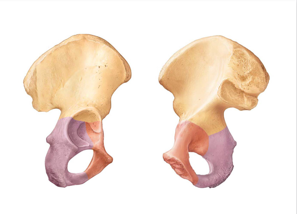
What are the components of the acetabulum (label the diagram
Illium on superior
Ishium and pubis
What are the 3 major parts of the pelvis
Ilium
Ischium
Pubis
What are the major joints of the pelvis and where do they articulate
Os coxae meet anteirorly at the pubic symphysis
Os coxae articulate posteirorly with sacrum
Oburator fromamen, obisturator membrane
hylain cartilage cover the ends of the pubic bones
fibrocartilagnous disc makes up the pubic symphysiss
sacroiliac joint = hylain cartilage

What are the different major joints in the pelvis: what are the different sacroiliac joints. What are the other joints Hint there are 4 - 6
Sacroiliac
Anteriror and posterior sacroiliac ligaments
Attach to the ilium and sacrum laterally
iliolumbar ligament
superiro/ inferior attachment between ilia and lumbar region
Sacrospinous
attaches sacrum to
ischial spine
Sacrotuberous
attaches sacrum to
ischial tuberosities
Regular joints
Anterior and posterior sacroccoygeal ligaments attach to the sacrum and coccyx
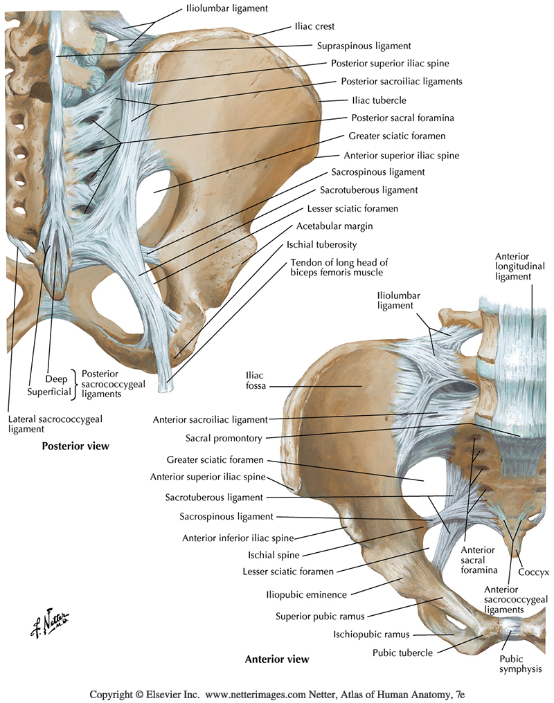
Which joints are the most stable in the pe
The sacroiliac joints
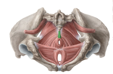
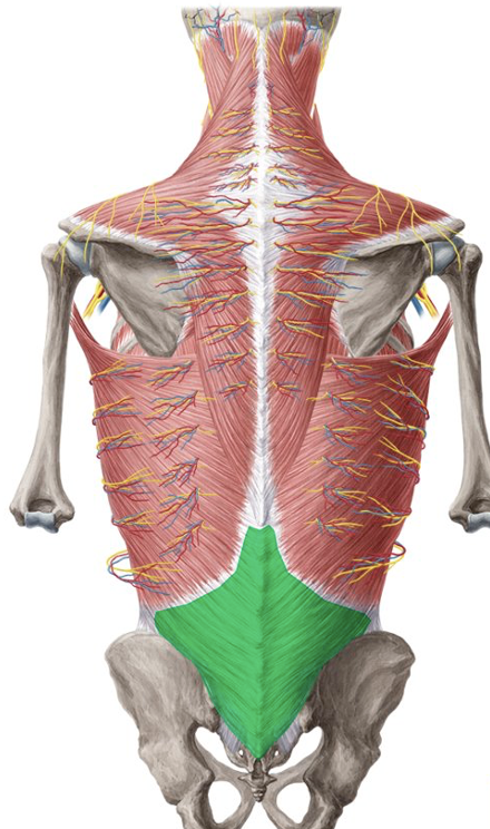
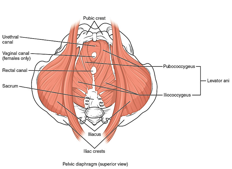
What are the different pelvic floor muscles and label ethem on the diagram. What do they do:
Ureatrhal canal
Vaginal canal
Rectal canal
Puboccoygeus
Iliococcygeus
Psoas
iliacus
Ureatrhal canal
Vaginal canal
Rectal canal
Puboccoygeus
Iliococcygeus
Psoas
iliacus
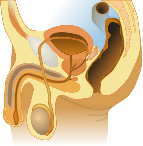
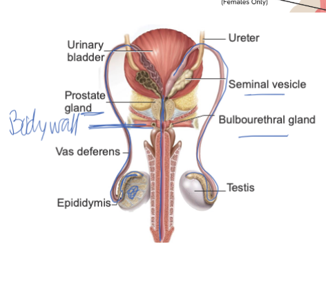
What organs are apart of the sperm path and describe their use and order
Testis: Produces sperm and testosterone.
Epididymis: Stores sperm and allows them to mature.
Vas Deferens: Transports mature sperm to the urethra during ejaculation.
Seminal Vesicle: Adds fructose-rich fluid that nourishes sperm (major part of semen).
Prostate Gland: Adds alkaline fluid that protects sperm from vaginal acidity.
Bulbourethral (Cowper’s) Gland: Releases pre-ejaculate to lubricate and neutralize the urethra.
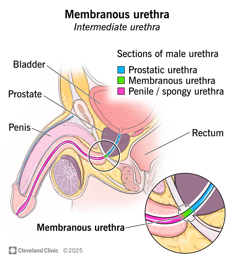
What are the 3 sections of the penis uretra
Prostatic ureathra
Extends throught the prostate gland
Membraneous urethra
External urethral sphincter
Spongy urethra
Encased with erectile tissue
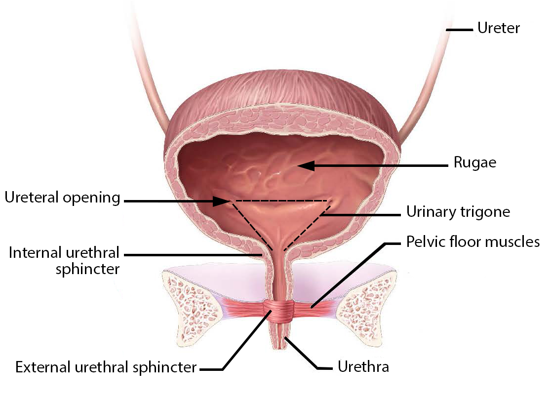
What are the parts ofthe female uritrha and why are women more prone to UTIS
Internal urethral sphinxer
External urethral sphinxer
Trigone
Stretch receptors to urge urination
bc the tract is so short women are more prone to utis thenmen
What are the layers to the uteral wall and lining
Endometrium: Inner lining of the uterus; thickens for possible pregnancy and sheds during menstruation.
Myometrium: Middle, muscular layer of the uterus; contracts during labor and menstruation.
Perimetrium: Outer protective layer of the uterus.
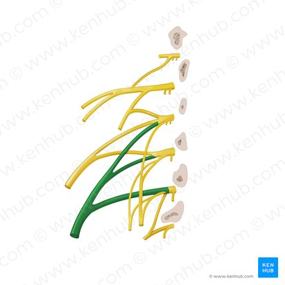
Draw out the lumbrosacral plexis and what are the functions of each muscle nerve (prayers)… i feel like you should work on the pats of the pelvis and lumbral section first then move onto the other info:
Iliohypogastric
Ilonguinal
Genitofemoral nerves
Lat. Fem. Cut n
Femoral
Obturator
Sciatic
pudental (S2-4)
Lumbar plexus L1-4
Innervates the
Abdominal wall muscles
Anterior thigh muscles
Medial thigh muscles
Components
Iliohypogastric (T12-L1)
Innervates internal oblique and transversus abdominus
Sensory innervation inpubic region
Ilonguinal (L1)
Innervates: internal oblique and transversus abdominus
Sensory innervation to pubic region
Genitofemoral nerves (L1-L2)
Innervates: genitalia and upper anteiror thigh
Lat. Fem. Cut n (L2-L3)
Sensory (only) for the skin of the anteiro lateral thigh
Femoral (L2-L4)
Muscle inneravation to hip flexors. Knee extensors (anteriror component)
Sensory innervation to anterior thigh and medial leg
Obturator (L2-4)
Innervates the adductor muscles and the sun over the medial thigh
Sacral plexus
Sciatic (l4, 5, S1-3) (largest nerve in teh body)
Innerates teh posterior thigh muscles
All leg and foto muscles
All joints in the lower limb
Mde of the common fibular and timbial nerves joind with connective tissue
pudental (S2-4)
Main nerve of the perinumen and genetalia
Exis the pelvis posterirorly through the gsf and reithers anterilory throught he lesser sciatica foramen
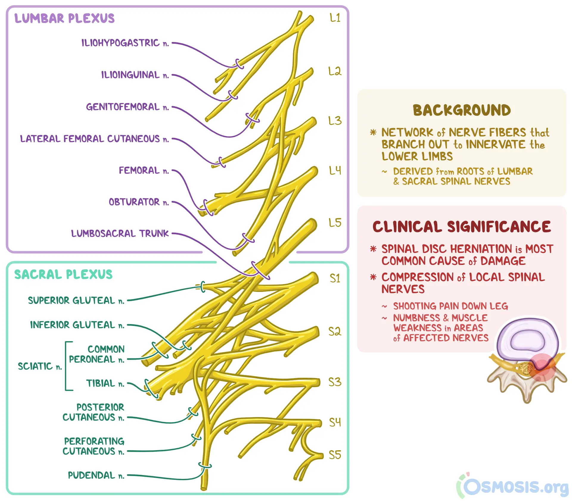
Where do most muscles of the sacral plexis pass through
the greater sciatica foramen
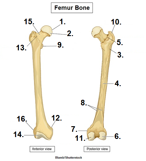
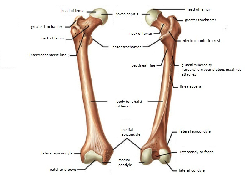
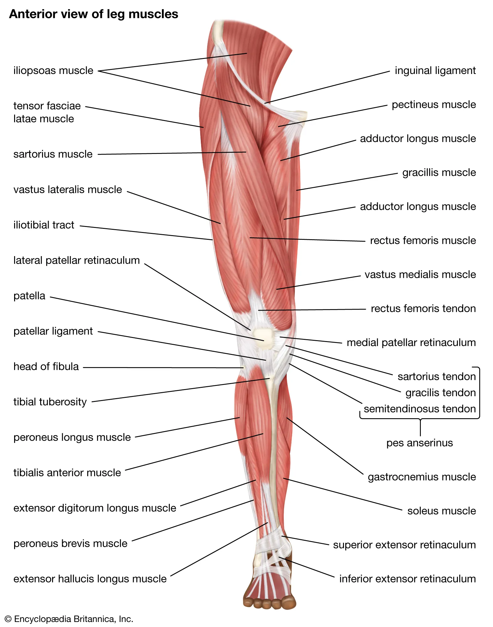
What are the components of the femur bone.. be able to label based on the diagram
Greater tochanter,
Head
Intertrochatic crest
Gutleal tuberosity
Lesser trochanter
Linea aspera
Lateral condile
Medial condile
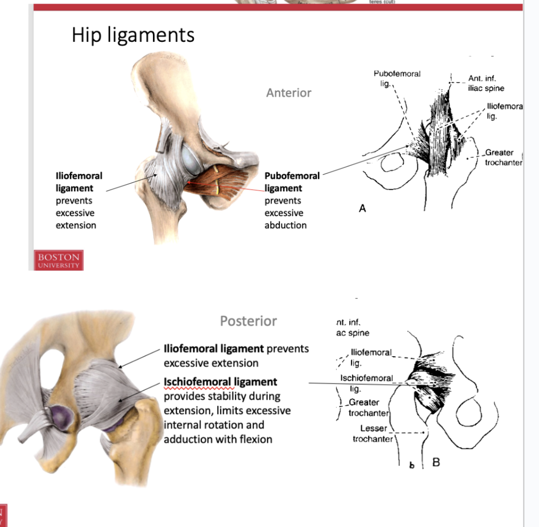
Why are hip joints so much more stable than other joints. What is the purpose of the acetabular labrum
a ring of fibrocartilage that surrounds the hip socket, called the acetabulum. It acts as a gasket to create a suction seal, which increases the stability of the hip joint
What factors increase the stability of the hip. What ligaments are invovled
Shape of the articulating surfaces
Acetabular labrum
Joint capsle
3 ligaments
Iliofemoral
Pubofemora
Ischiofemoral
Ligamentum teries
Transverse acetabular ligament
What are the functions of the ligaments in the hip. What are they and what are their functions
Anterior
iliofemoral (also posterior)
prevents excesive extension
pubofemoral
precents excessive adduction
posterior
ischiofemoral
provides stability during etension and limits excessive intrnal rotation and adduction with flexion
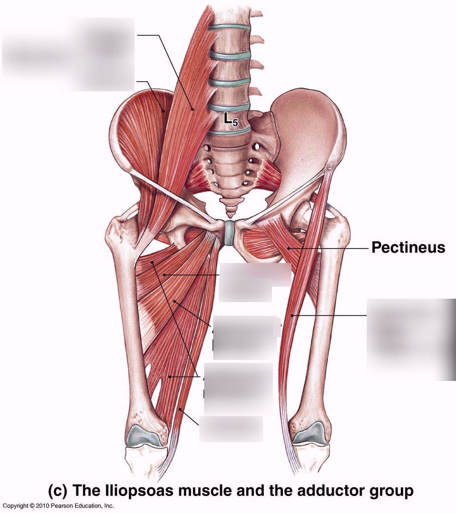
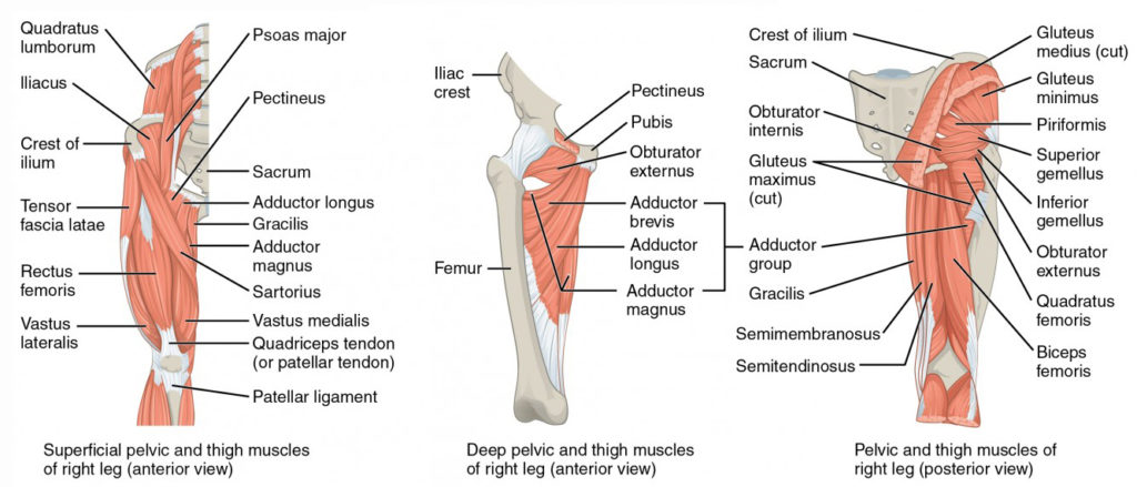
What thigh muscles medial rotate the hip hint there are 9
Ilopsoas
Sartorius
Tensor fasciae latae and the IT band
Pectianius
Adductor longus, brevus, magnus
Gracilis
Gluteal medius, minimus, maiumusWhat
Piriforms, inferior and superior gemelus, obturator internus
Obuturator externus, quadrucepts femorus
What are the hip flexor muscels of the hip
Illiopsoas
Sartorius
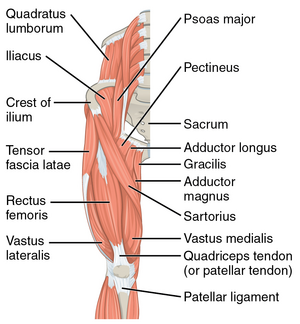
What are the adductors of the hip
Dductor longgus, brevis, magnus
Gracillis
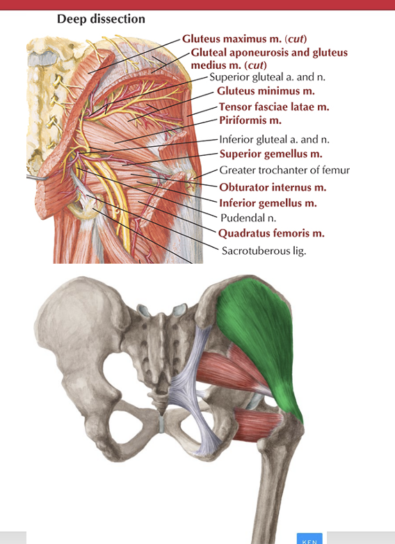
What are the externors of the hip
Gluteus medius, minims, maximus
what are the lateral rotators of the hip
Piriformus, inferior and superior gemelllus, obturater internus, obturator externus, quadracepts femorsi, gluetus maximus
Waht is the origin, insertion, and innervation of the tensor facia latae
Origin: Anterior iliac crest
Insertion: Lateral condyle of tibia via IT band
Action: Stabilizes knee, flexes hip, abducts hip, internally rotates hip
Nerve: superior gluteal nerve
What is the origin and insertion of thegluteus minimus and medius (hip abductors)
Gluteus minimus
Origin: Inferior posterior ilium
Insertion: Anterior surface of greater trochanter of femur
Action: Abducts hip, internally rotates hip
Nerve: superior gluteal nerve
Gluteus Medius
Origin: Middle posterior illium
Insertion: Greater trochanter of femur
Action: Abducts hip, Internally rotates hip
Nerve: superior gluteal nerve
What makes hip stabilizers hip stabilizers
they run along the lateral and posteiror sides of the hip
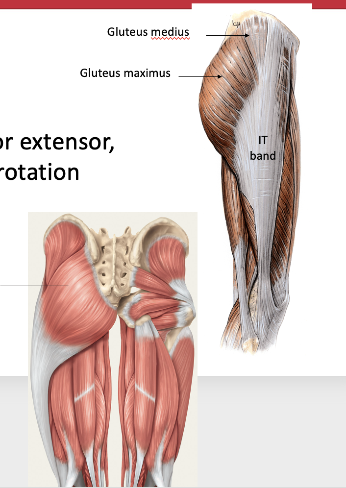
What is the origin, sinertion, and nerve innervation, and obv actino of the gluteus maximus
Gluteus Maximus
Origin: Posterior sacrum and sacrotuberous ligament, superior posterior ilium
Insertion: Gluteal tuberosity of femur, lateral condyle of tibia via IT band
Action: Extension of hip, external rotation of the hip
Nerve: inferior gluteal nerve
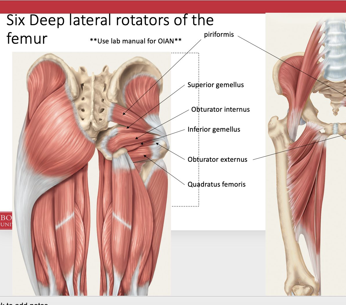
What are teh 6 deep lateral roaters of the femur
Piriforms
Superior gemellus
Obutrator internus
Inferior gemellus
Obturarter externus
Quaduratus femoris
Label all of the muscles of the hip joint and thier useage
What are the hip extensors
Gluteus maxiumus
Biceps femoris (hamstrings)
Semitendinosis
Semimembranosis (deep to the semitendinosisi
What is the origin and insertion of the iliopsoas (iliacus and psoas)
Iliacus
Origin: Iliac fossa
Insertion: Lesser trochanter of femur
Action: flexes hip, flexes lumbar vertebral column
Nerve: Femoral nerve
Psoas major
Origin: T12-L5 vertebral bodies and discs, L1-5 transverse processes
Insertion: Lesser trochanter of femur
Action: flexes hip, flexes lumbar vertebral column
Nerve: Anterior rami – spinal nerves L2-3
What is the origin and insertion of the sartorius
Origin: Anterior superior iliac spine
Insertion: Pes anserinus
Action: flexes hip, abducts hip, externally rotates hip, flexes hip, flexes knee, internally rotates knee
Nerve: Femoral nerve
What makes a hip flrxor a hip flexor
if they pass over the hipjoint on the anterior side
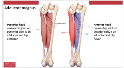
What is the difference between the posterior head and anterior head of the abductor magnus
Posterior head
Crosses the hip joint on the posterior side and is an addu tor an dhip extnsor
Anteirro head
Crosses the hip joint on the anterior side and is an adductor and hip flexor
What is the difference and use of the Q angle
The q angle is larger with a wider pelvis due to more estrogen and stuff
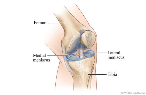
What are teh unique properties of the knee. What is the purpose of the menisci
It is weight bearing ands hock absorbing.
They fibrocartilage (menisci) to help cushion
They also dissipate forces and increase the congruency of the femur and tibia
The femur has rounded ends and teh tibia is flat.. = increase congruency of the joints
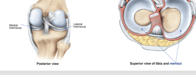
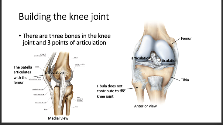
What are the 3 joints of the knee and their articulation points
Femur, tibia and fibula (note teh fibula doesnt contribute to knee joint
The patella articulates with the femur
Articulation of the femur and the tiba
What type of joing capsule is the knee joint
it is a synovial joint with a synovial membrane lining the inside of the fibrous couple
subdivided joints pace by lining intra-articular ligaments
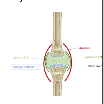
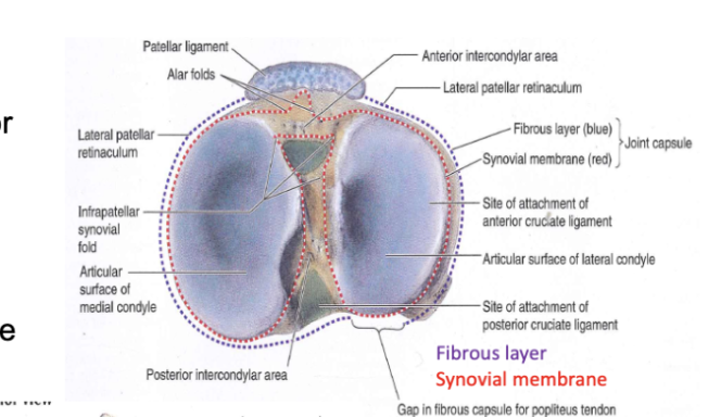
Where is the fibrous layer and synovial membrane and what is the purpose ofthe fibrouse casplul gap
Fibrous layer is the outer part surrounding the knee
Inner layer is the synovial membrane
Gap allows tendons to go through
What is the fibrous layer of the joint capsle. Where is it fully formed and not fully formed
The fibrous layer is complete in the posterior poplital region except where the popliteus passes through
Anteirrorly fibrous layer is incomplete . the quadricepts tendon, patella, and ptellear ligament replace the fibrous lay in the middle fo each knee
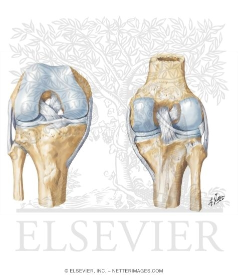
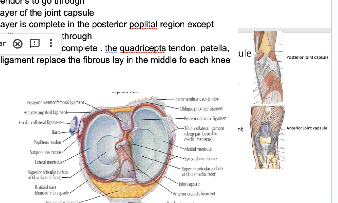
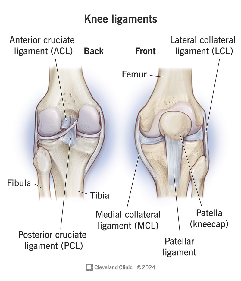
What are the 4 major ligaments of the knee and what differentiates them label them on the diagram. What is their use
Anterior cruciate ligaments (ACL)
Posterior cruciate ligament (PCL)
tibial/medial collateral ligament (TCL)
Fibular (lateral) colalterla ligament (FCL)
Collateral ligament = in parallel
Cruciate ligaments = cross shaped
ACL - prevents hyerextnesion and anterior shear of the tibia
Passes posteirrorly to anteriorly
pcl - prevents posterior shear of the tibia
Only found posteriorly
Collateral ligametns taught in extension but more important in slight flexion to increase stability
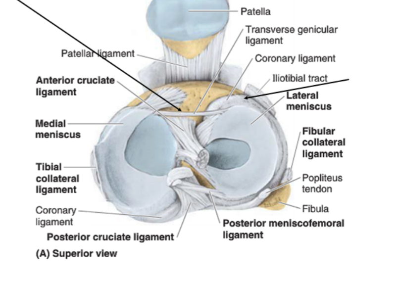
What are the following fuctures of these ligaments. what does it articulate to? what are their shapes
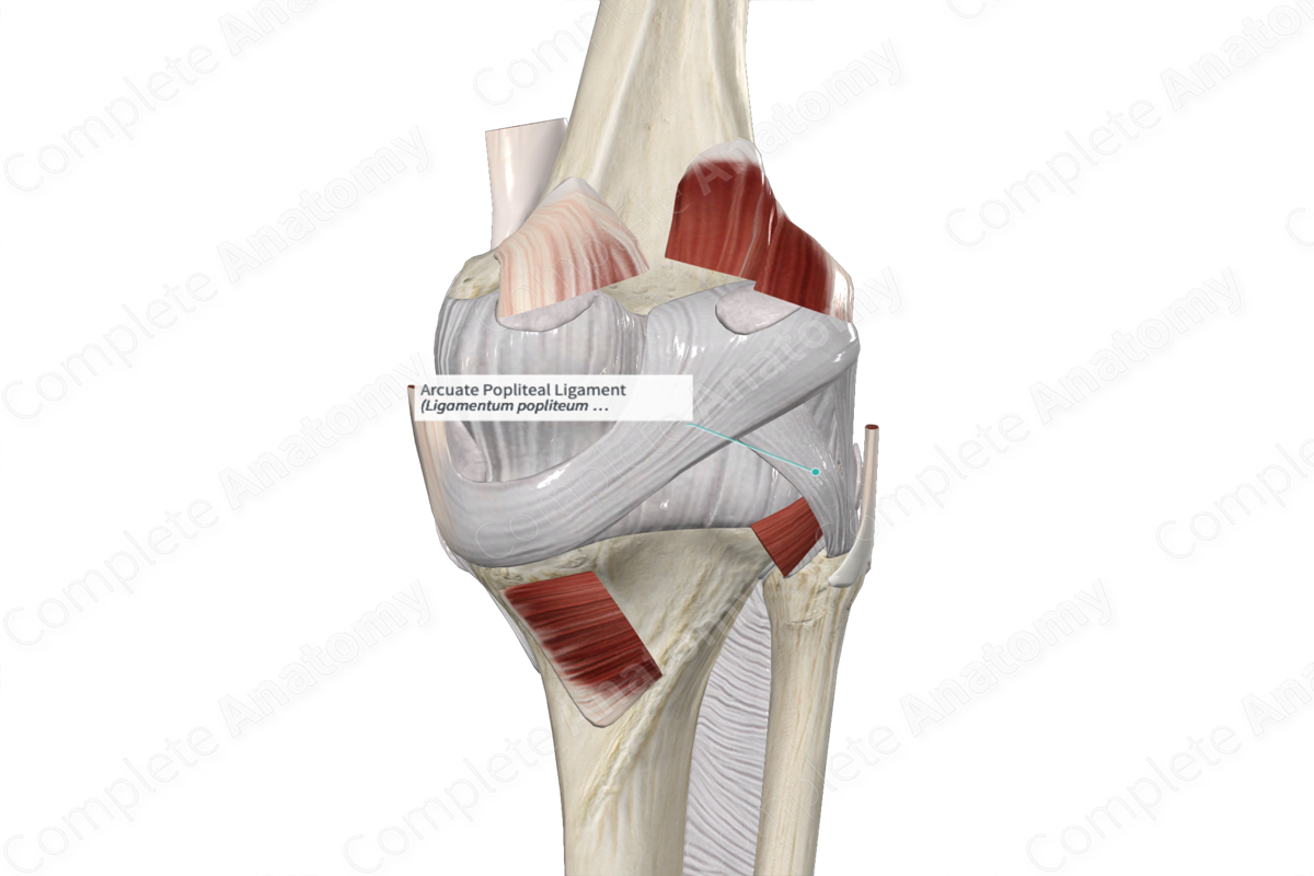
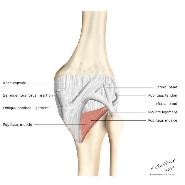
Oblique popliteal – From semimembranosus; reinforces posterior capsule; limits hyperextension.
Arcuate popliteal – From fibular head; supports posterolateral corner; resists varus + external rotation.
Coronary (meniscotibial) – Anchor menisci to tibia.
Transverse (intermeniscal) – Connects anterior horns of menisci; coordinates meniscal motion.
ALL (anterolateral) – Lateral femoral epicondyle → anterolateral tibia; controls internal tibial rotation / pivoting.
Patellar ligament – Patella → tibial tuberosity; knee extension; used in ACL grafts.Extension of the quadricepts tendon and the patella
Arcutate = shaped like a bow
Oblique = slanted
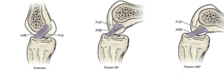
What are the function of the following ligaments during extension and flexion: anterirmedial band and the posterior lateral band
During extension, the bands are not fully taught . the plb is taught wile the amb is relaxed
During flexion the amb is taught witle the plb is relaxed ish
Further you go into flexion = the more taught the amb = more instability since the knee is less articulated with the
What are the degress of motion in the knee
Flexion/extension
internal/external rotation
how does rotation work in the knee. Full extion of internal rotation (work on this stuff0
Internal rotation = lateral plateau moves anteriroly, textenral rotation when the lateral plateau moves posteriorly
Because the medial articular surface of the tiba has more surface area, the tibia must externally rotate to reach full extension
To move from extension ot flexion the tibia must internally rotate
Full extension is where the joint is most stable, each femoral condyle is nestled within the meniscus
Why are the knees such a vulnerable joint
Much less surface area during flexion = makes it much weaker

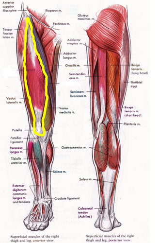
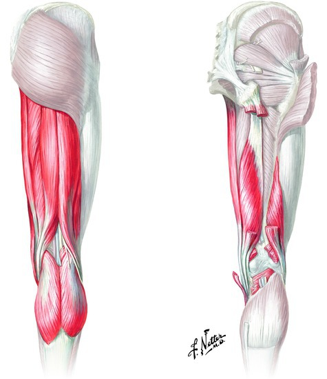
What are the extensors of the knee and flexors of the knee
Extesnors (nateriro )
Rectus femoris
Vastus lateralis
Vastus medialis
Vastus intermediaus
Flexors
Semimembranosus*
Biceps femoris*
Sartorius*
Popliteus*
Gastrocnemius
Plantaris (when present)
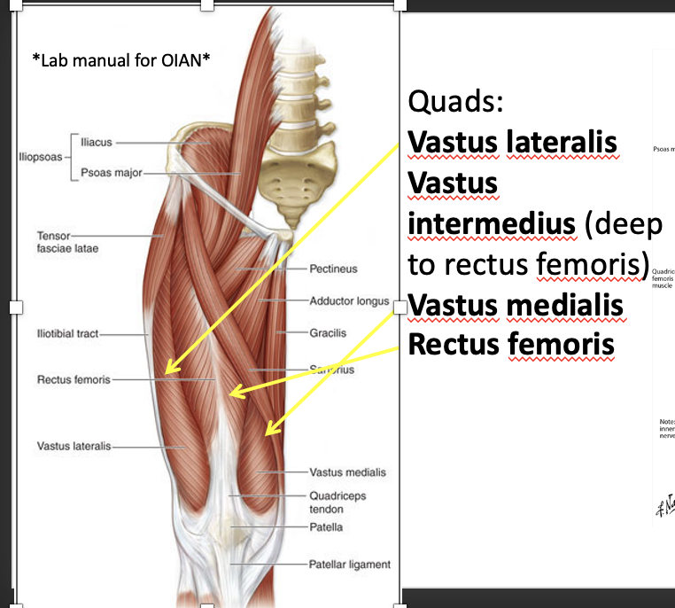
What are the muscles of the quads origin and insertion
Vastus laterallis, intermedius nmedalism and rectus femorsi. Know their locations aon a chrt
Rectus femoris = most superficial
Know their origin insertion and stuff
Rectus Femoris
Origin: AIIS + superior acetabular rim
Insertion: Tibial tuberosity (via patellar ligament)
Innervation: Femoral n. (L2–L4)
Function: Knee extension + hip flexion
Vastus Lateralis
Origin: Greater trochanter + lateral linea aspera
Insertion: Tibial tuberosity
Innervation: Femoral n. (L2–L4)
Function: Knee extension
Vastus Medialis
Origin: Intertrochanteric line + medial linea aspera
Insertion: Tibial tuberosity
Innervation: Femoral n. (L2–L4)
Function: Knee extension, stabilizes patella (VMO)
Vastus Intermedius
Origin: Anterior & lateral femoral shaft
Insertion: Tibial tuberosity
Innervation: Femoral n. (L2–L4)
Function: Knee extension
What is the origin, insertion, innervation, and function of the Gracilis
Origin: Body & inferior ramus of pubis
Insertion: Pes anserinus (medial proximal tibia)
Innervation: Obturator nerve (L2–L3)
Function:
Hip adduction
Knee flexion
Medial rotation of leg when knee is flexed (the tibia)
spands the knee joint

What is the origin insertion innervation and function of the semimembranoss
Origin
ischial tuberosisty
Insertion
postterir medial tibial condile
action
flexion of knee
internal rotation of flexed knee
extension of the hip
Nerve supply
tibial division of sciatic nerve
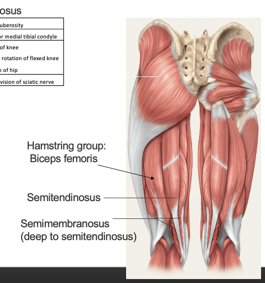

What is the origin insertion innervation and function of the semitendonosus
Origin
ischial tubersity
Insertion
pes anseinus
Action
flexion of the knee
internal rotaiton of flexed knee
extension of the hip
Nerve supply
tibial division of sciatic nerve
What is the origin insertion innervation and function of the bicepts femorus
Origin
long head - ischial tuberosity
short head- linea aspera
insertion
lateral surface of head of fibula
lateral condyle of tibia
action
both heads
flexion of knee
external rotation of flexed knee
long head
extension of the hip
Nerve supply
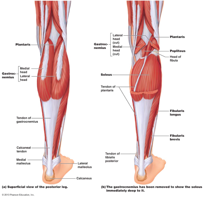
What are the lower leg msucles that act on the knee
Gastronemius - weak knee flexor when the ankle is not in plantarflexion
popliteus - internal rotaiton of the tibia to unlock the knee form a locked extended position
plantaris - absent in many people and a weak contribitor to knee flexion

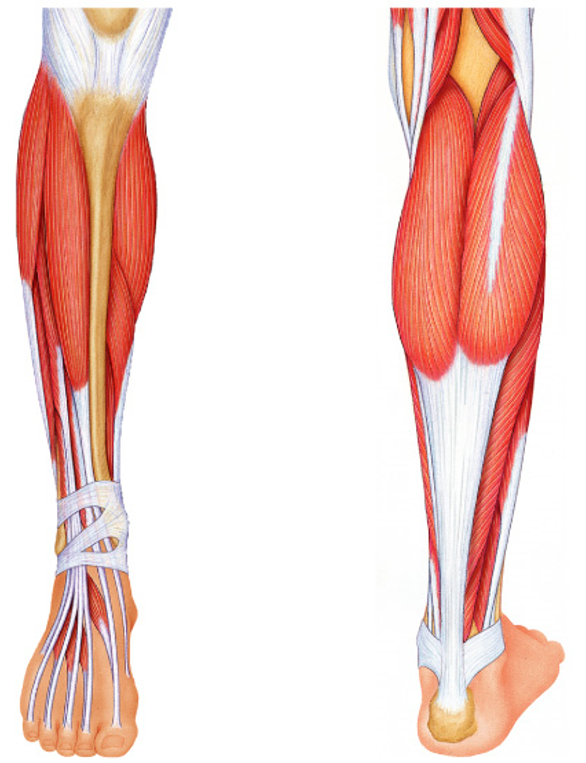
What is the origin, insertion, innervation, and action of the gastronemius
Origin
Medial heald - medial epicondile fo the femur
lateral head -alteral epicondile of the femur
Inserition
calcaneus via calcaneal tendon
Action
lantar flexion of ankle
flexion of knee
Nerve supply
tibial nerve


What is the origin, insertion, innervation, and action of the popliteus
Origin
lateral condyle of femur
Inserition
proximal postrior tibia
Action
unlocks extended knee by internal roation of tibia (normal)
or external rotation of the femur (reverse
Nerve supply
tibial nerve


What is the origin, insertion, innervation, and action of the gastronemius
Origin
lateral supracondyler rige of femur
Inserition
posteiror calcaneus
Action
plantar flexion ofth eankle
flexion of the knee
Nerve supply
tibial nerve
Abdominal muscle innervation. what are they innervated by and which sections
most antieror = intercostal nervees t7-12
lower fibers are innervated by 2 lumbar lexus nervs: iliohypogastric and ilioingunal
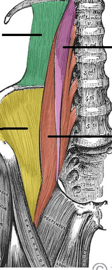
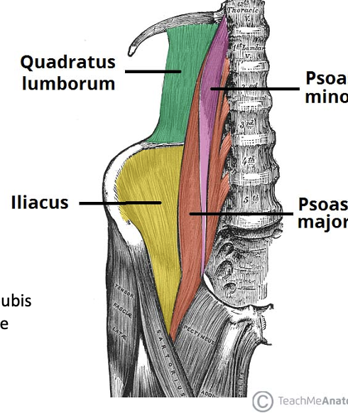
What are the muscles shown here, their origin, insertion, action, and inervation
Quadratus Lumborum
Origin: Iliac crest
Insertion: 12th rib, transverse process of L1-4
Action: Lateral flexion of spine, elevates hip
Innervation: Ventral rami T12-L4
Psoas Minor
Origin: Body of T12-L1
Insertion: Superior ramus of pubis
Action: Flexion of lumbar spine
Innervation: Spinal nerves L1
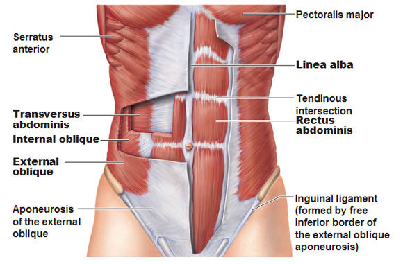
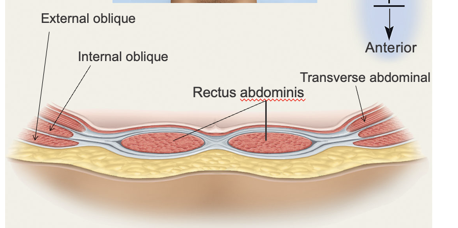
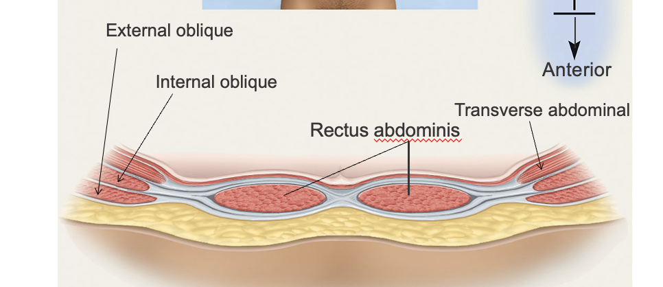
What are the 3 layers of the abdominal muscles and their order from deepest to most superficial and their actions. origin insertion and innervation
deepest
transverse abdominius - compresses the abdomen
abdominal aponeuros in the mindline
T 7-12, iliohypogastric & ilioinguinal n.
stability and immobility
intermediate
internal ovlique - flexion of spine, compress the abdomen, ipsilateral flexion and roation of the spine
also has an aponerosis in the midlene that forms the anterior rectus sheath (thhoracolumbar fasica
Internal Oblique
Origin: Inguinal Ligament, iliac crest, throracolumbar aponeurosis
Insertion: Costal cartilage of lower ribs, abdominal aponeurosis/linea alba
Action: flexion of spine, compresses abdomen, ipsilateral flexion and rotation of spine
Innervation: T 7-12, iliohypogastric and ilioinguinal
Superficial
Rectus abdominus- runs vertically oriented
Origin: Pubic symphysis and crest
Insertion: Costal cartilages of ribs 5-7, xiphoid process
Action: Flexion of spine, compression of abdomen
Innervation: T 7-12,
extenral oblique
originates on the ribs but not the thoracolumbar faica: inserst on th eiliac creset and aponeurosis
Origin: Lower 8 ribs
Insertion: Abdominal aponeurosis/linea alba, pubis, anterior iliac crest
Action: flexion of spine, compresses abdomen, ipsilateral flexion and contralateral rotation of spine
Innervation: T 7-12, iliohypogastric and ilioinguinal
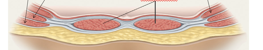
Cross section of the abdomen. which muscleis which point it out and hwat are the viewpoints
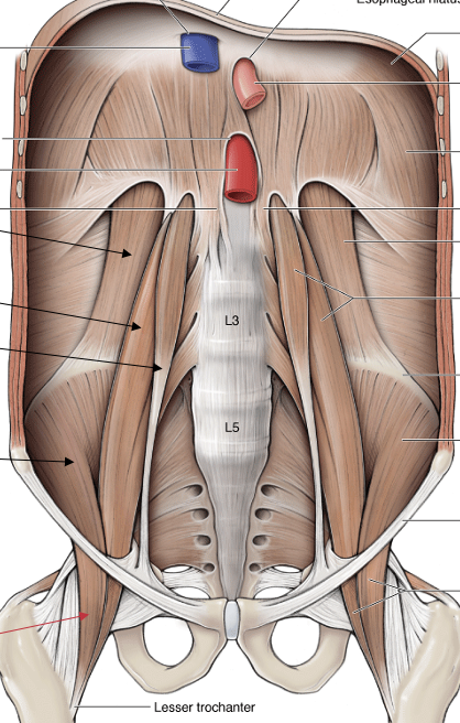
What are the 4 muscles of the posteriro abdomen. Where are they found and what do they do
•quadratus lumborum - stabilizes the spine and contributes to lateral flexion and extension
•psoas major
•psoas minor
(only present in some individuals)
•iliacus
Iliacus and psoas major both pass deep to the inguinal ligament and insert on the femur, they are hip flexors
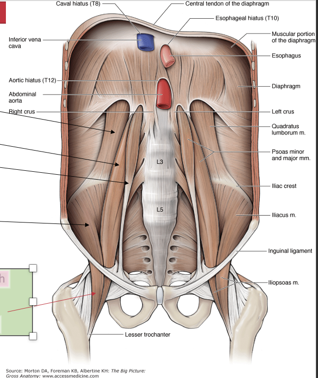
What is the thoracolumbar fascia
•Critical structural connection, connects force between trunk and leg- Origin of transverse abdominal and internal oblique, also latissimus dorsi
Separates into several layers
Wraps around quadratus lumborum