Looks like no one added any tags here yet for you.
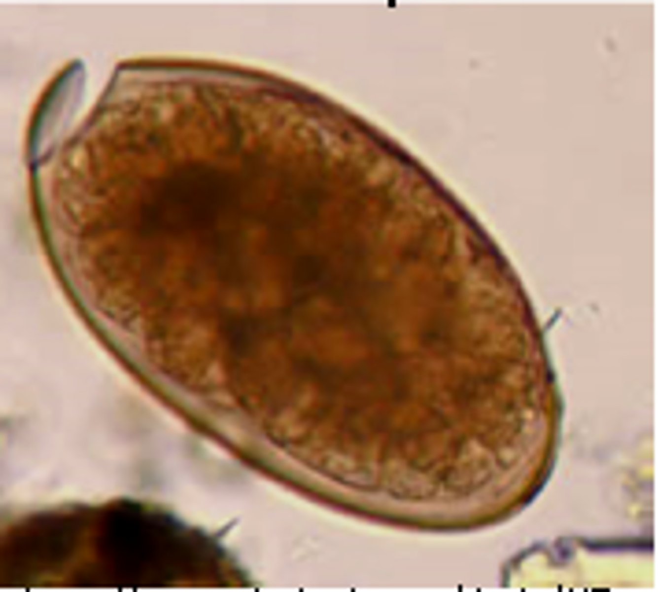
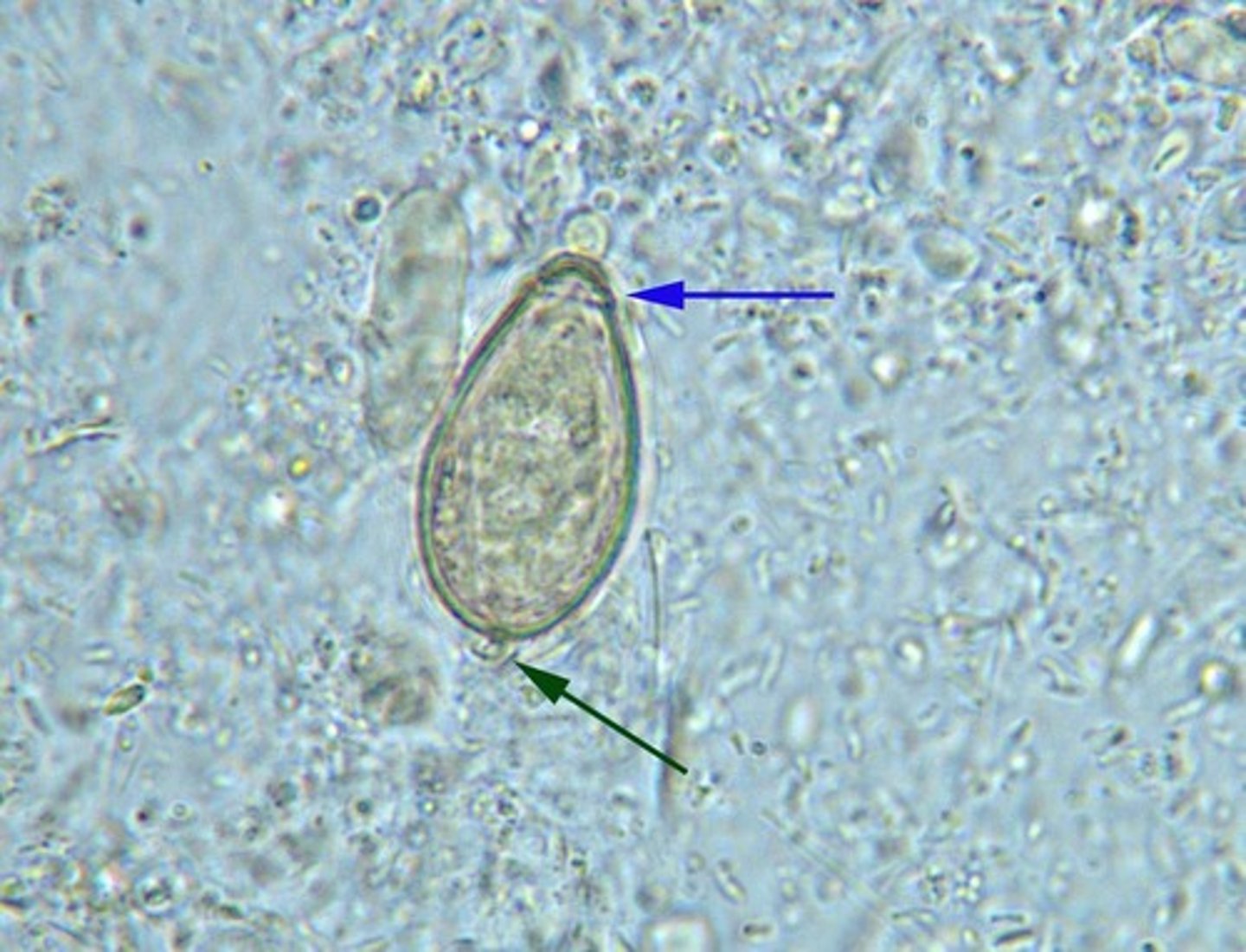
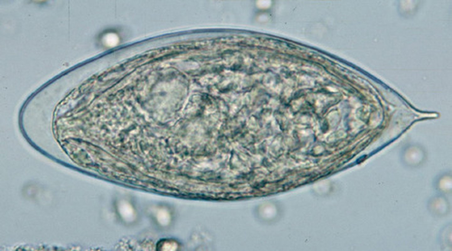
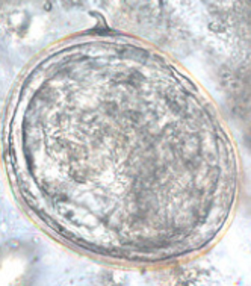
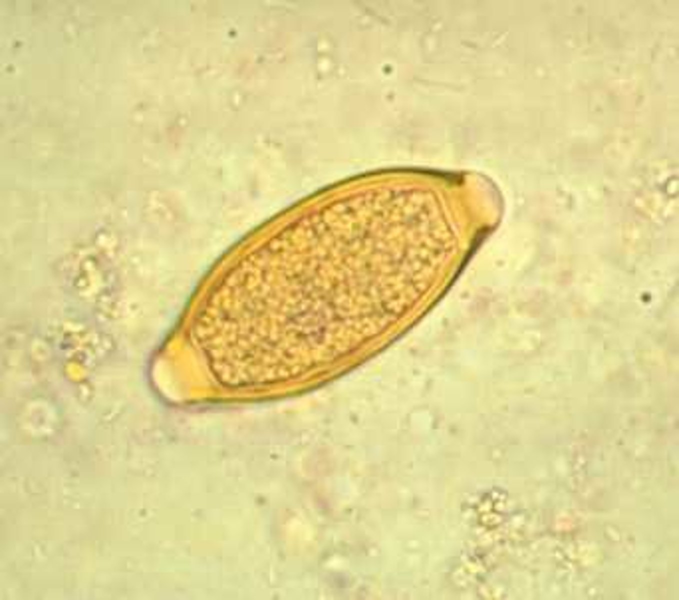
Fasciola hepatica (egg)
*operculum is falling off
Paragonimus westermani (egg)
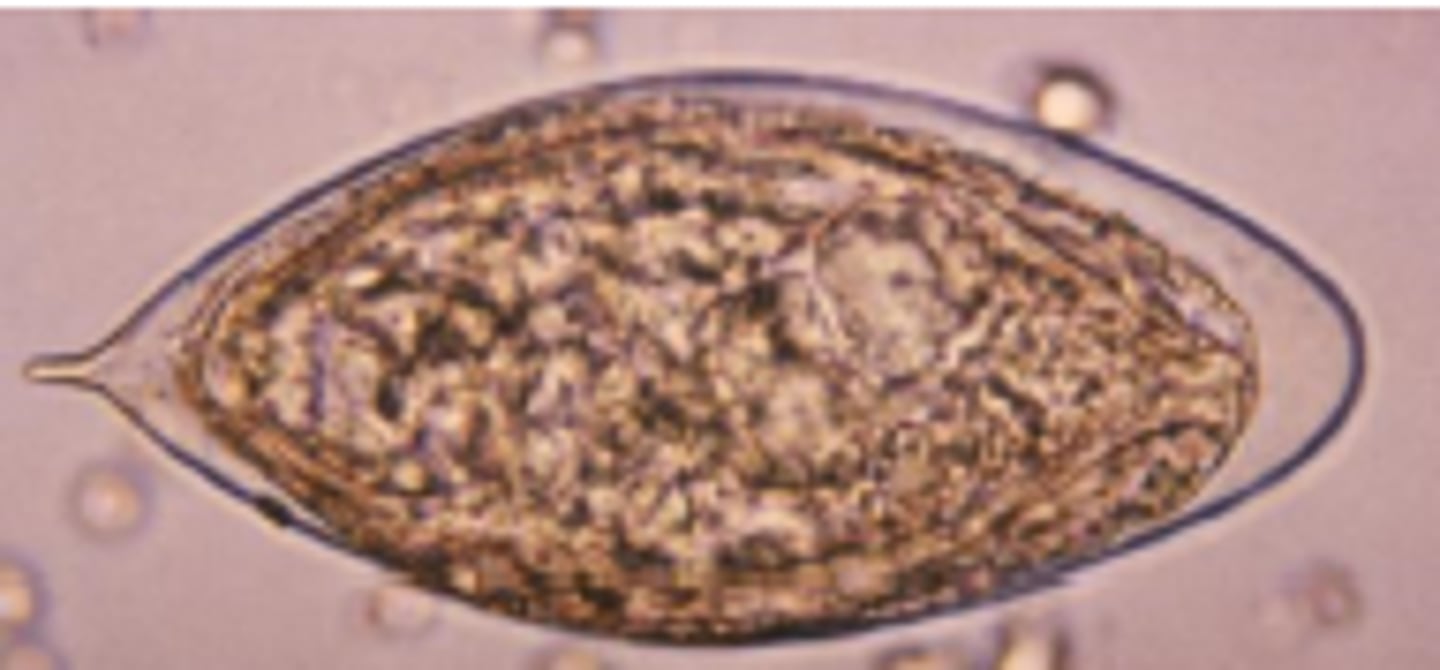
Schistosoma haematobium (egg)
-trematode
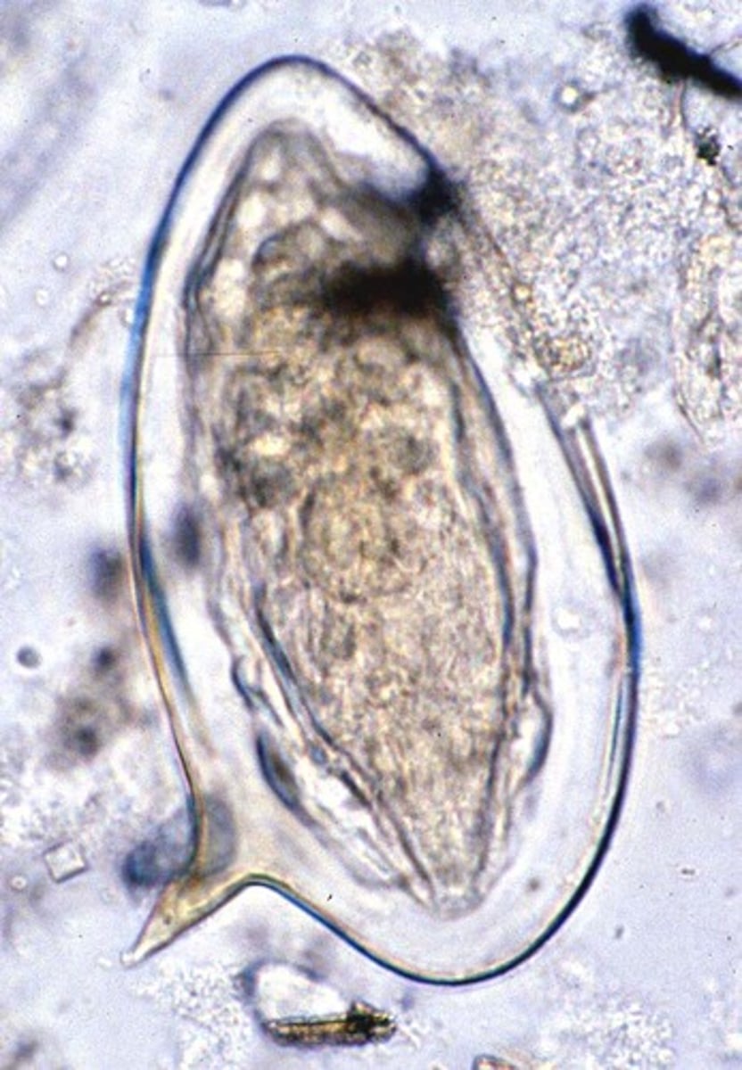
Schistosoma mansoni (egg)
-trematode
NOTE: the big hook
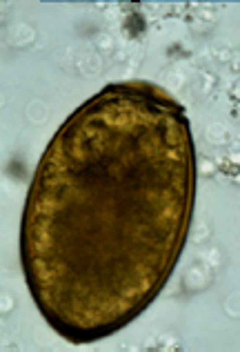
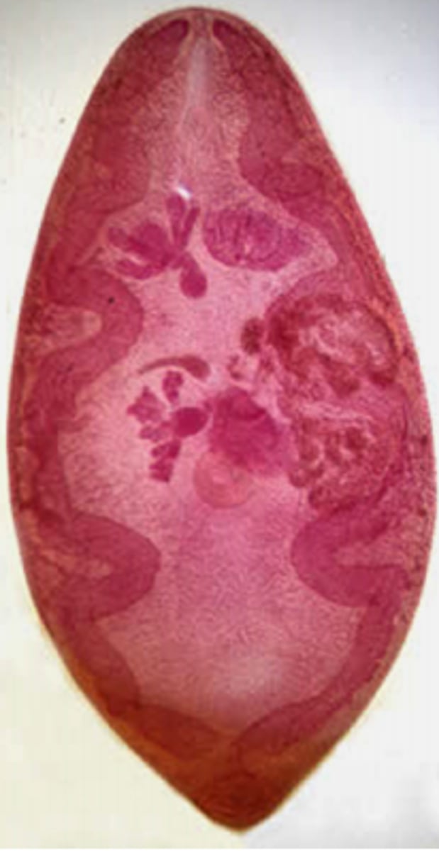
Clonorchis sinensis (adult)
Clonorchis sirensis (egg)
NOTE: knob and operculum
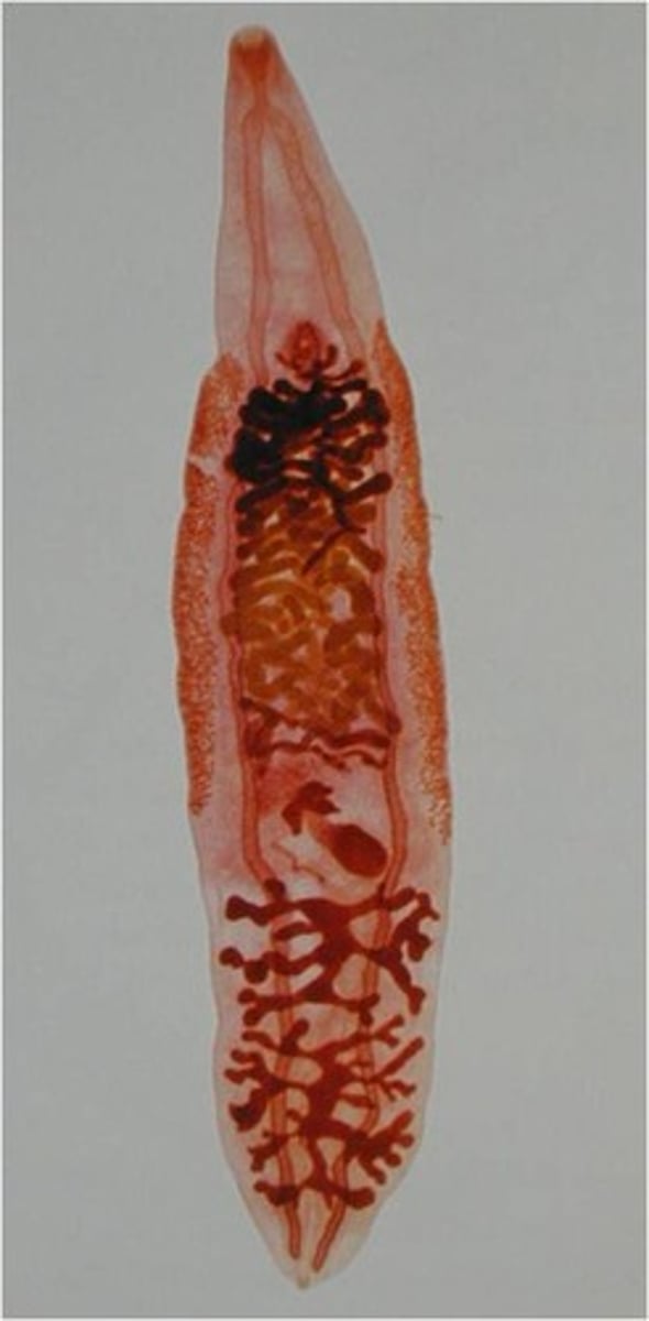
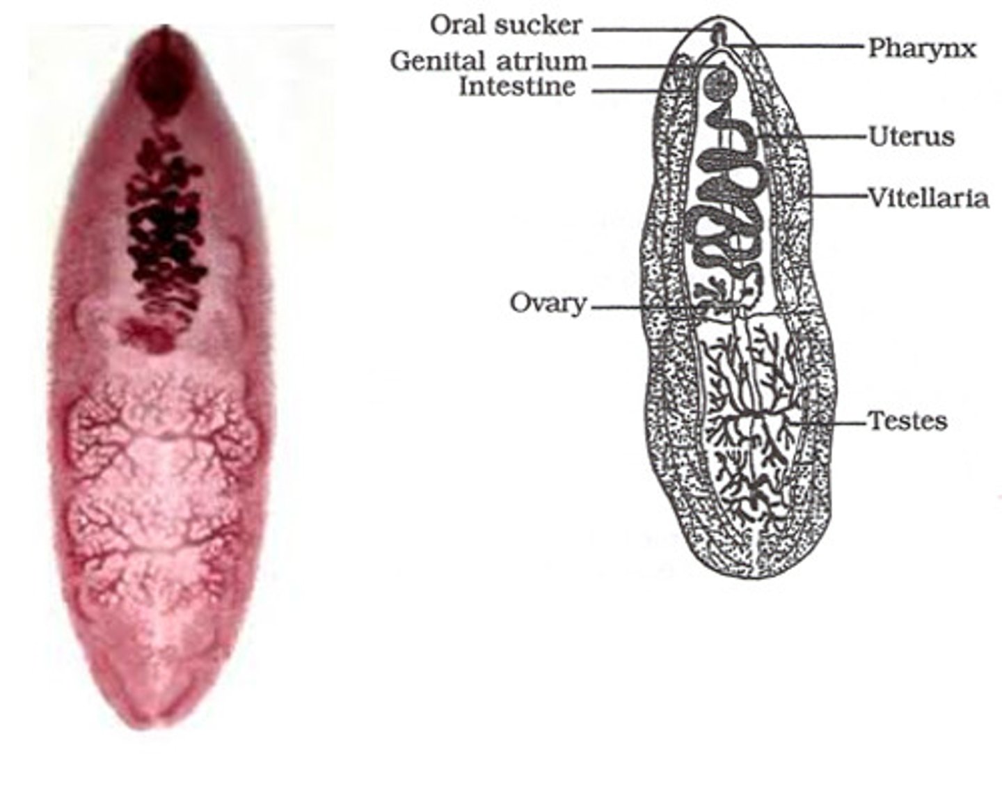
Fasciola hepatica (adult)
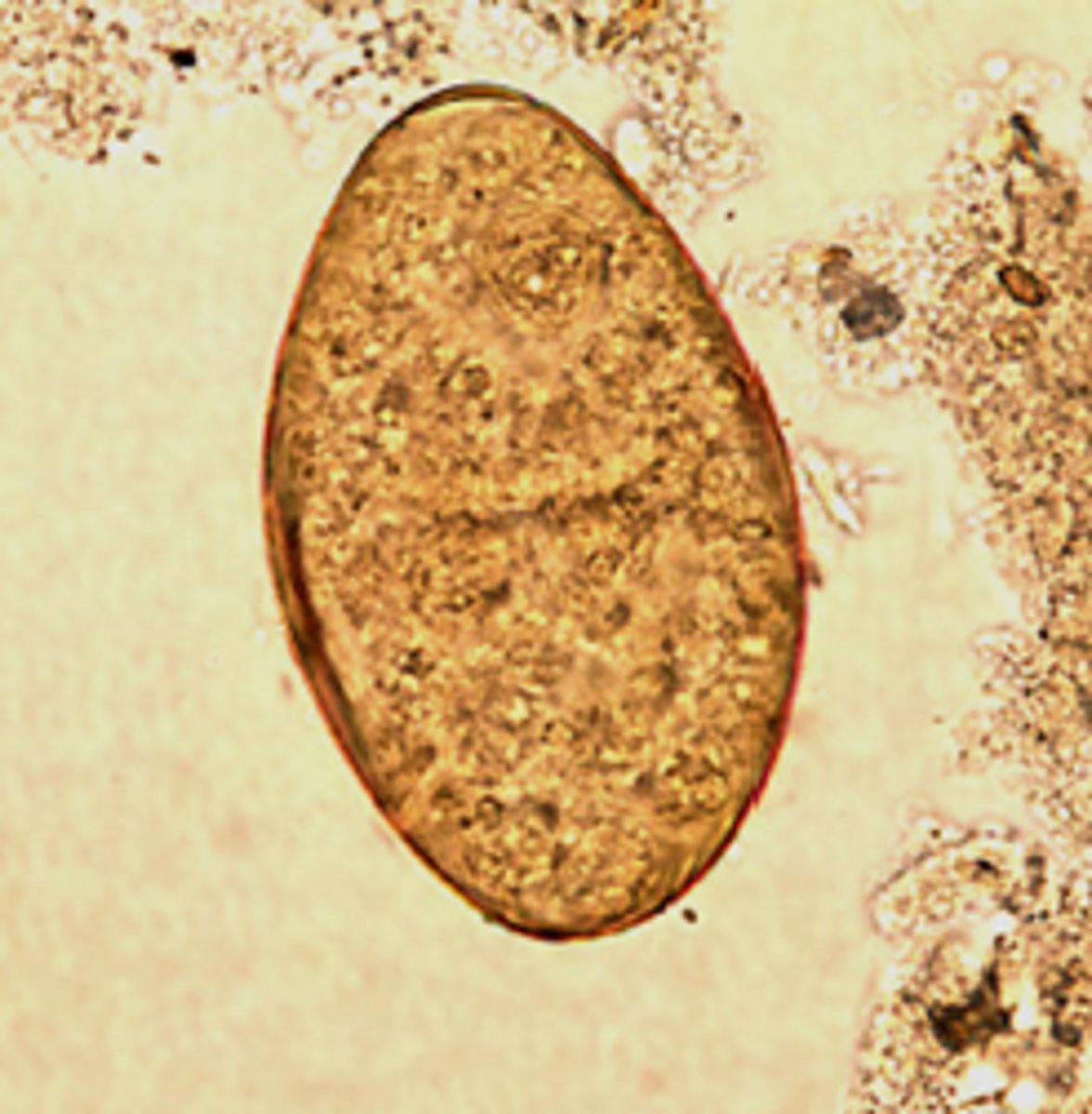
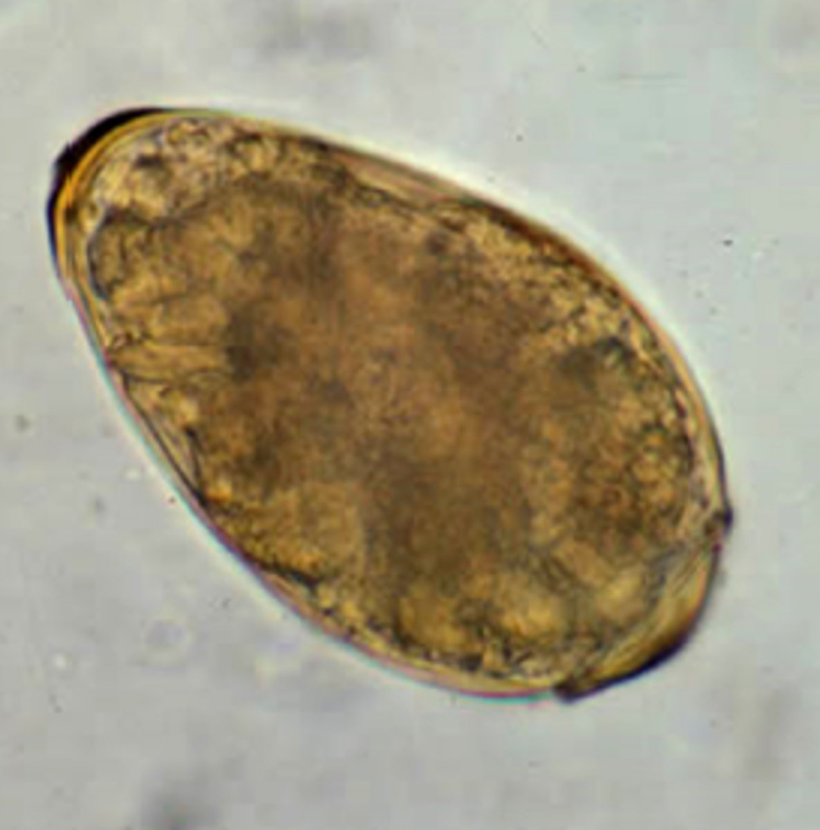
Fasciolopsis buski (egg)
Fasciolopsis buski (adult)
-trematode
-largest intestinal fluke of humans
Paragonimus westermani (egg)
Paragonimus westermani (adult)
Schistosoma haematobium (egg)
-trematode
*the little pointed end
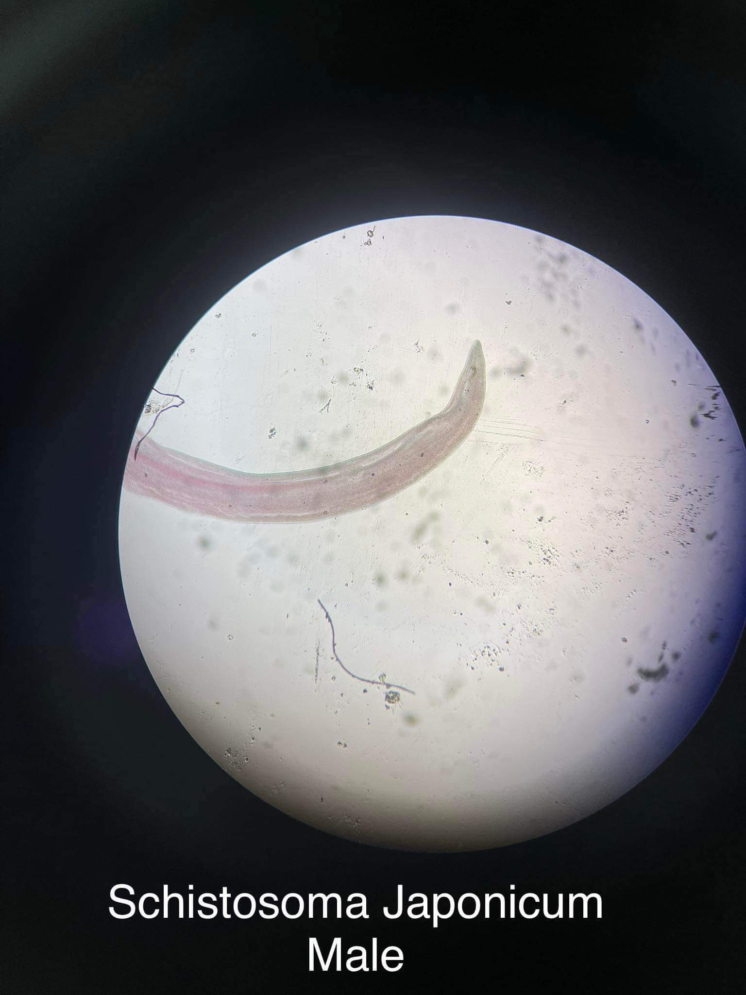
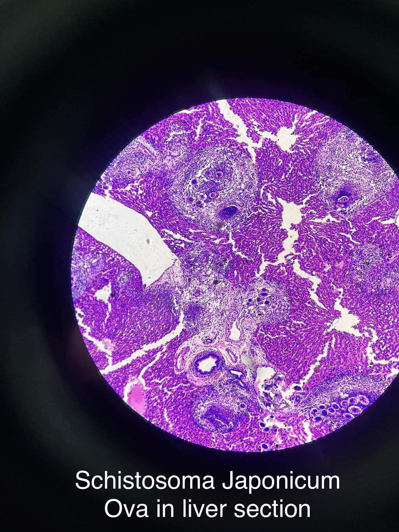
Schistosoma japonicum (egg)
-trematode
NOTE: the little hook
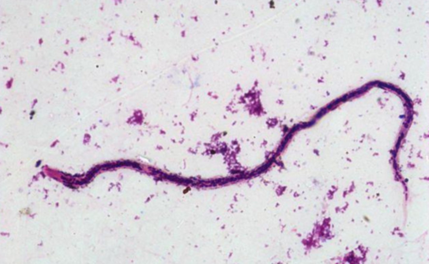
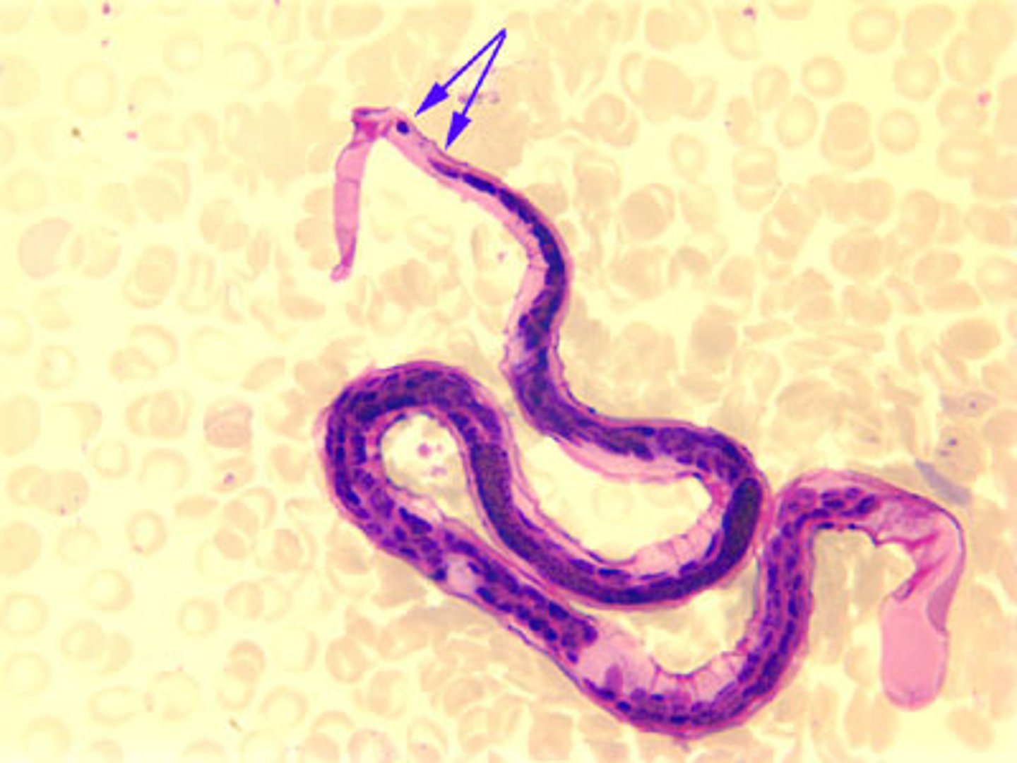
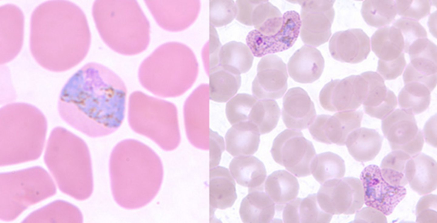
Loa loa
-nematode
-microfilaria
NOTE: giemsa stain, deep blue
Loa loa
-nematode
-microfilaria
*notice the converging nuclei into one row
*sheath is present
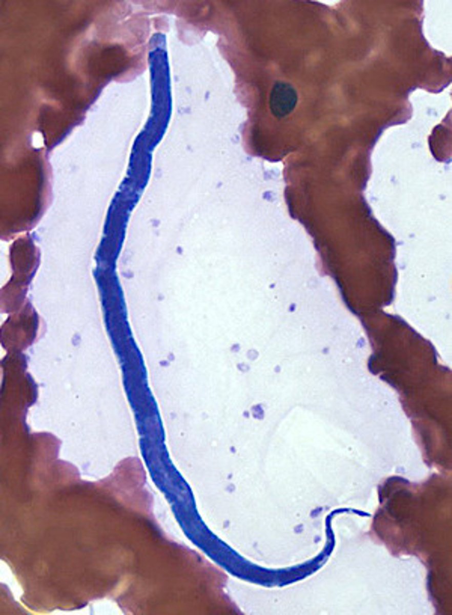
Onchocerca volvulus
-nematode
-microfilaria
*no sheath
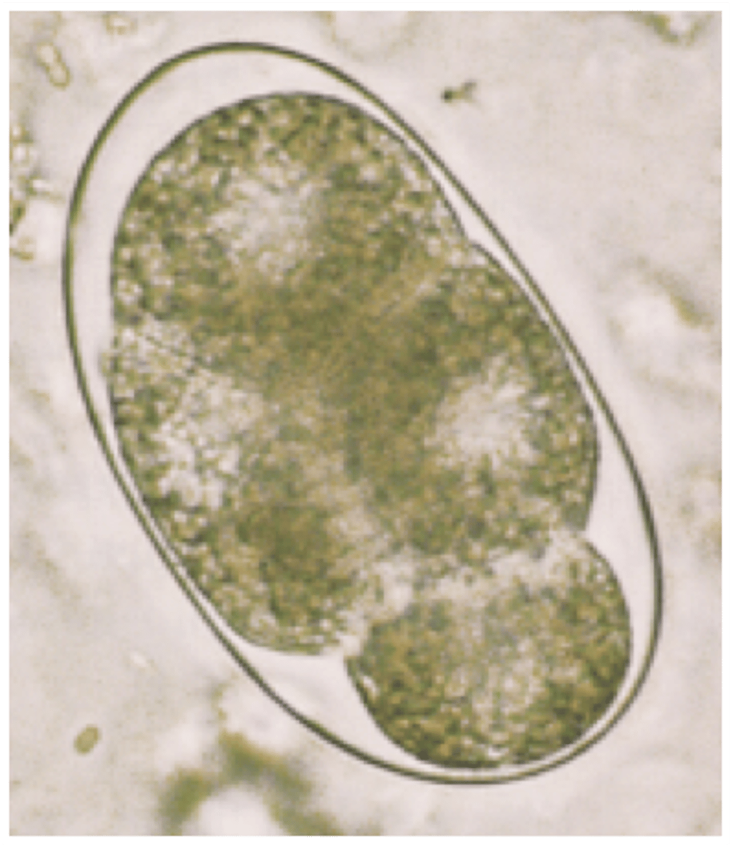
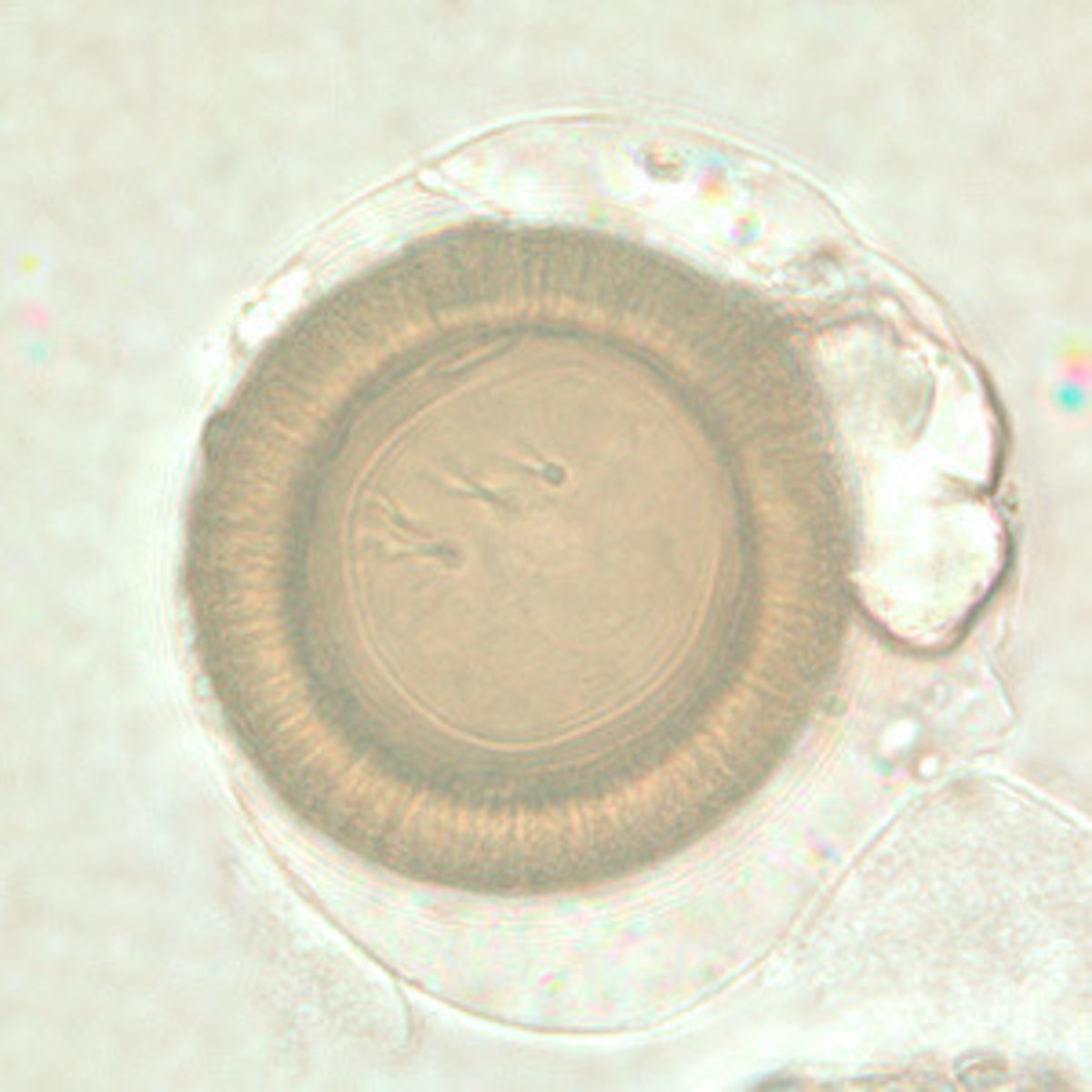
Hookworm egg
-Necator americanus
-Ancylostoma duodenale
-nematode
*2-8 cell cleavages
*thin-smooth colorless shell
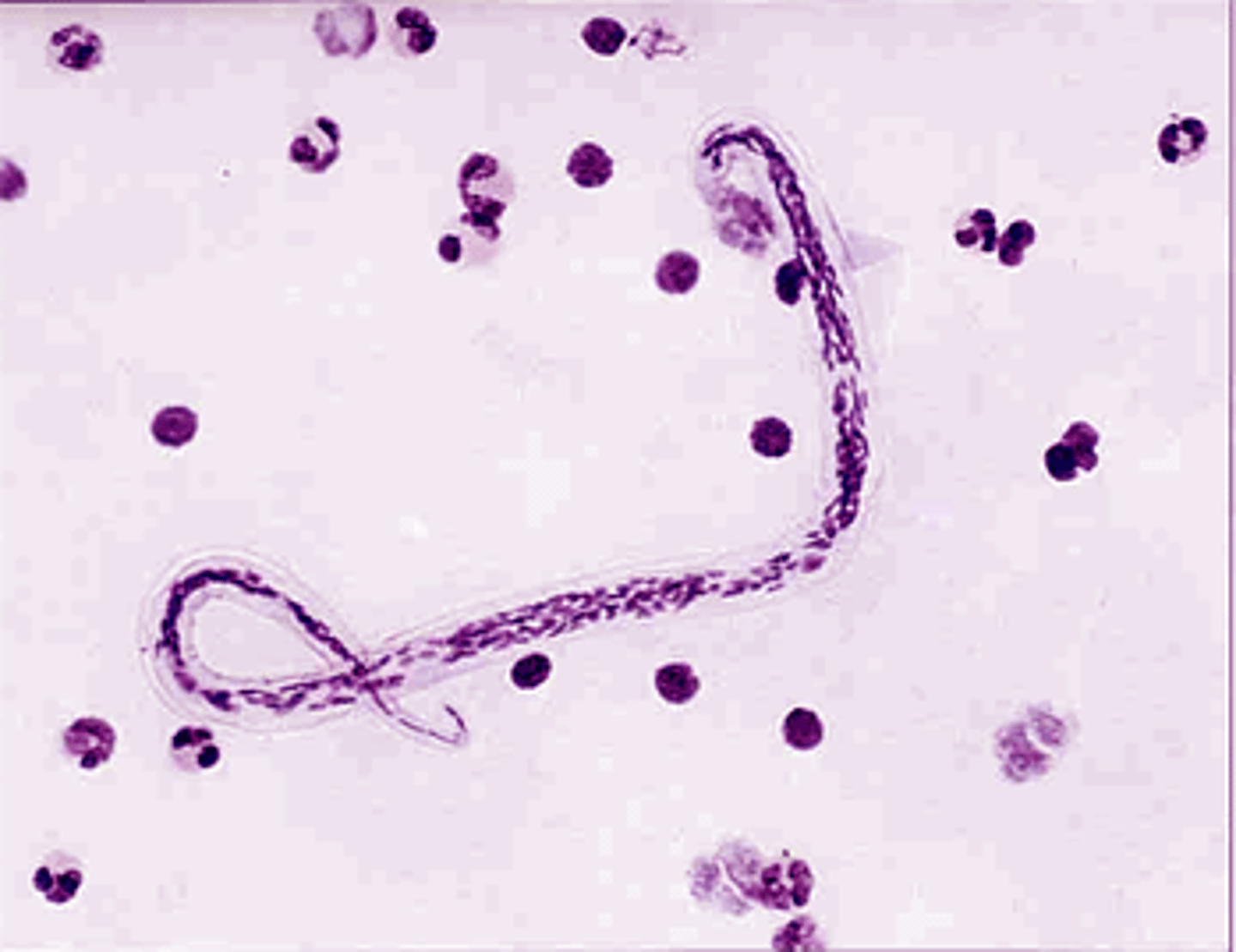
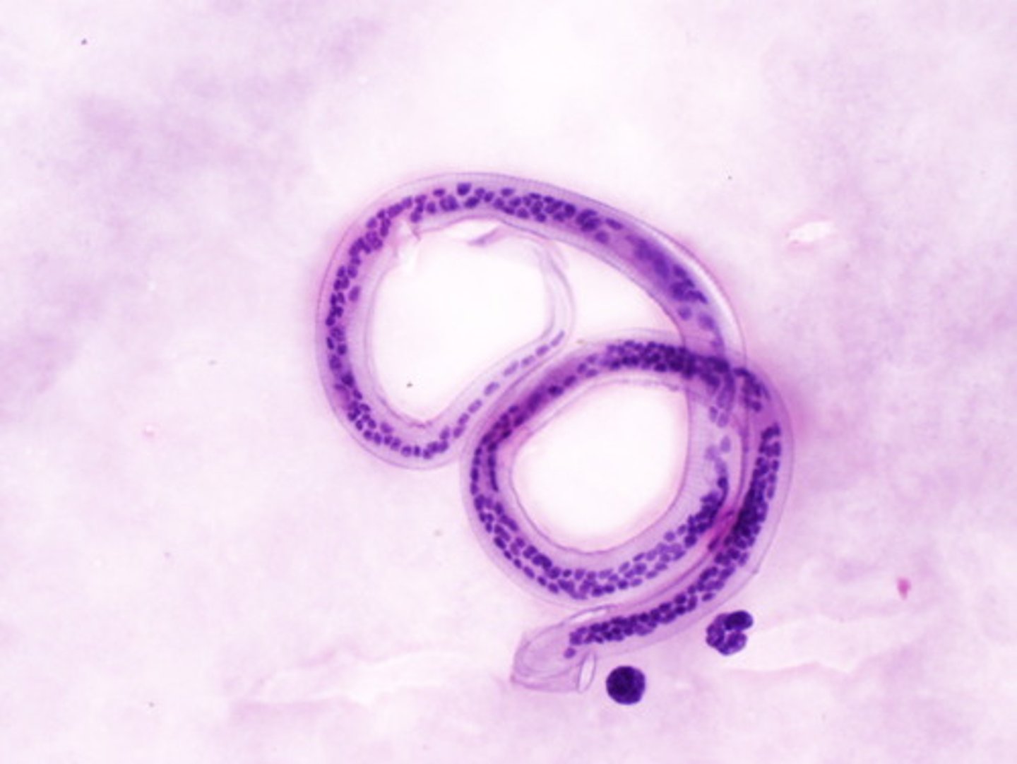
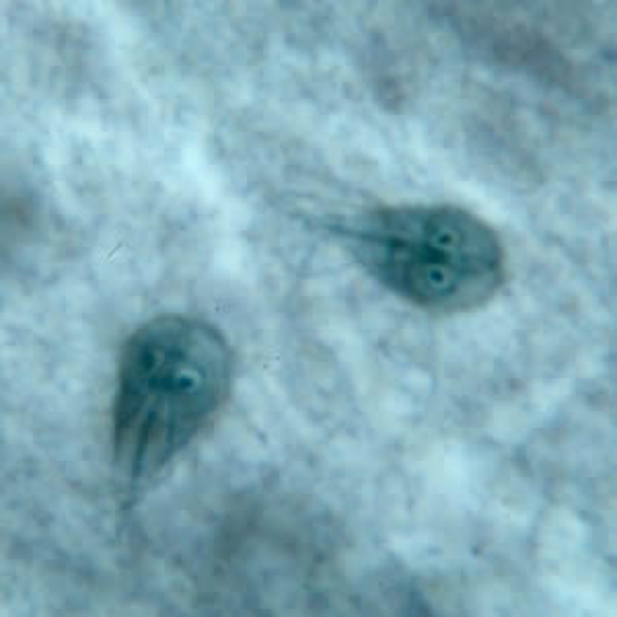
Wuchereria bancrofti
-nematode
-microfilaria
*the nuclei doesn't go up to the tip
*sheath present
Brugia malayi
-nematode
-microfilaria
*two nuclei at the tip of the tail
Enterobius vermicularis (egg)
-intestinal nematode
*notice the one flatter side
Trichuris trichiura (egg)
-intestinal nematode
*bipolar plugs
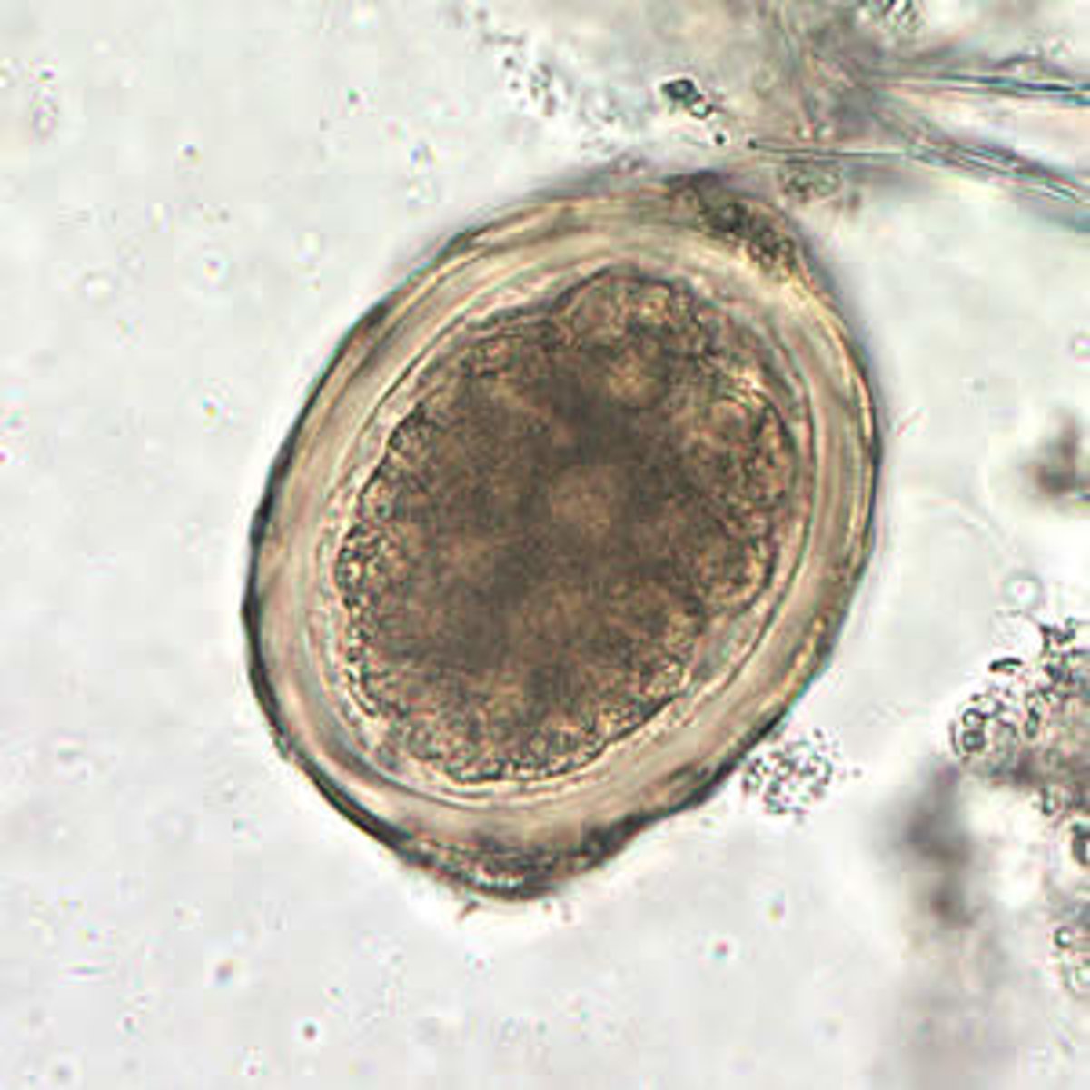
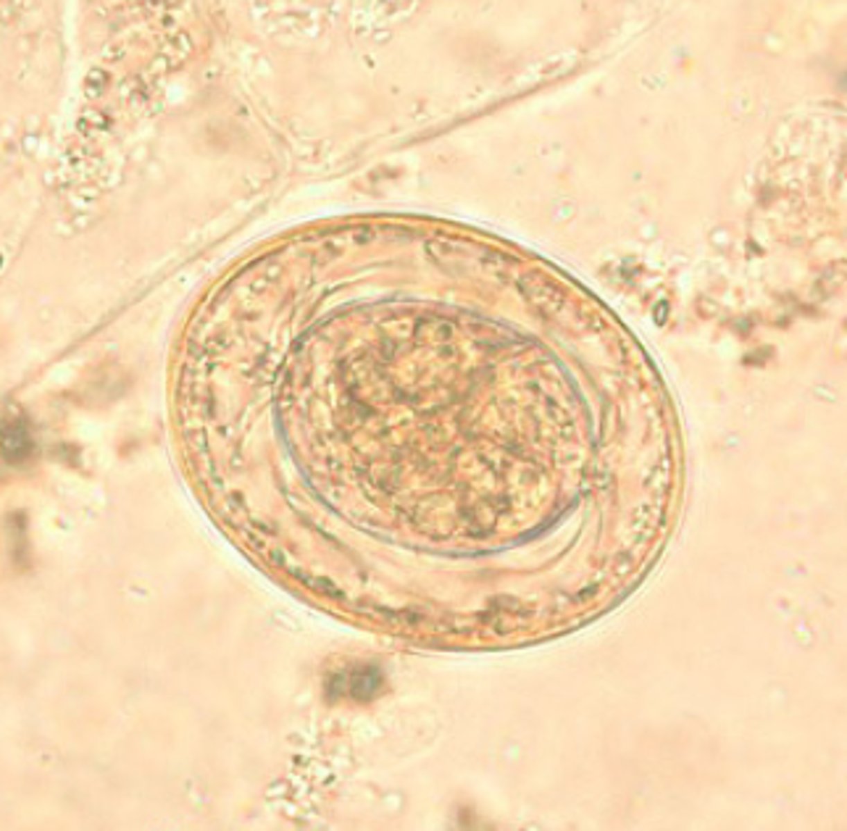
Ascaris lumbricoides (fertilized egg)
-intestinal nematode
*has a thick wall
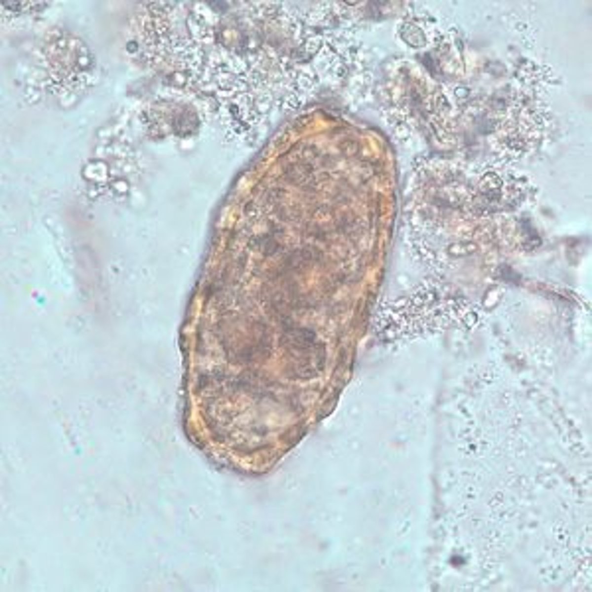
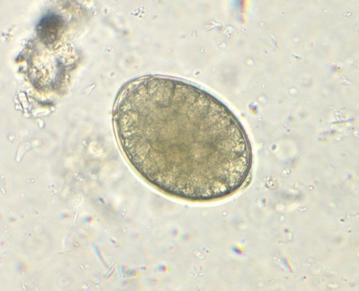
Ascaris lumbricoides (unfertilized egg)
-intestinal nematode
*thinner shell than fertilized
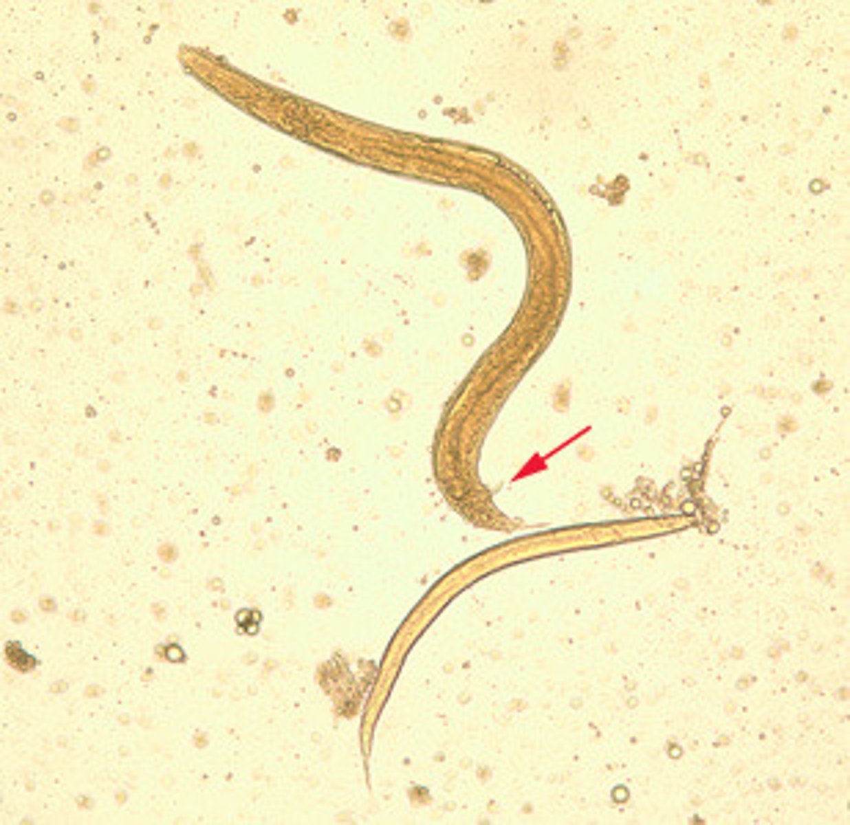
Strongyloides stercoralis (adult and rhabditiform)
-intestinal nematode
*arrow is pointing to a spicule
Trichinella spiralis (adult)
-intestinal nematode
-affects muscles
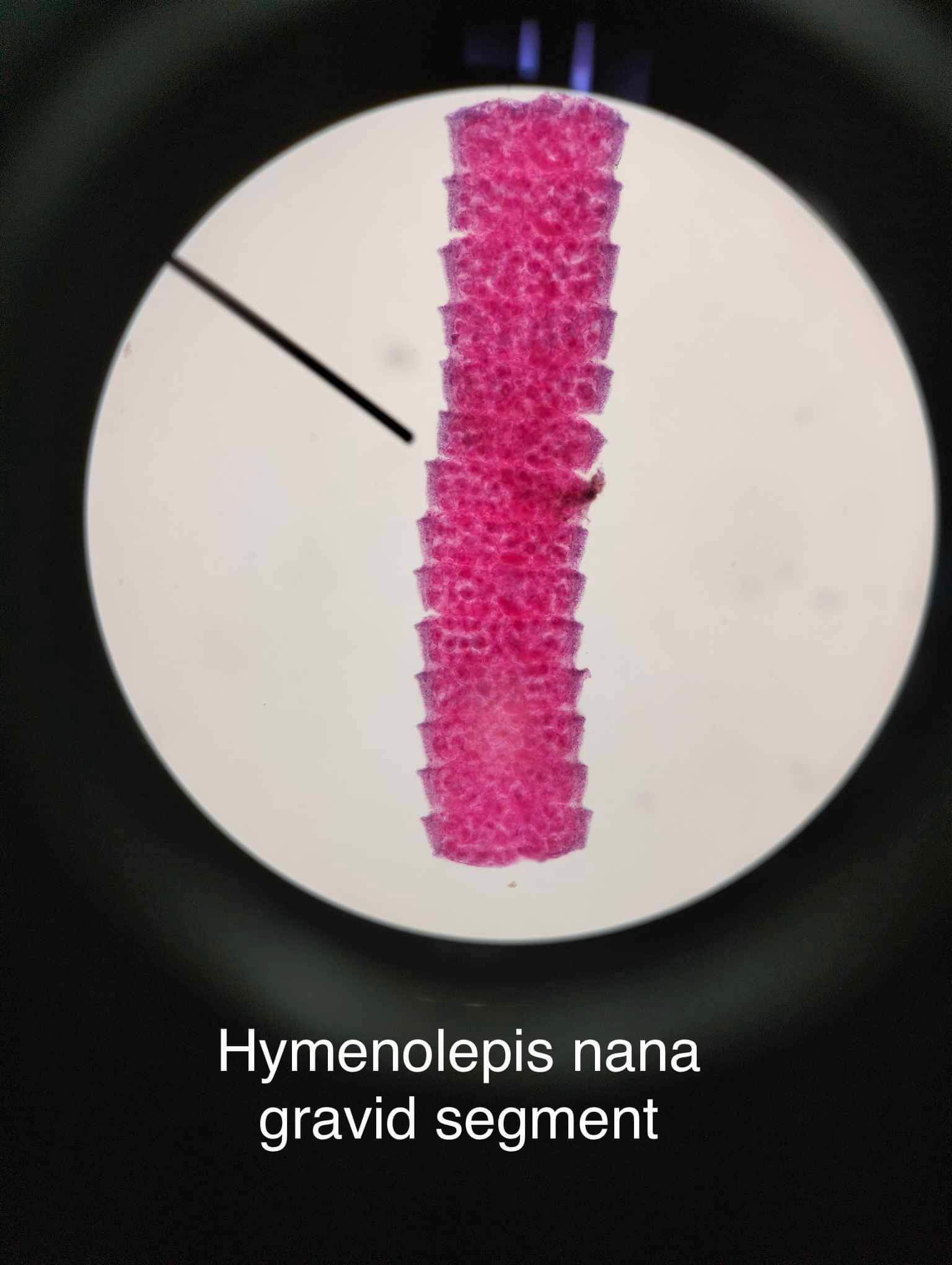
Hymenolepis nana (egg)
*colorless shell
*filaments are emerging from the polar thickenings
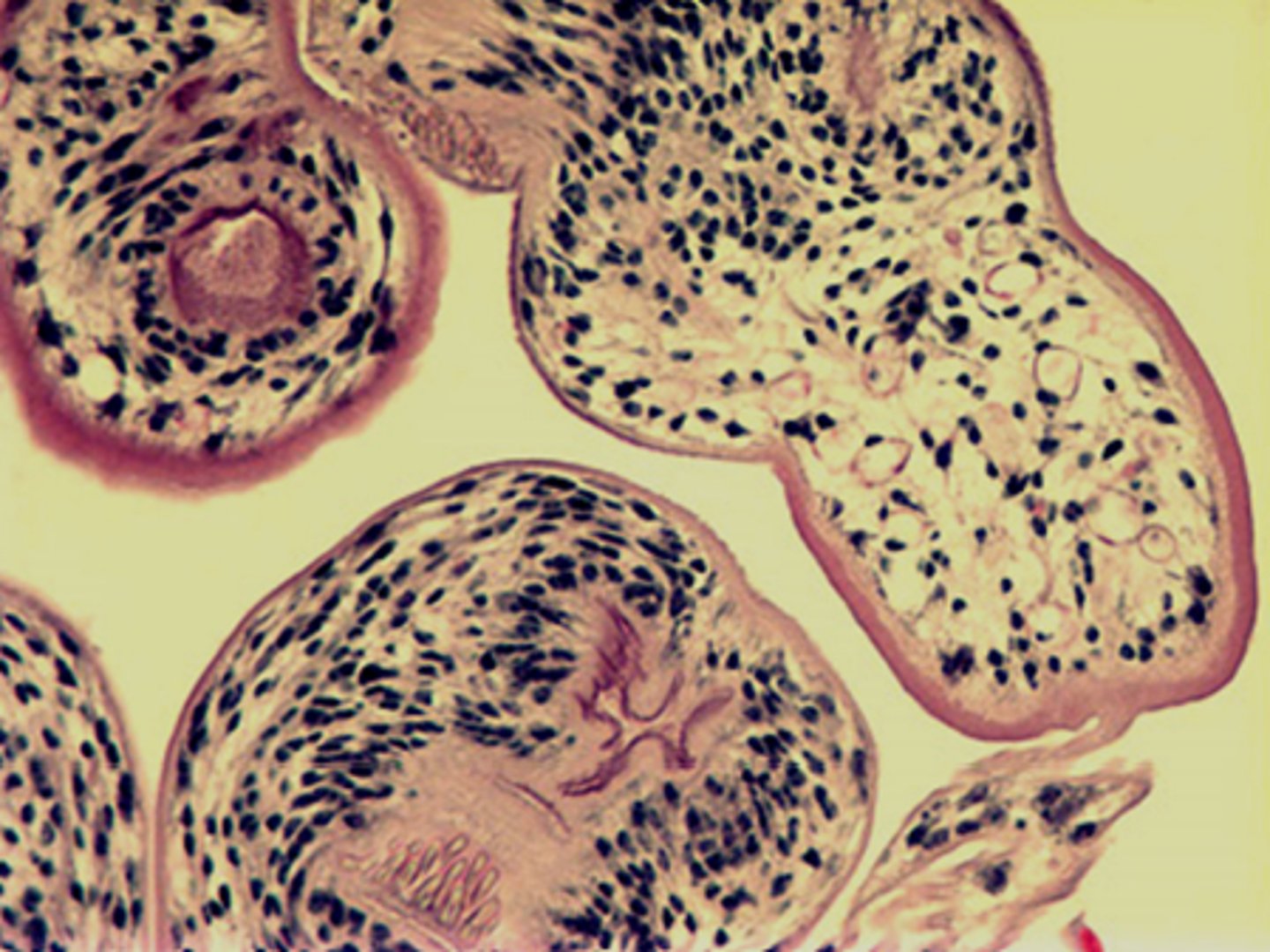
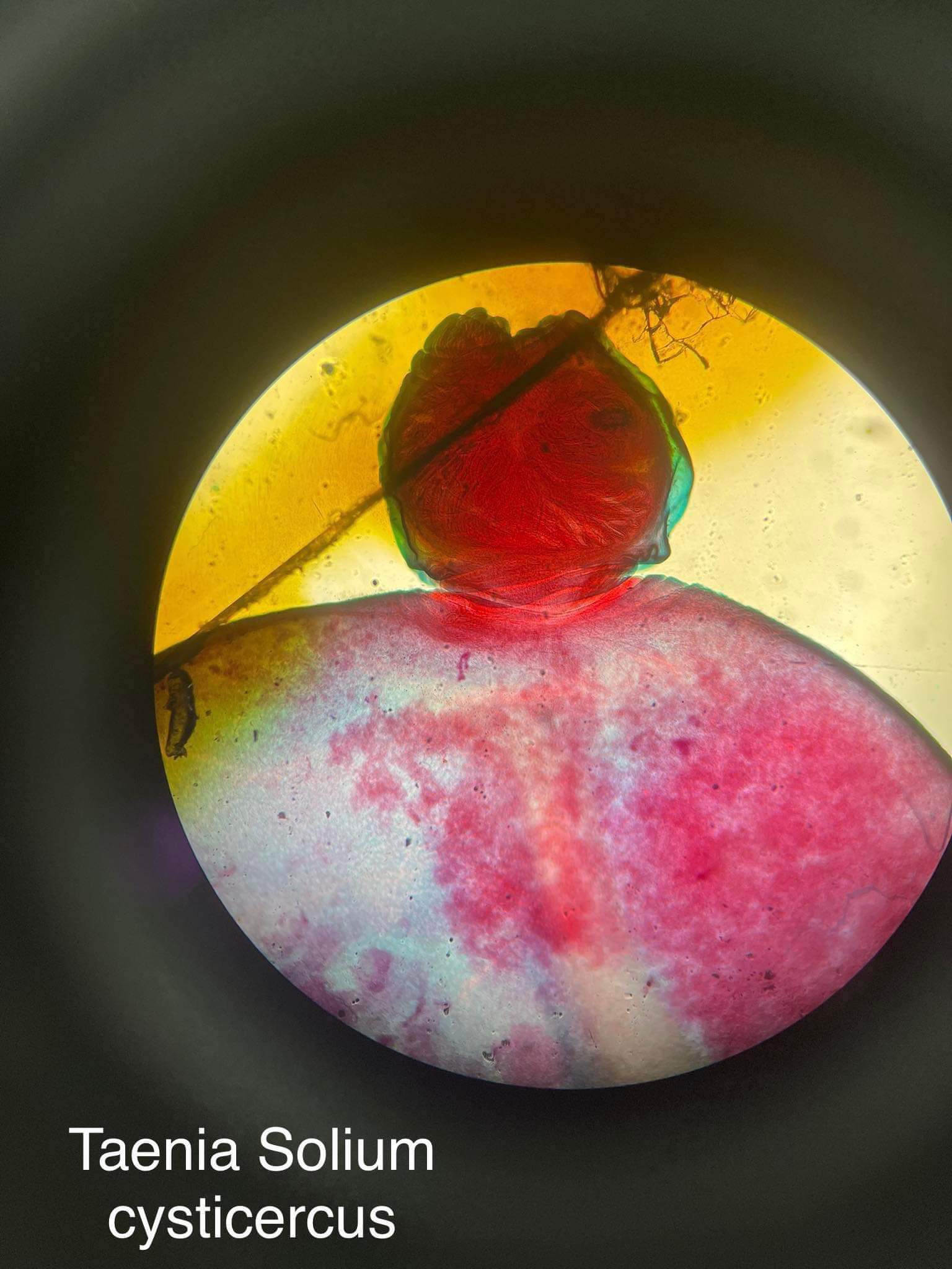
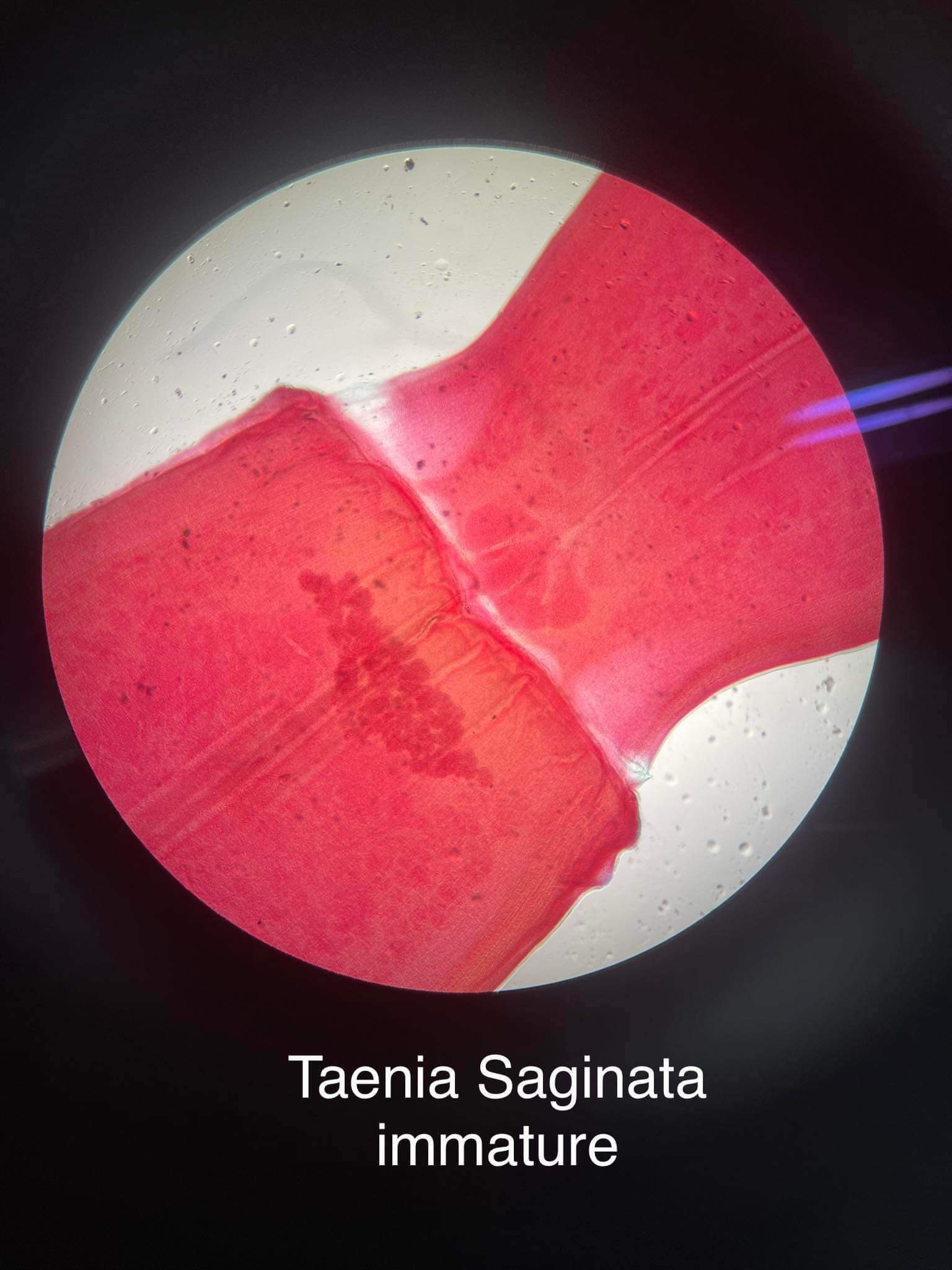
T. saginata + T. solium (egg)
-cestode
*3 slightly visible hooks in center
*yellowish striations in embryo
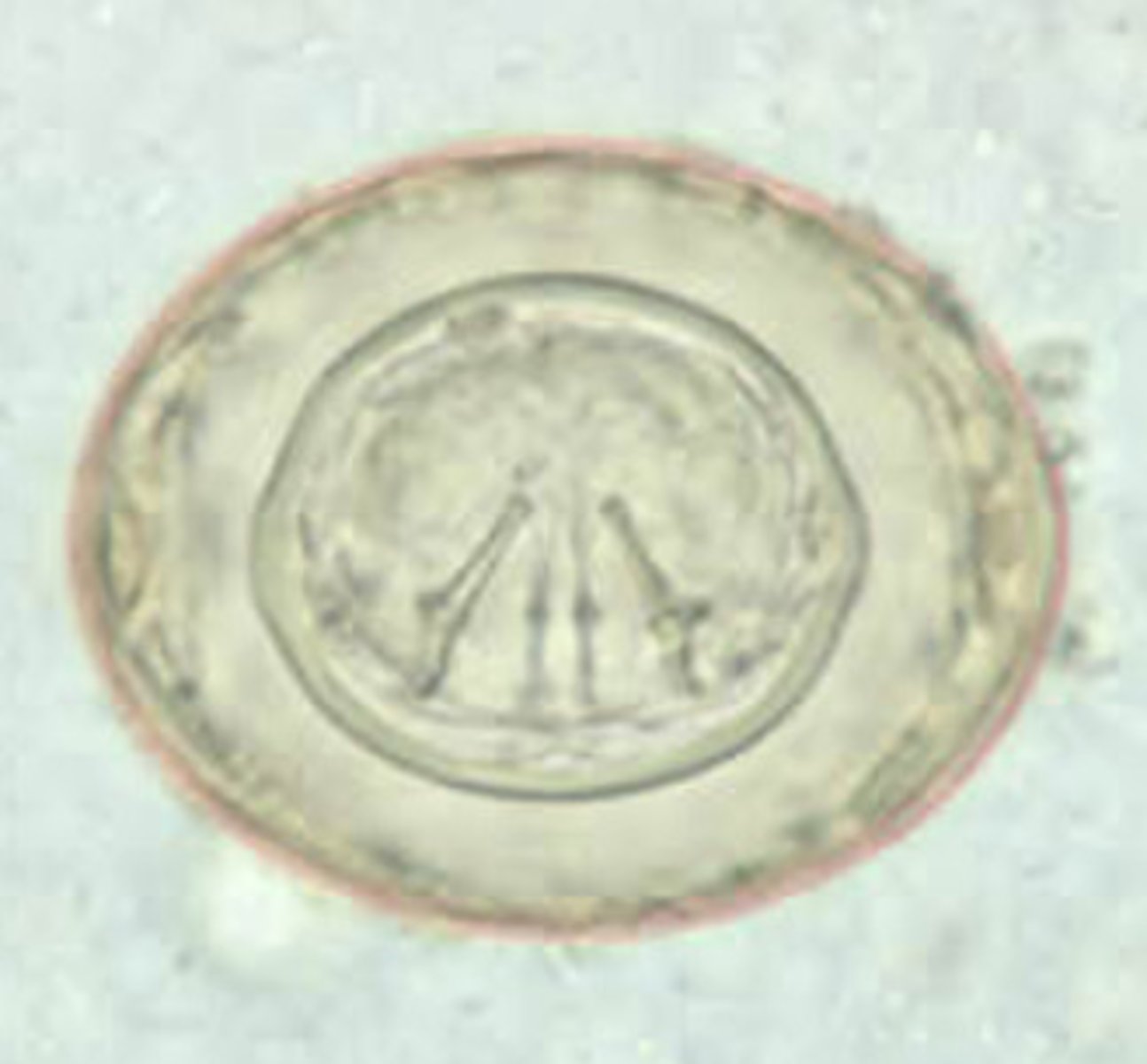
Diphyllobothrium latum (egg)
*operculum on one end
*also a small knob on the edge
Echinococcus granulosus (cyst)
-cestode
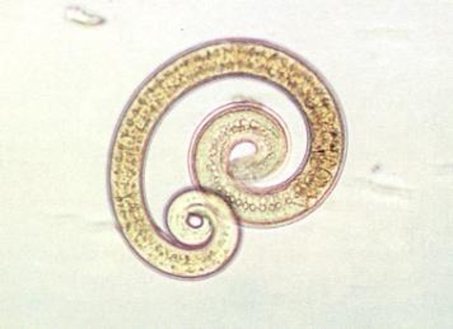
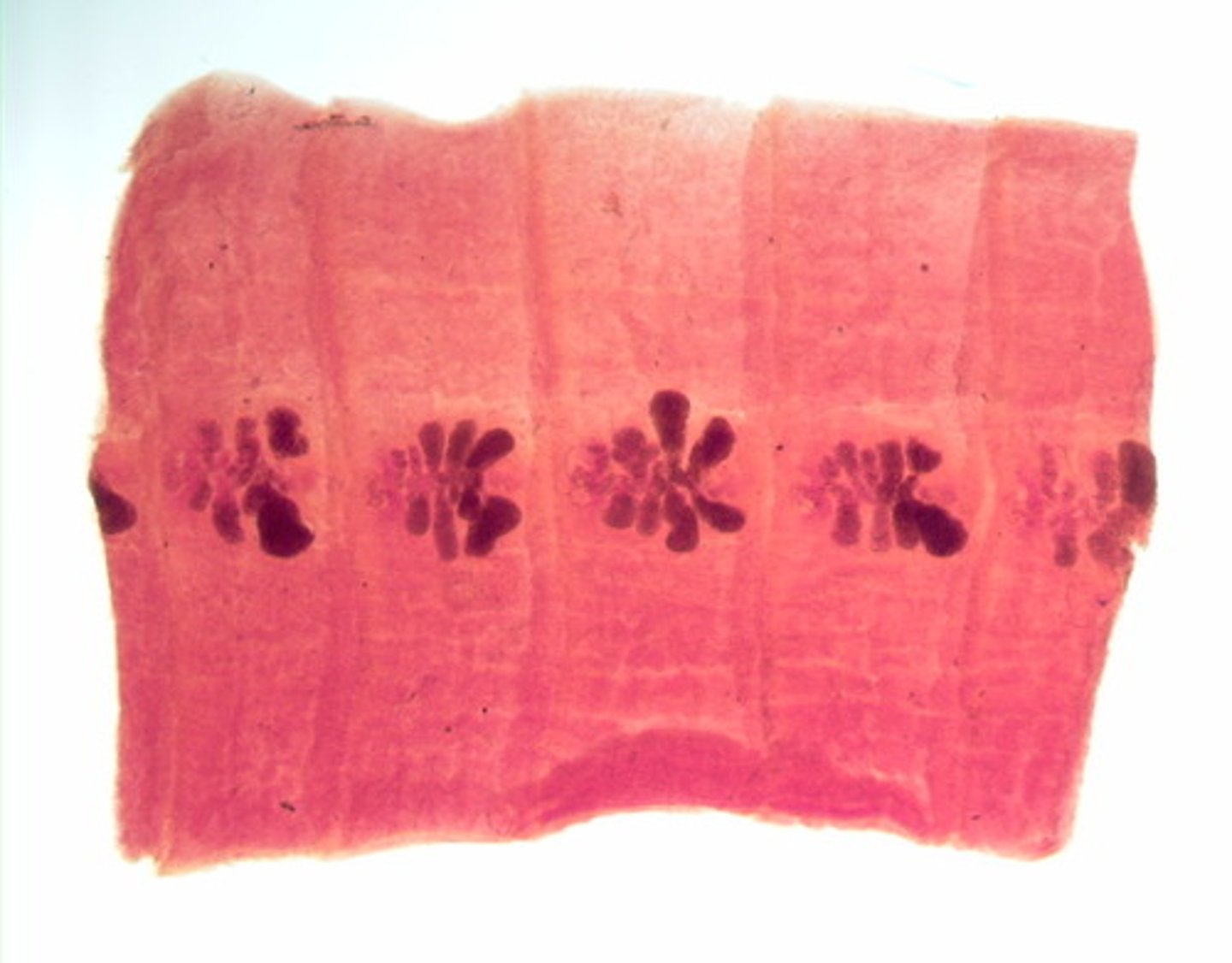
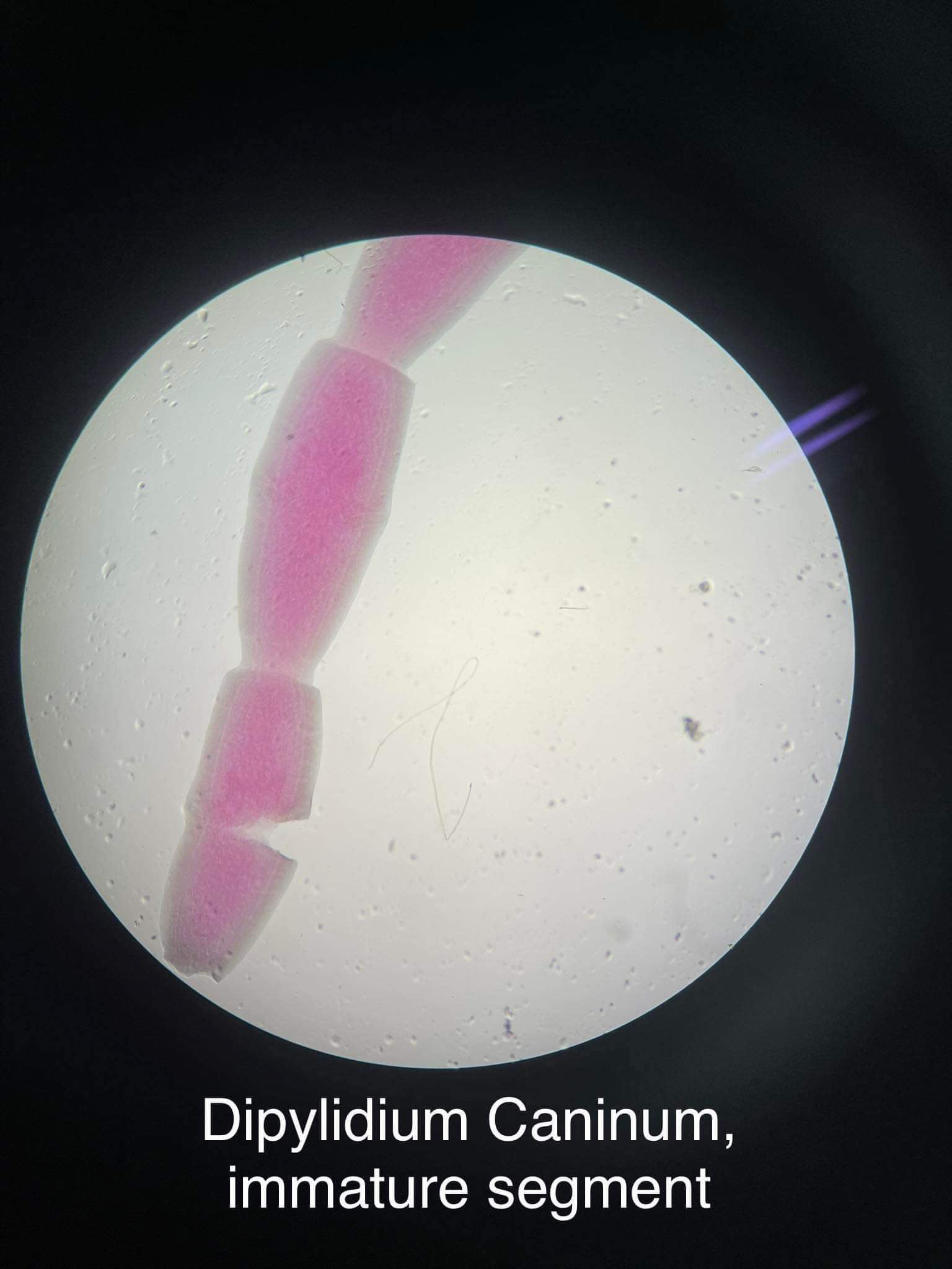
Diphyllobothrium latum (proglottid)
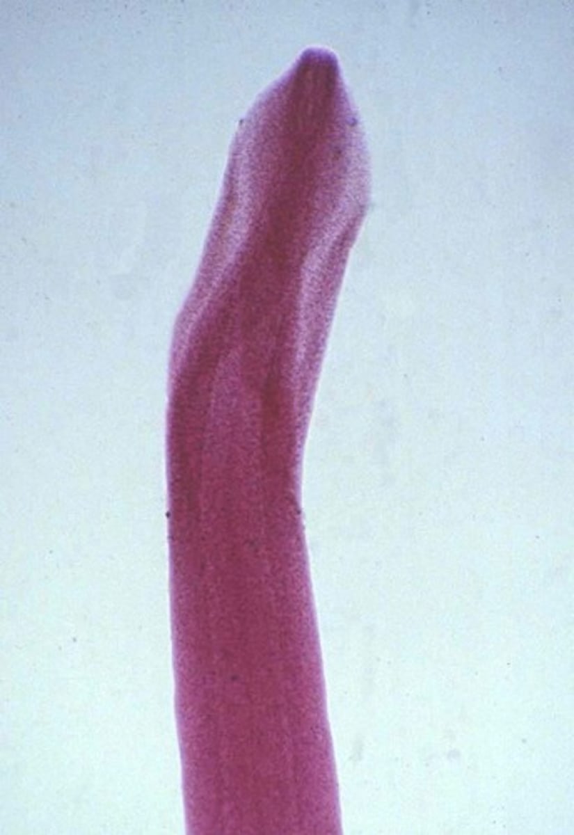
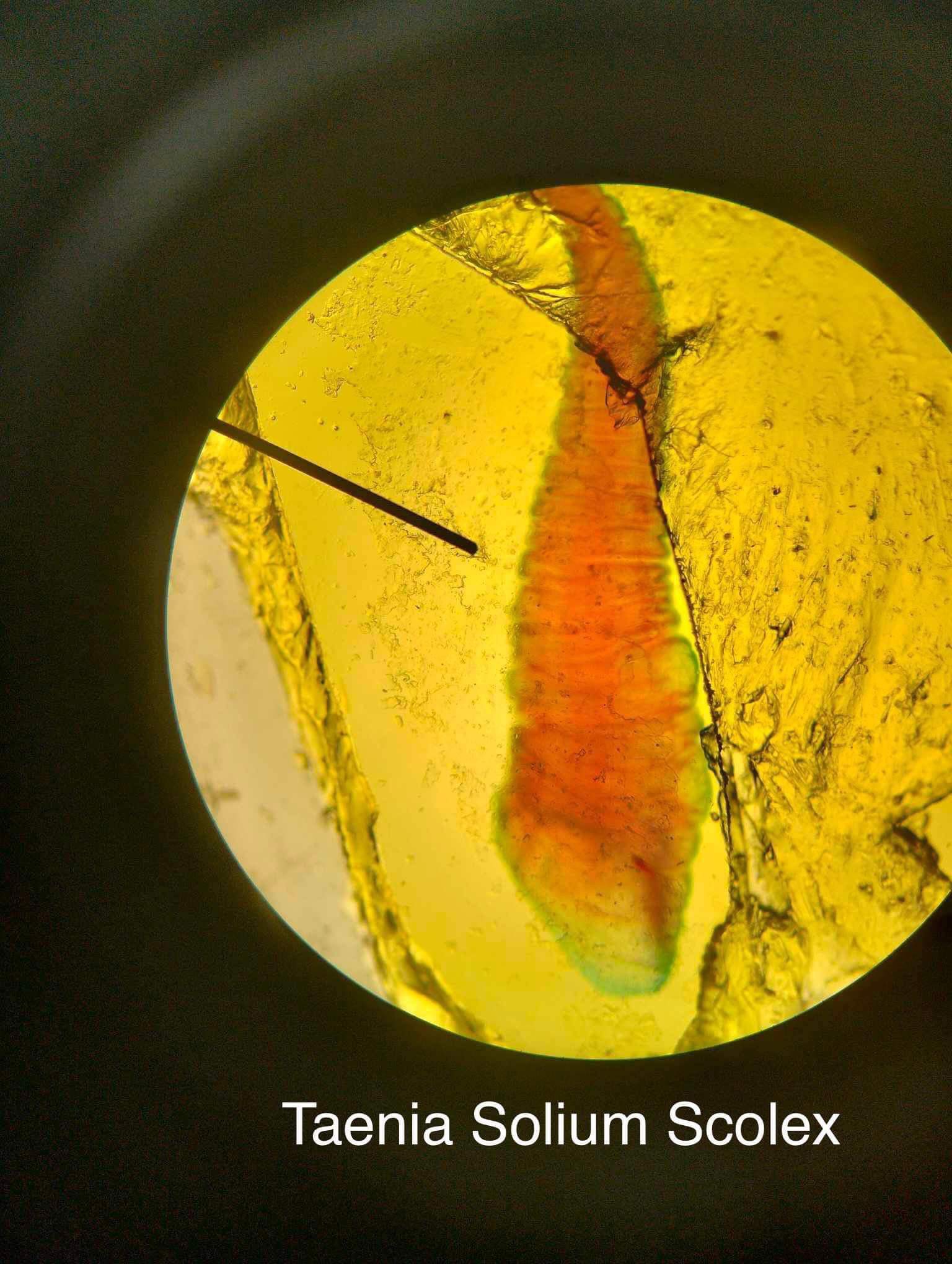
Diphyllobothrium latum (scolex)
-cestode
*no cup-shaped suckers
Hymenolepis diminuta (egg)
*polar thickening on both sides
*visible hooks
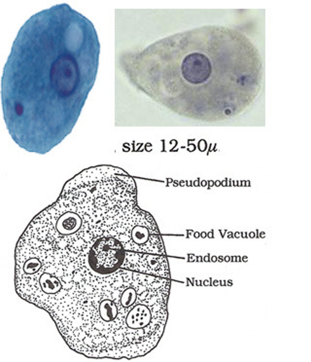
Entamoeba histolytica/dispar (trophozoite)
*ingested RBCs may be visible in histolytica
*active progressive motility in wet mount
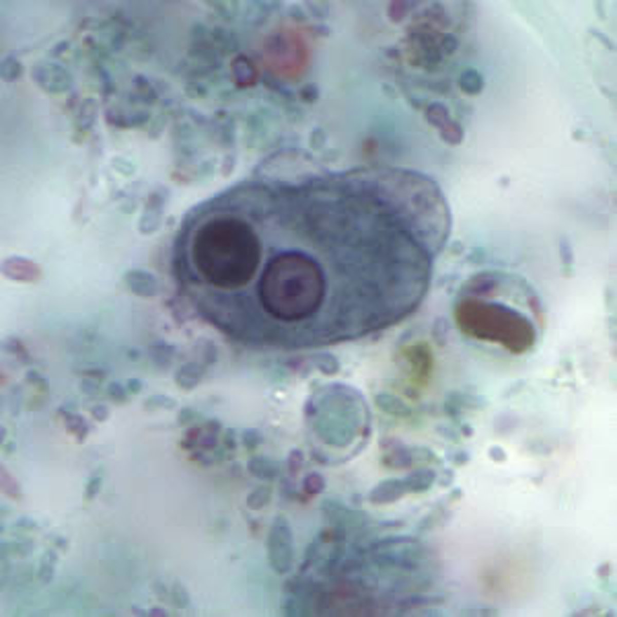
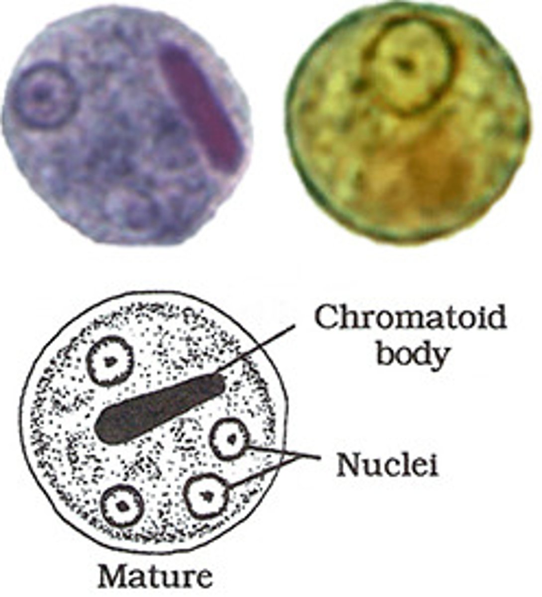
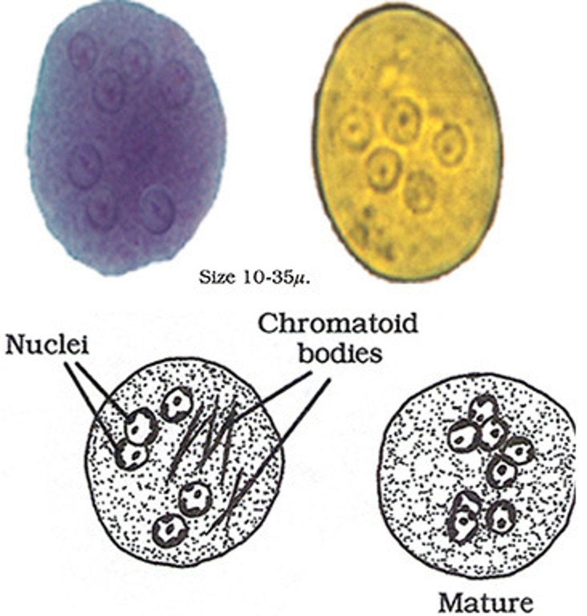
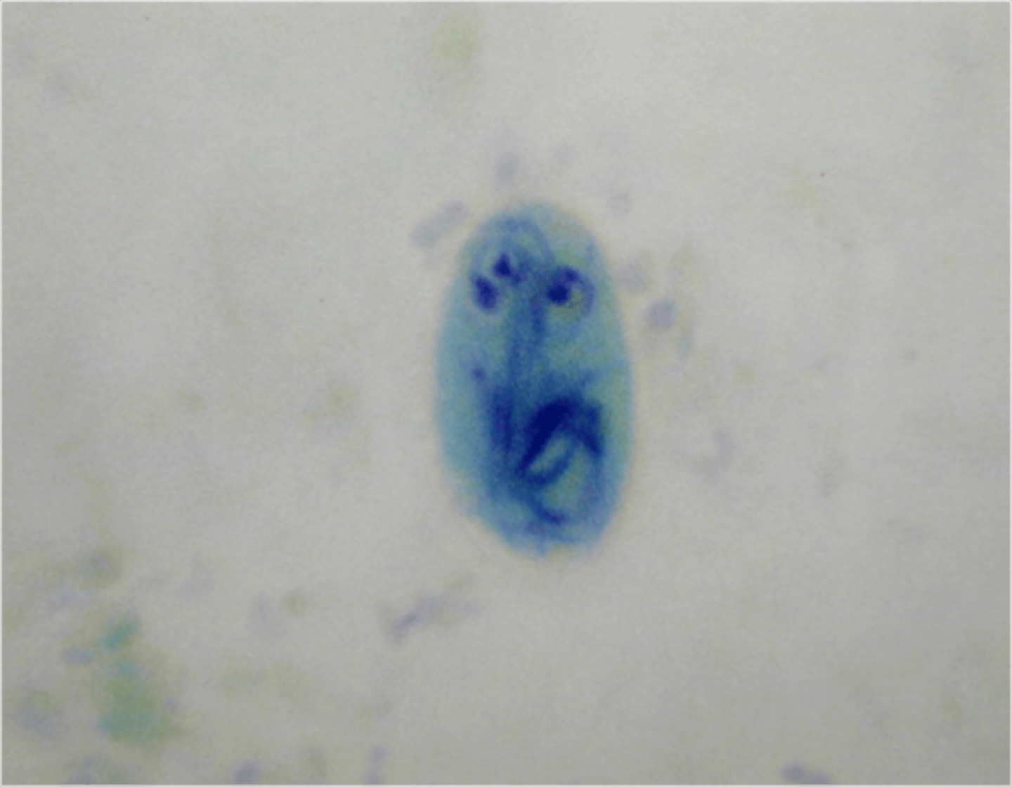
Entamoeba histolytica/dispar (cyst)
*1-4 nuclei
*may have a cigar shaped chromatoid bar
*similar to hartmanni cyst, but is much larger
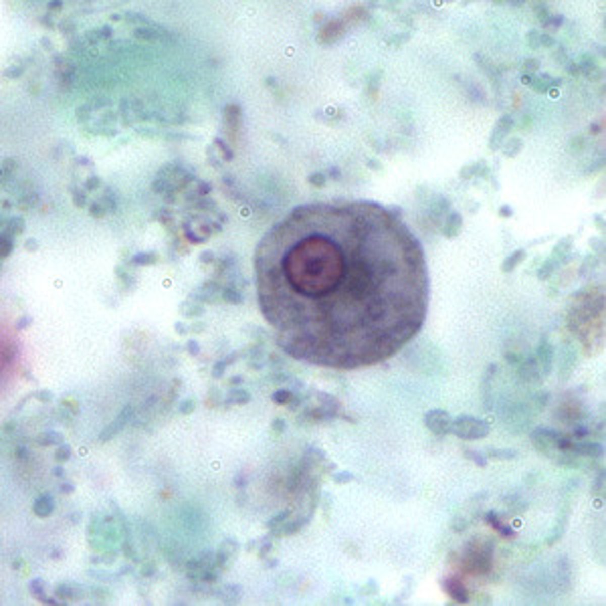
Entamoeba hartmanni (trophozoite)
*similar to histolytica, but much smaller
Entamoeba hartmanni (cyst)
*similar to histolytica, but much smaller
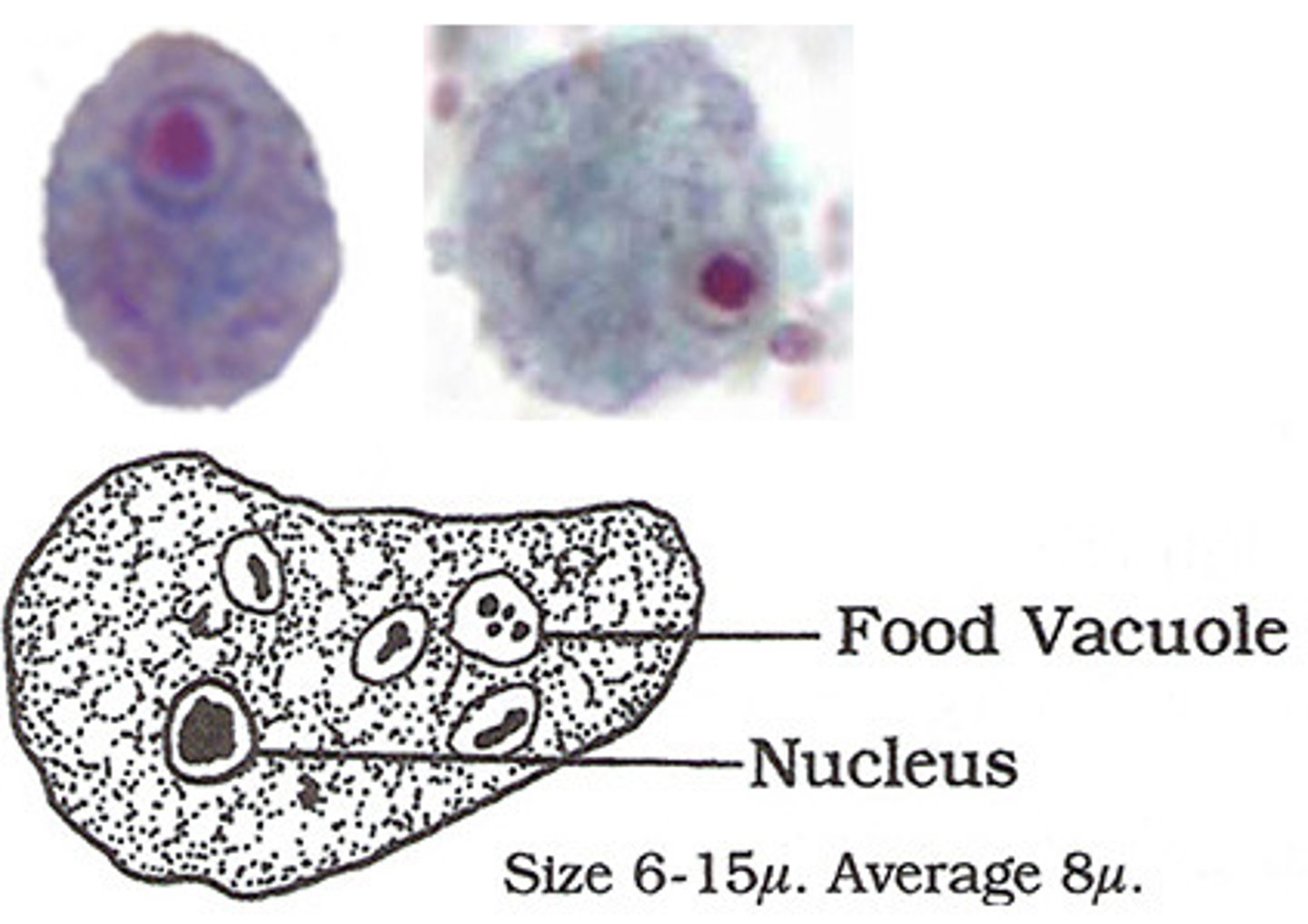
Entamoeba coli (trophozoite)
*note the dot...absent in histolytica
*may ingest bacteria
*granular cytoplasm, sluggish motility
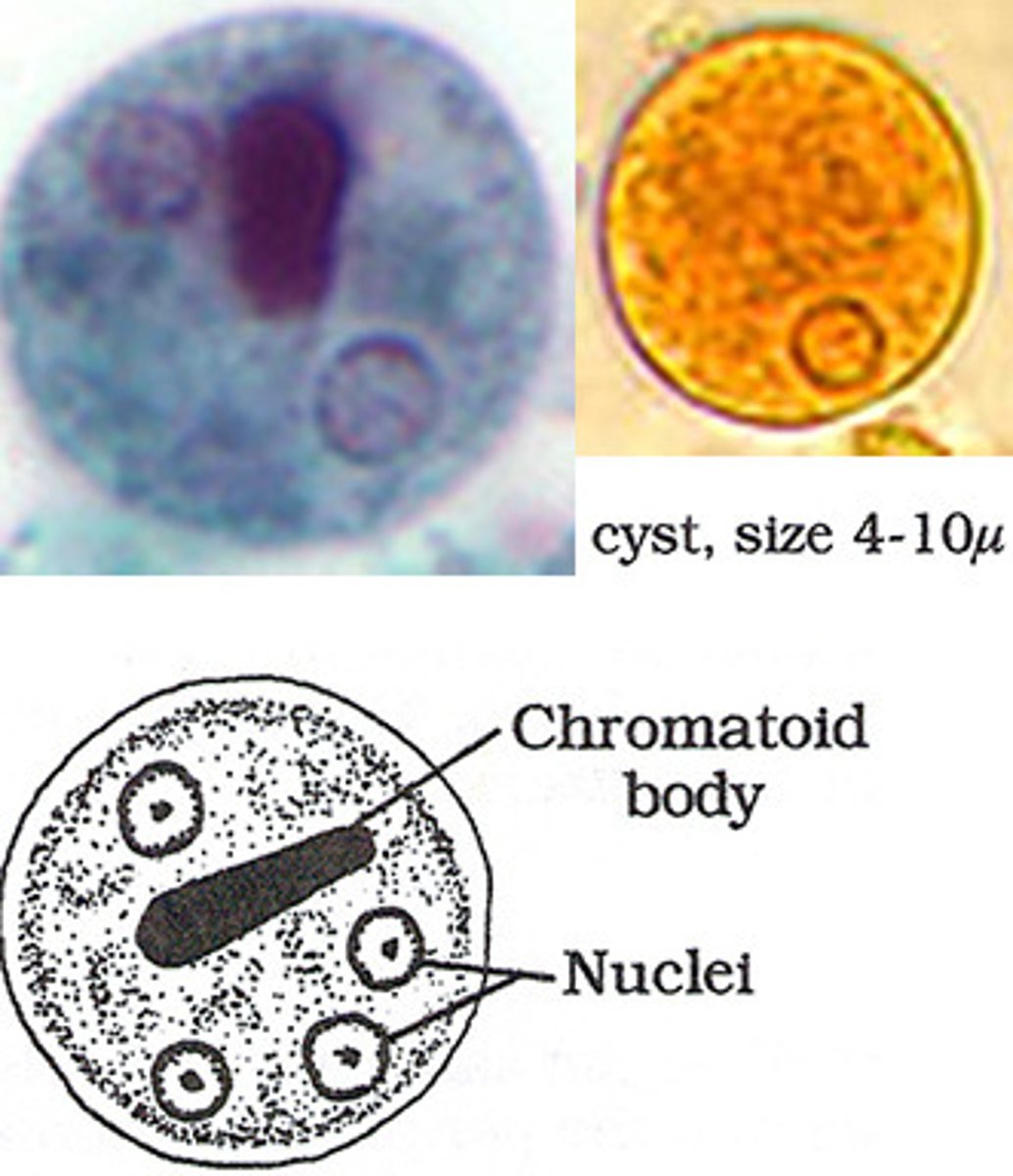
Entamoeba coli (cyst)
*similar to histolytica, BUT can have up to 8 nuclei
*chromatoid bars if present, have splintered/pointed ends
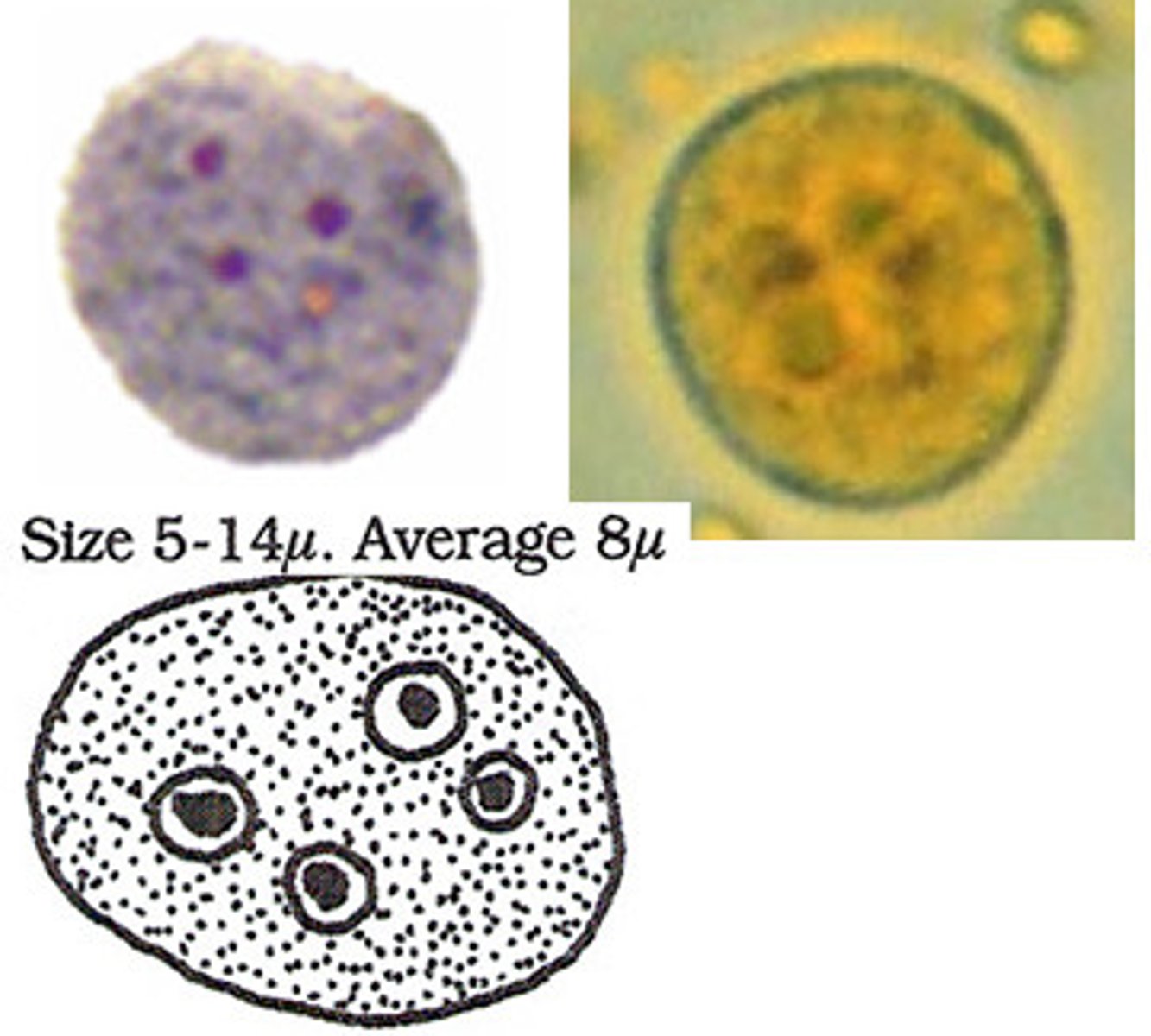
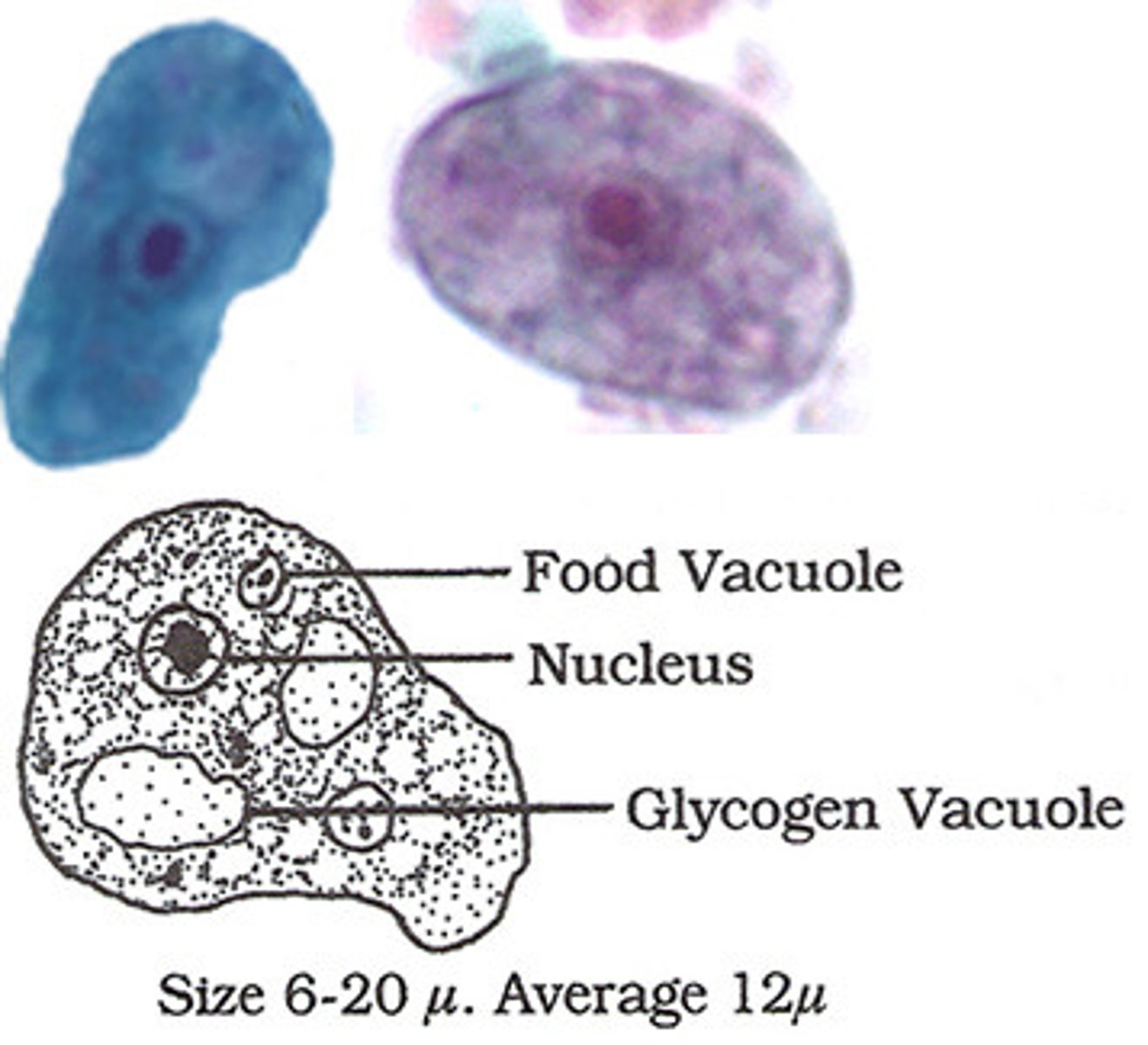
Endolimax nana (cyst)
*up to 4 nuclei
*ovoid shape
Endolimax nana (trophozoite)
*karysome is larger and deeper than E. hartmanni
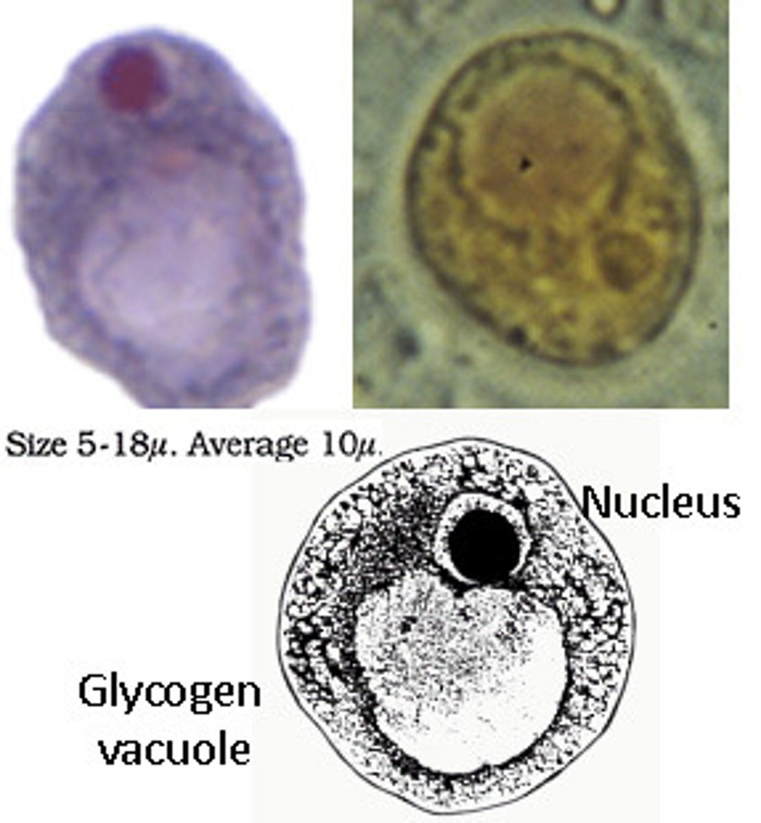
Iodamoeba butschlii (cyst)
Iodamoeba butschlii (trophozoite)
*has visible glycogen vacuoles
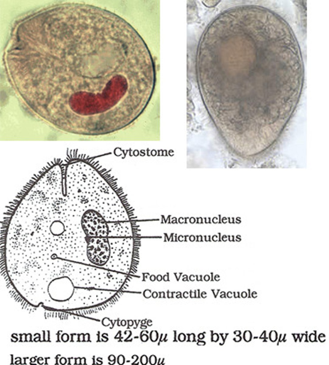
Balantidium coli (trophozoite)
*more oval than the cyst
*ciliated
*CYTOSTOME (the little asscrack looking thing)
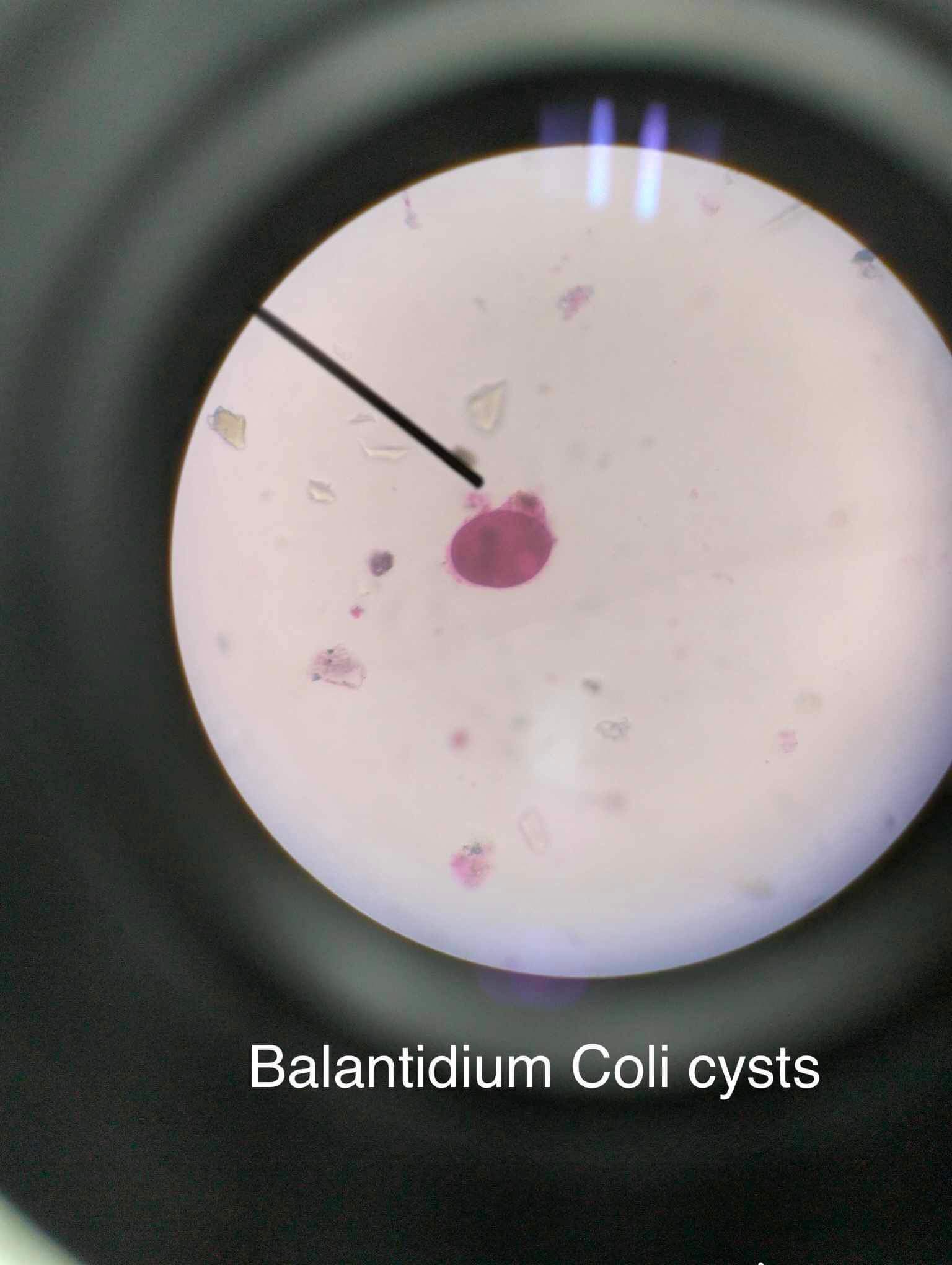
Balantidium coli (cyst)
*large circle with dumbbell looking nucleus
Chilomastix mesnili (trophozoite)
*4 anterior flagella
*curved posterior
Chilomastix mesnili (cyst)
*looks like it has fish hooks inside
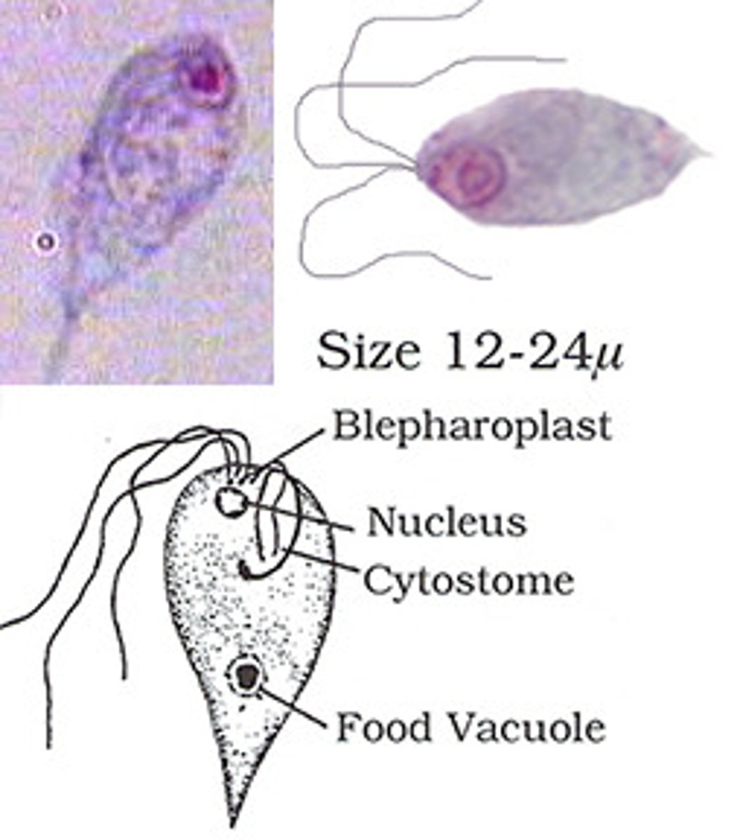
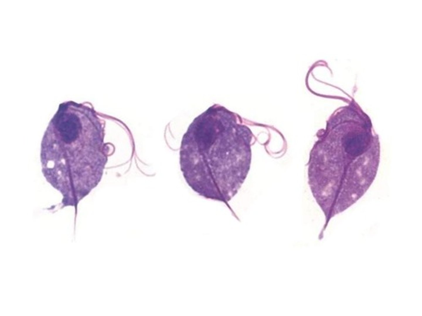
Giardia duodenalis/lamblia (trophozoite)
*flagella
*looks like old woman
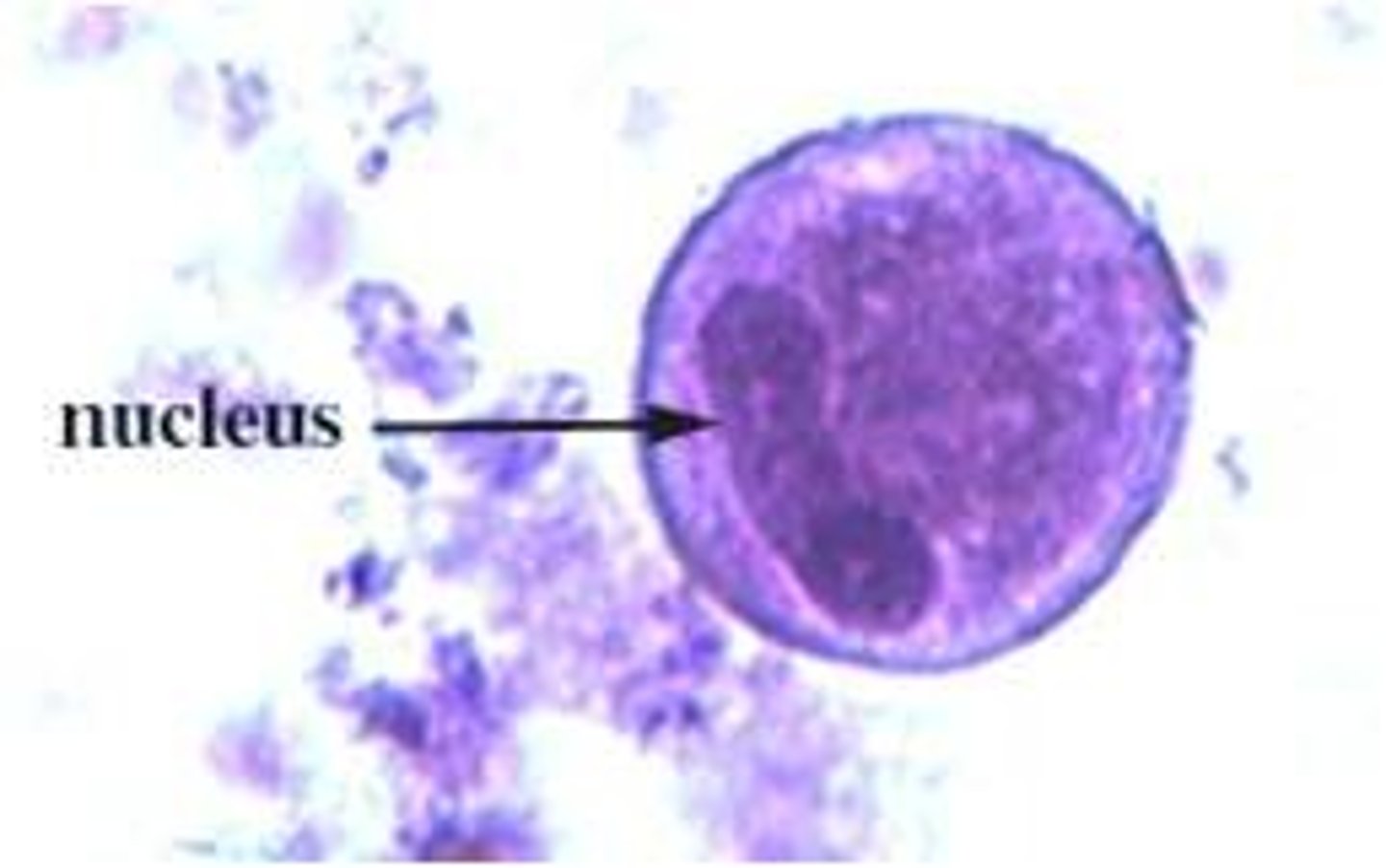
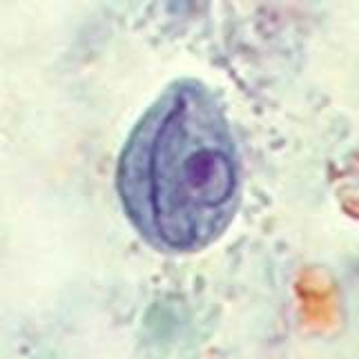
Dientamoeba fragilis (trophozoite)
*two nuclei
*no cyst
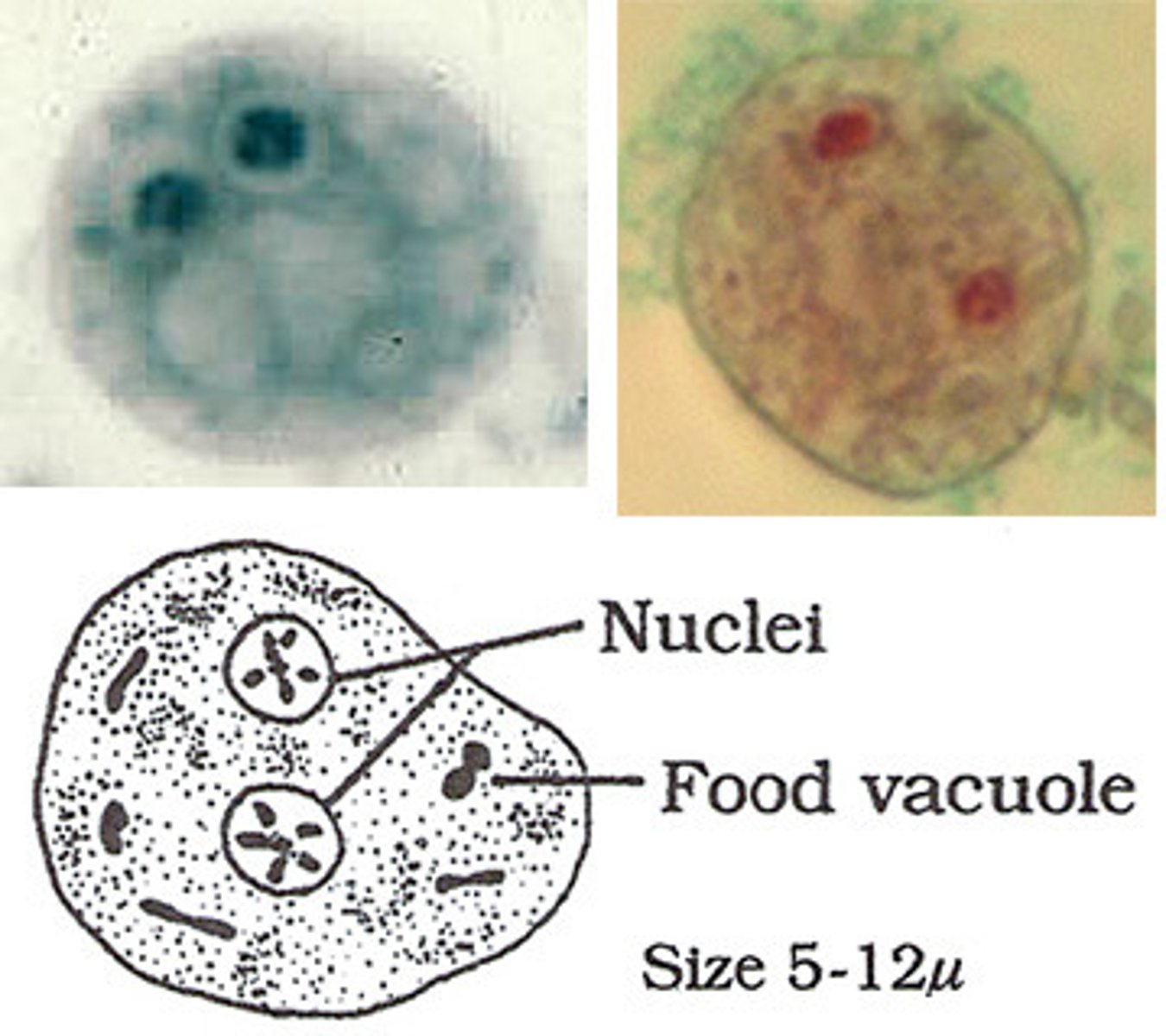
Giardia lamblia (cyst)
*no flagella, you can see them developing inside
*4 nuclei
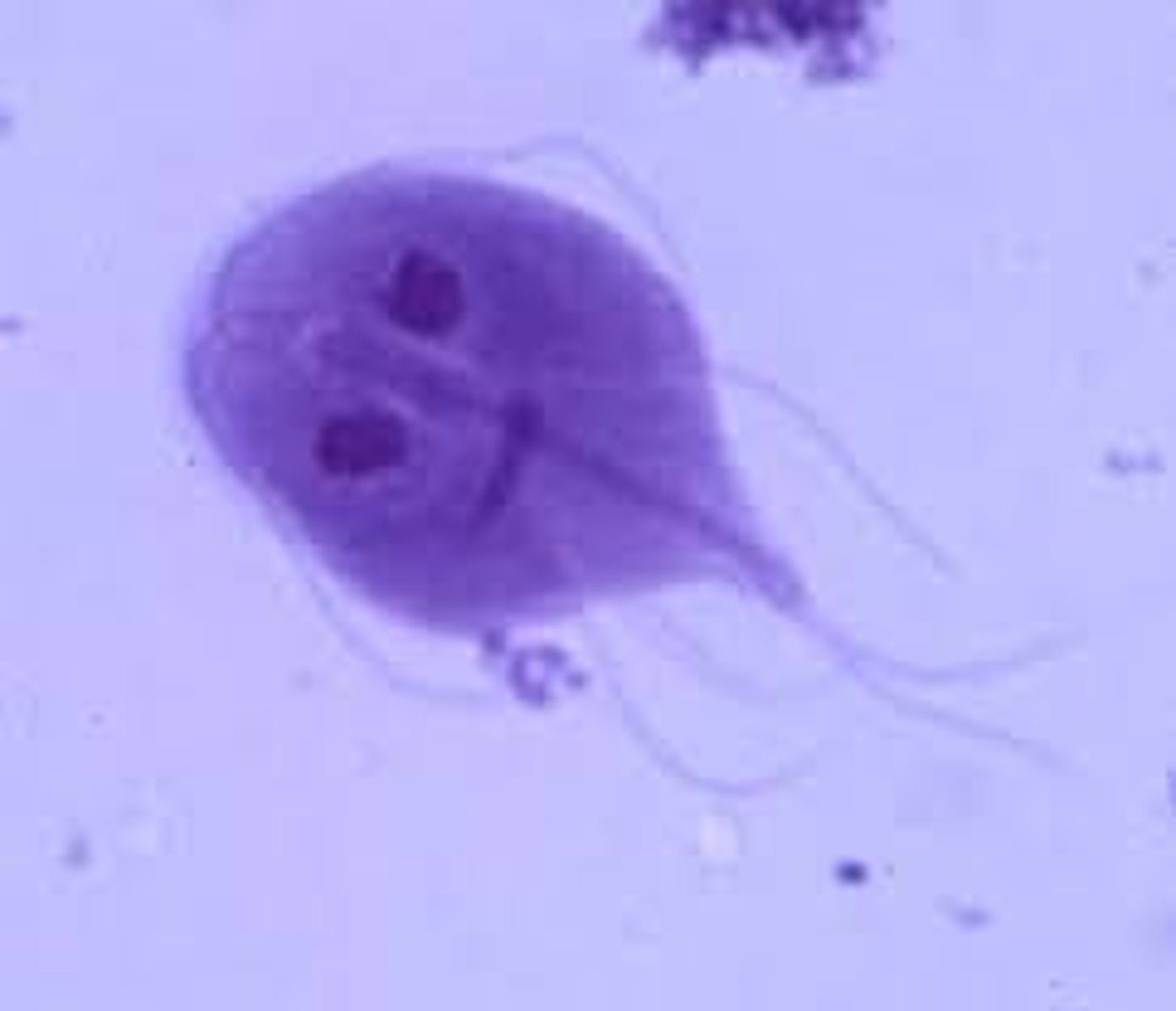
Giardia lamblia/lamblia (trophozoite)
*note flagella
*looks like old woman
Trichomonas vaginalis
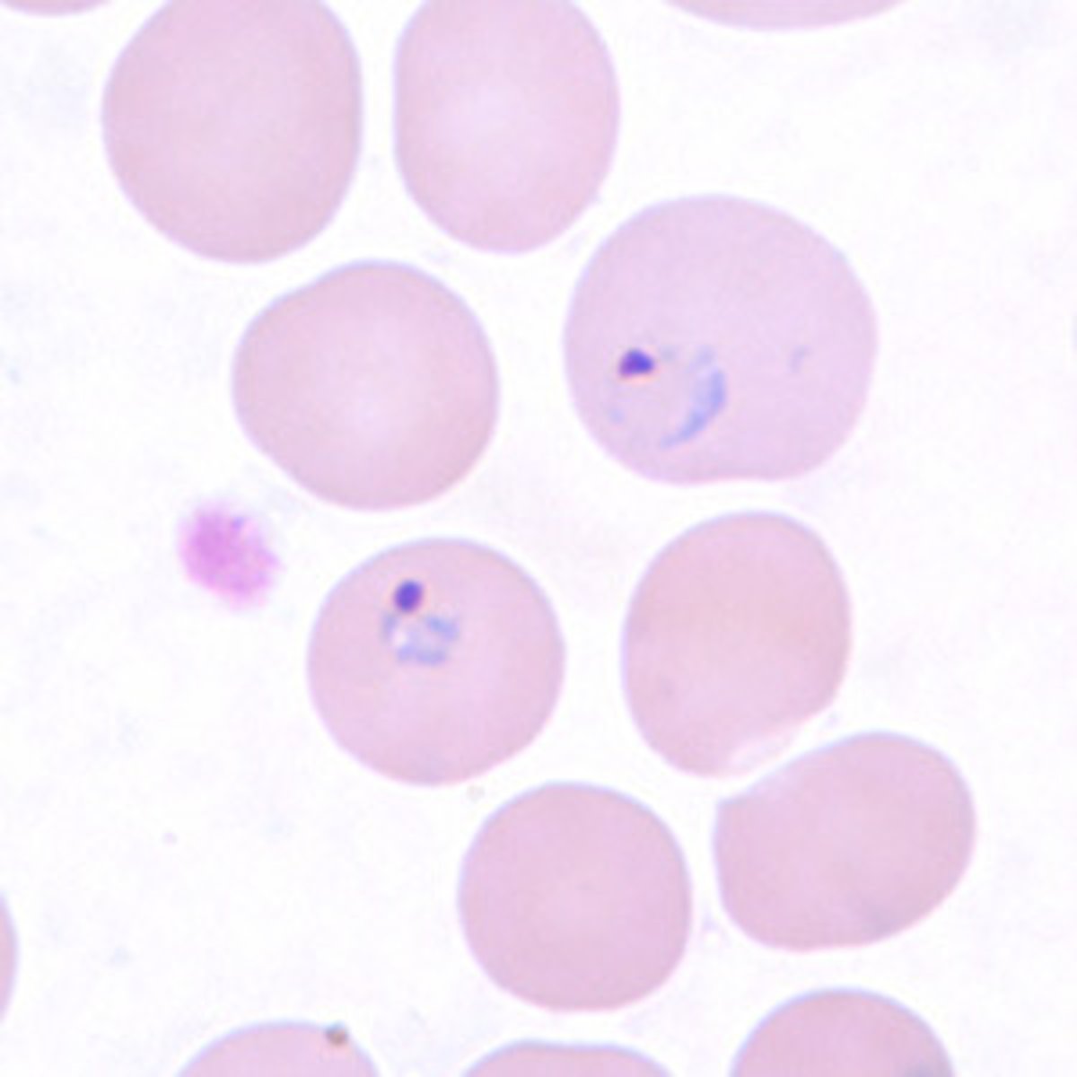
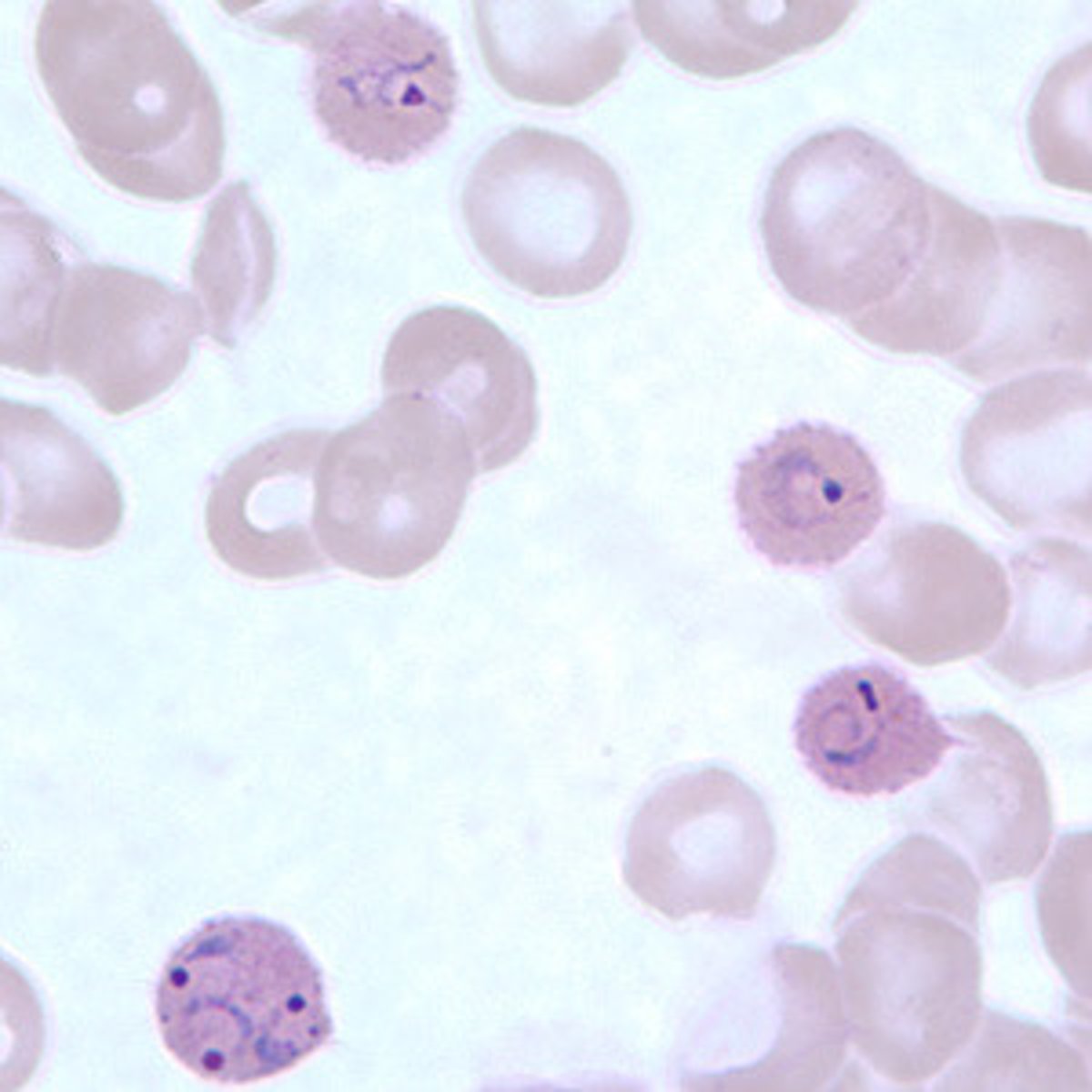
Plasmodium vivax (rings)
*RBCs become enlarged when infected, may be distorted
Plasmodium vivax (trophozoite)
*the one on the left is mature
Plasmodium vivax (shizont)
*12-24 merozoites, yellowish-brown pigment
*may stain darker as well
Plasmodium falciparum (rings)
*RBCs are not enlarged
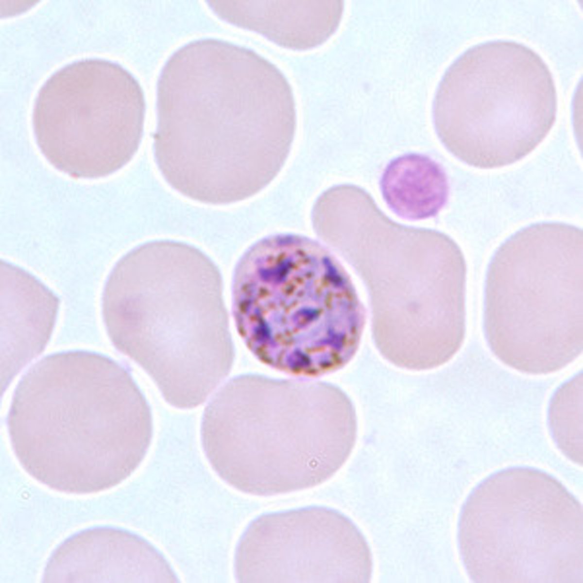
Plasmodium falciparum (schizont)
*rarely seen in peripheral blood
Plasmodium ovale (rings)
*multiplied infected rings
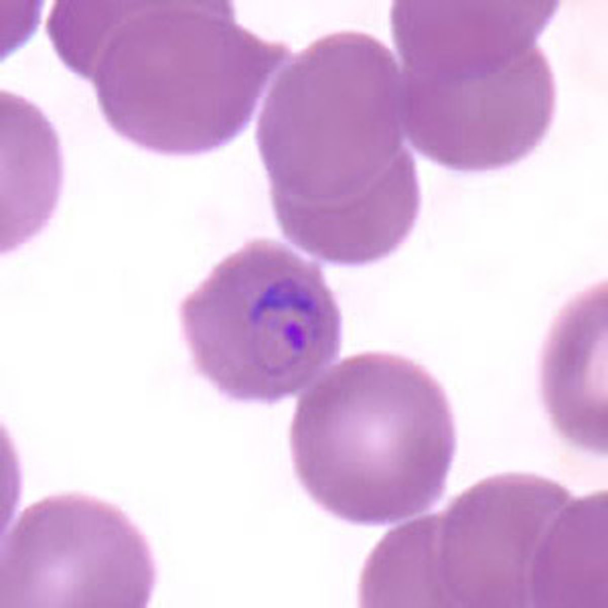
Plasmodium ovale (gametocyte)
*round to oval, pigment is brown and is more coarse compared to P. vivax
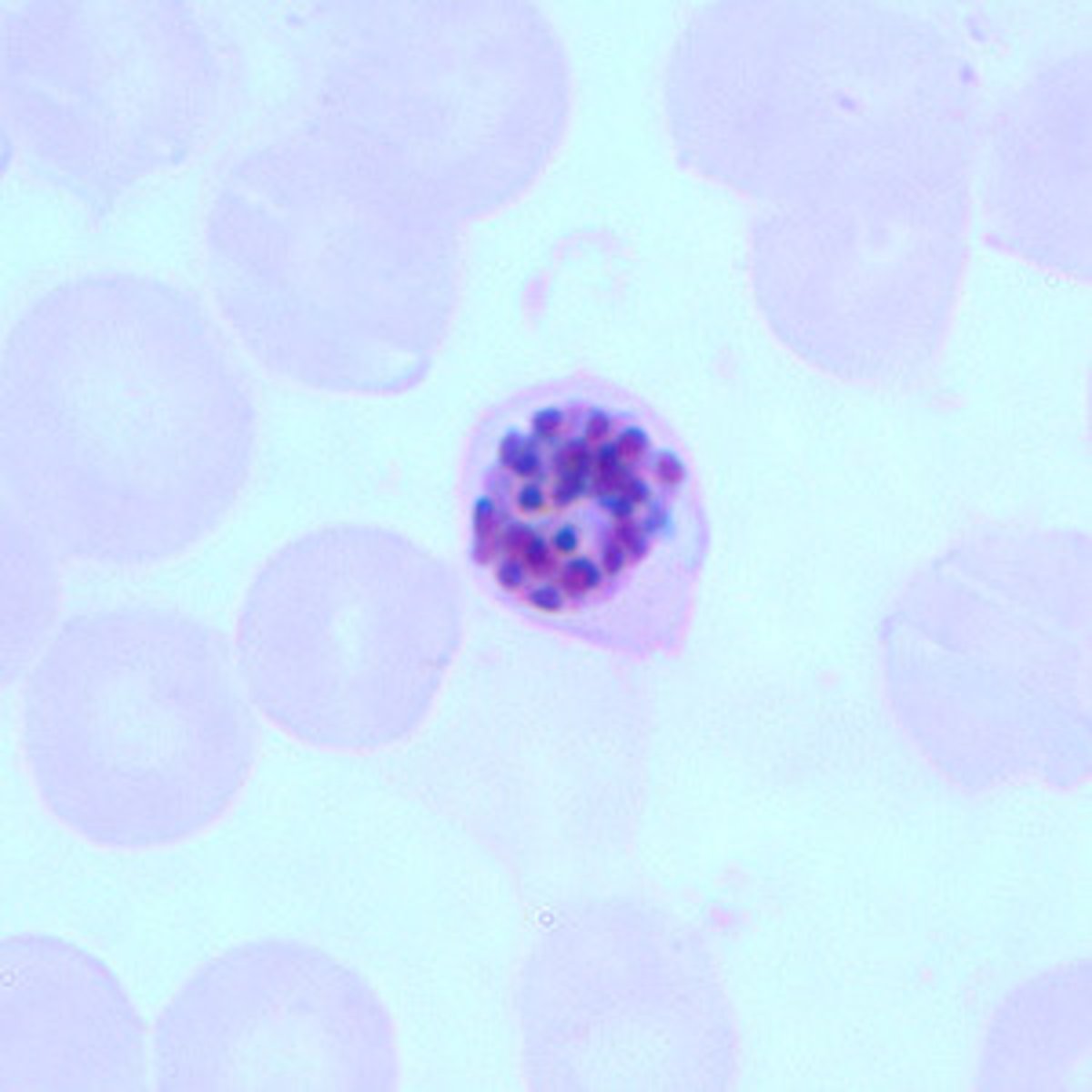
Plasmodium ovale (schizont)
*6-14 merozoites with large nuclei clustered in a mass of dark pigment
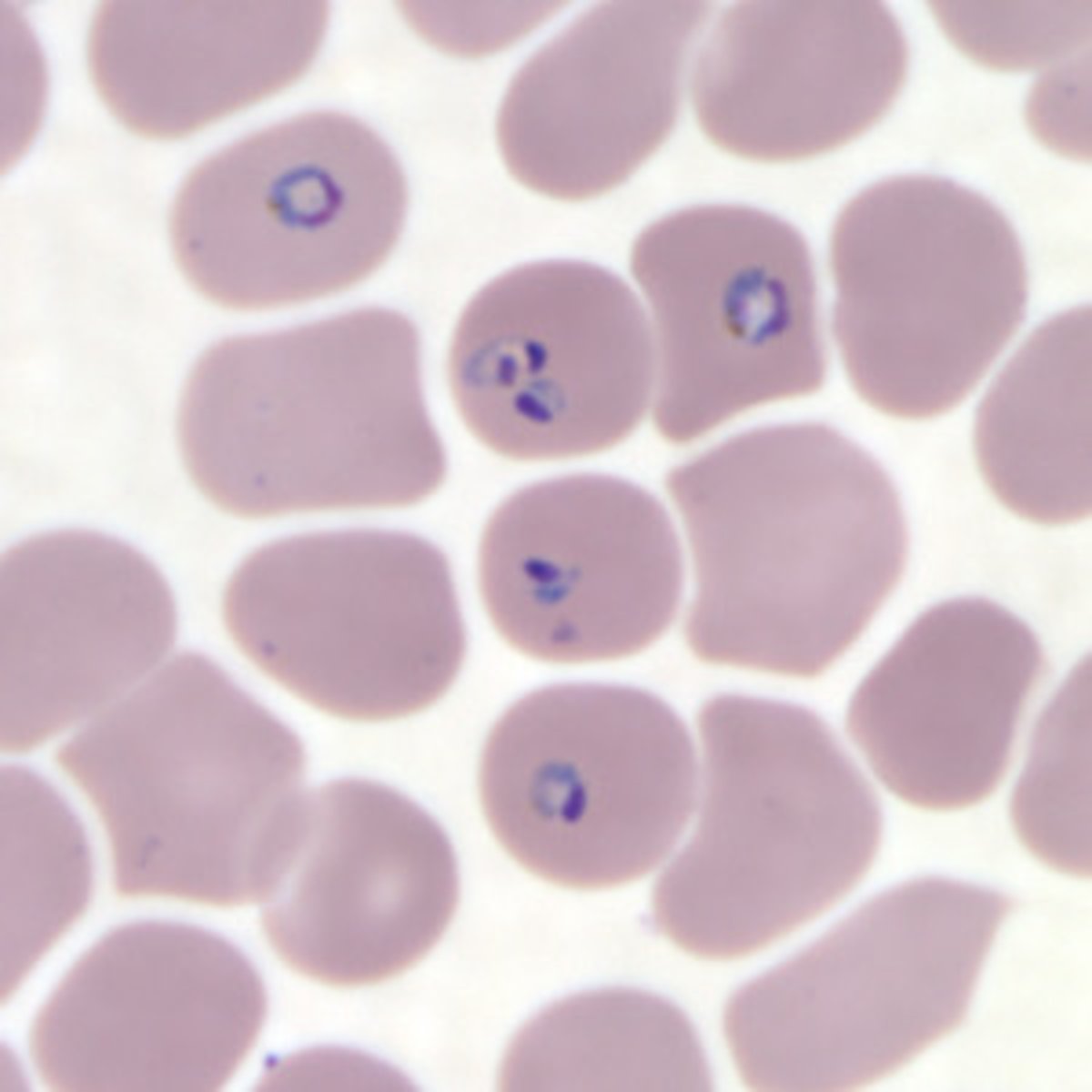
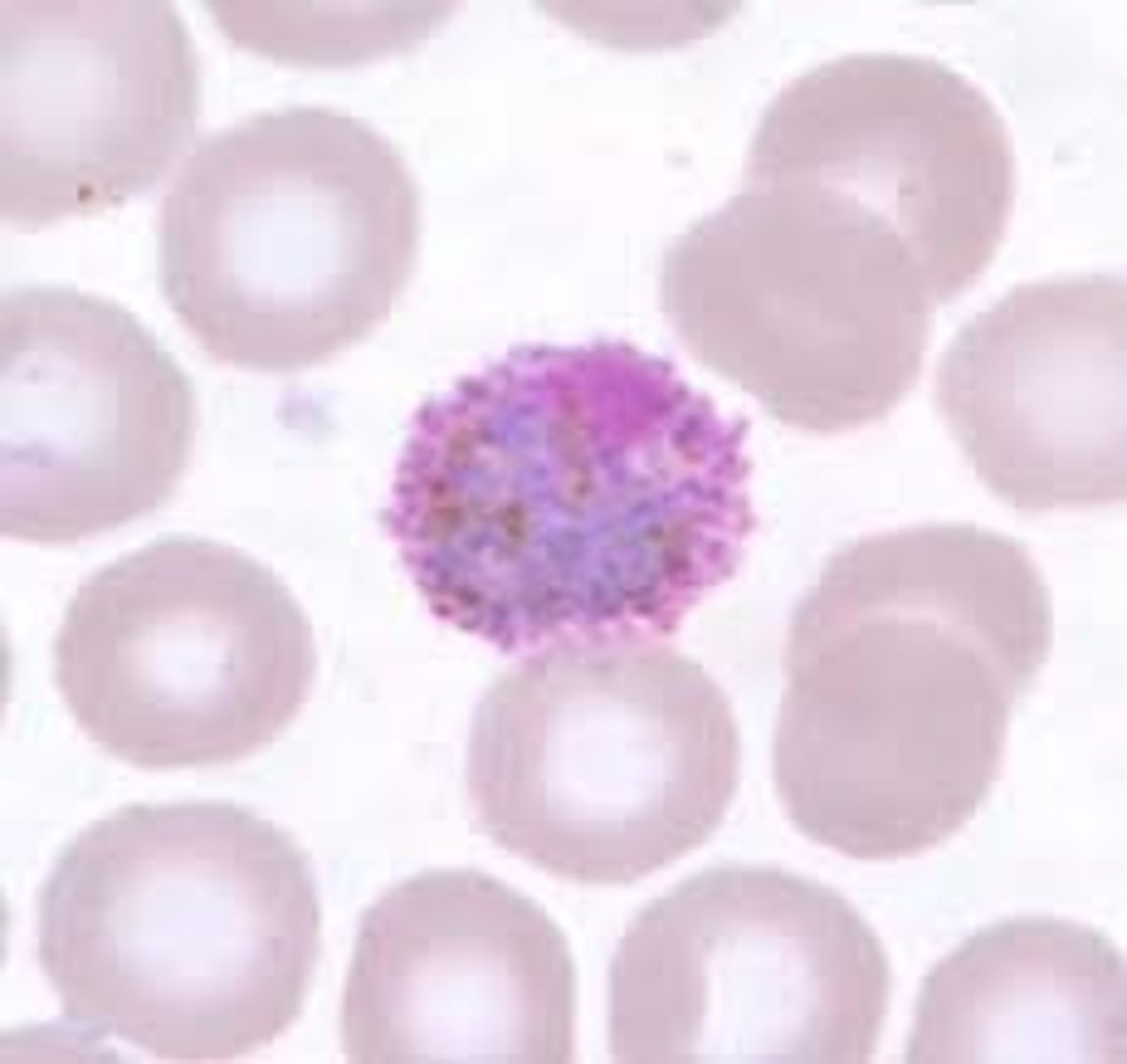
Plasmodium malariae (rings)
*large chromatin dot
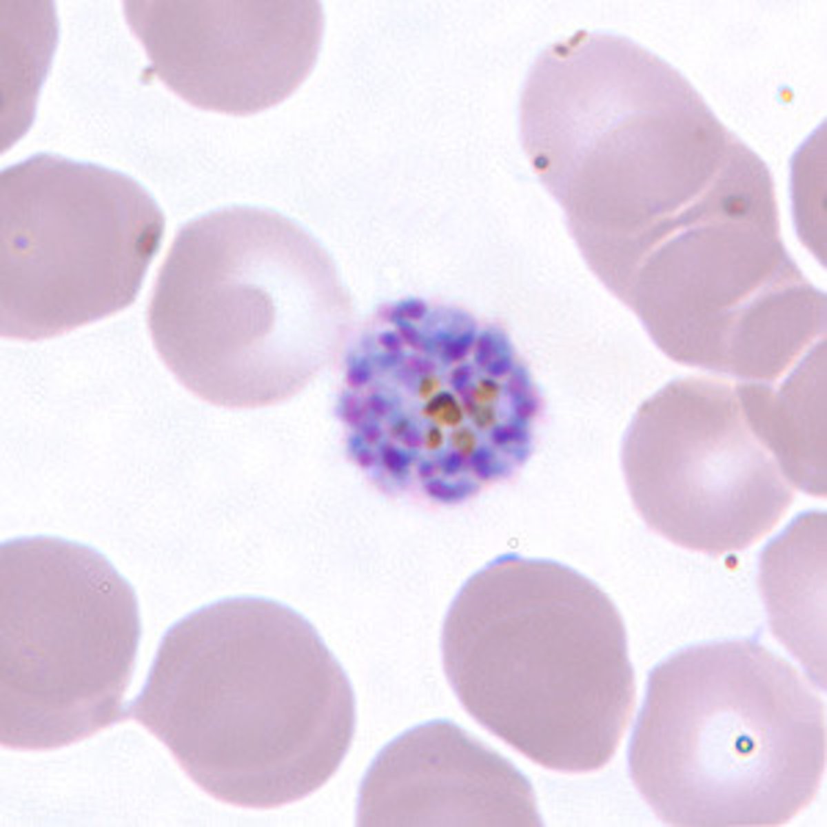
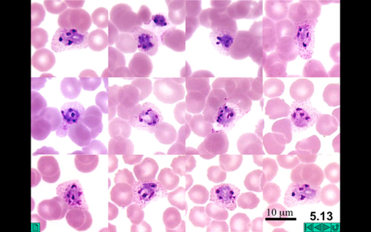
Plasmodium malariae (schizont)
*6-12 merozoites with large nuclei
*clustered around a mass of coarse dark-brown pigment
*sometimes arranged in a rosette pattern
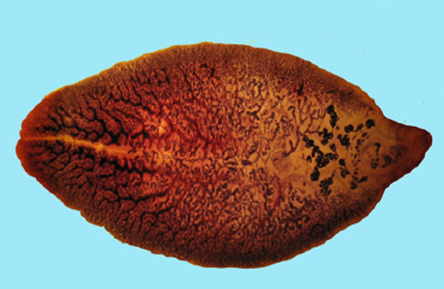
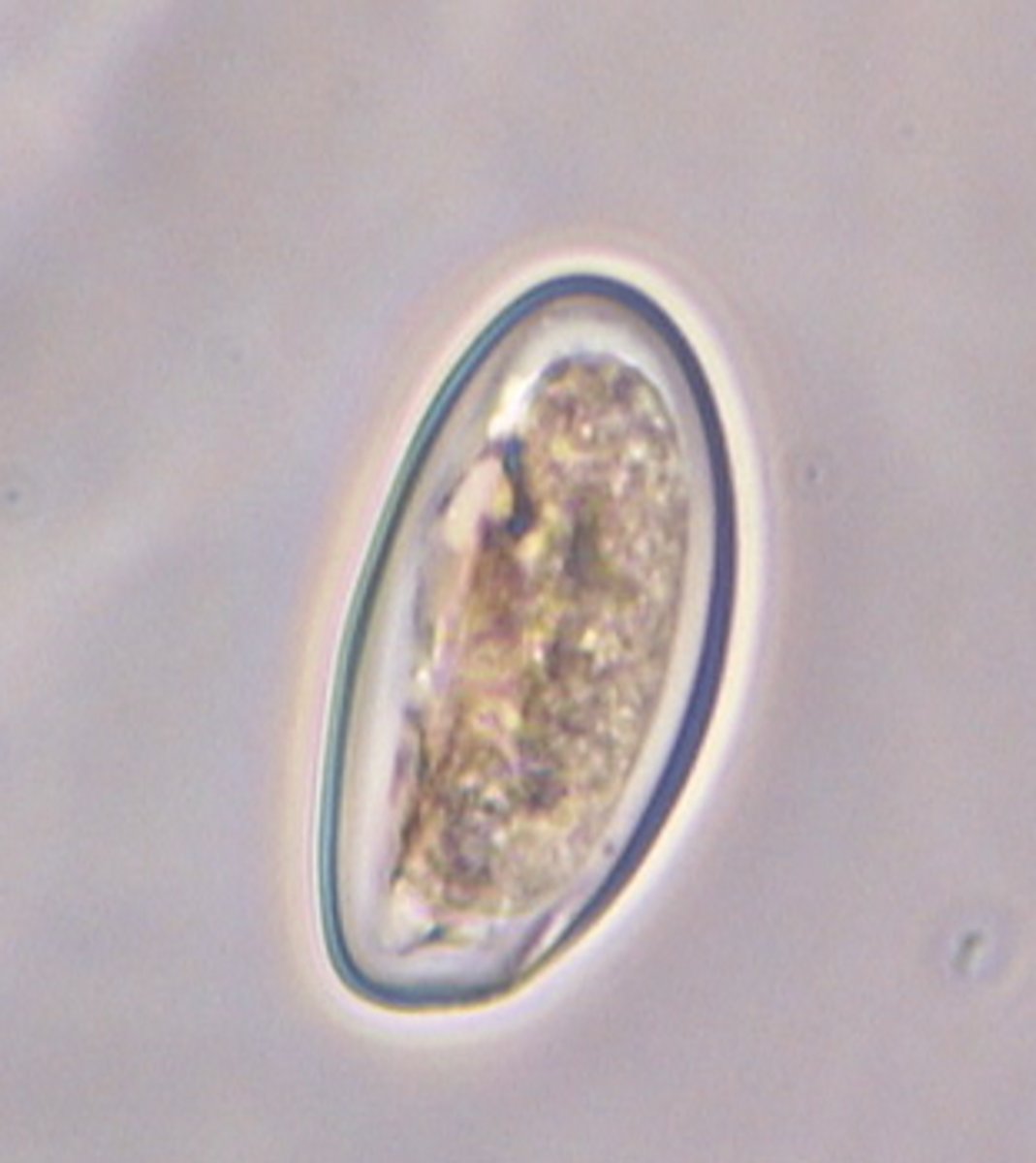
Fasciola hepatica ova