L3: Dendrites and dendritic integration
1/54
There's no tags or description
Looks like no tags are added yet.
Name | Mastery | Learn | Test | Matching | Spaced | Call with Kai |
|---|
No analytics yet
Send a link to your students to track their progress
55 Terms
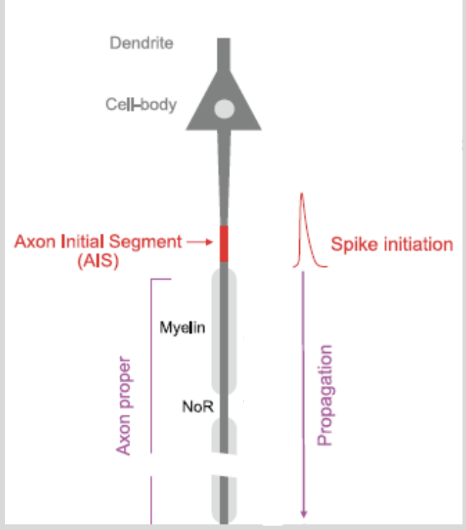
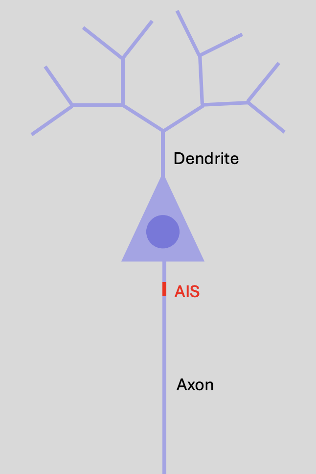
Dendrites and dendritic integration: Axon initial segment
site at which AP is initiated
between soma and first myelinated segment
has a lower threshold for excitation→ certain types of VG channels

Dendrites and dendritic integration: AIS features
has subtypes of voltage gated Na+ channels
some that are more sensitive to voltage
→ require less depolarisation to open
some higher density of voltage gated Na+ channels
→ contributing to a larger depolarisation in that region
Dendrites and dendritic integration: what determines whether the AIS is depolairsed sufficiently to inititate an AP?
Depends on the dendrites
Dendrites and dendritic integration: what do dendrites do
receive signals from other neurons:
some neurons receive >10,000 synapses in their dendrites
EPSPs and IPSPs
Propagate these to the AIS
Dendrites and dendritic integration: what is dendritic integration
how the dendrites process and propagate these signals to the AIS
How dendrite morphology and electrical properties influence the voltage at the AIS
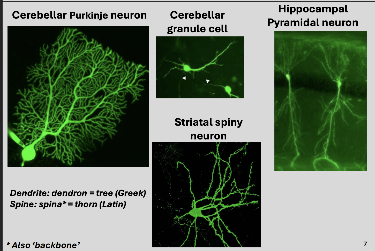
Dendrites and dendritic integration: Features or dendrites
can be simple of complex
different lengths (distance from AIS)
different thicknesses→
Thicker when closer to soma
more or less branched
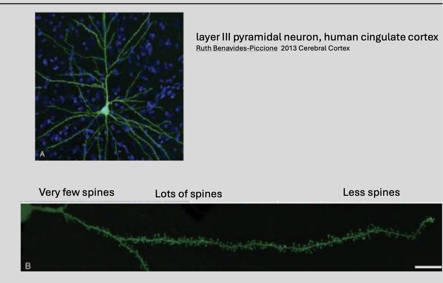
different numbers of spiney protuberances→ wwhere some synapses form (different distributions)
Even in a single neuron→ can be more than one dendritic tree
each having different properties
This can be in DIFFERENT neurons or even in the SAME neuron
More than one dendritic tree→ having different properties examples
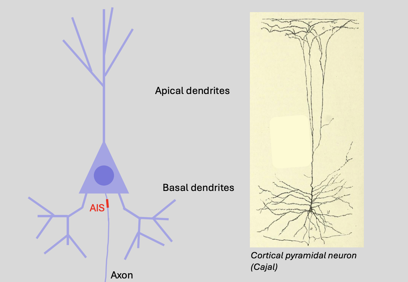
basal and apical dendrites of cortical pyramidal neurons
density of spines can vary along the dendritic tree
signal processing can be different between the apical and basal dendrites
a→ the axon in the image on the right
Therefore dendritic integration is affected by
morphology of dendrites
(also) functional influences
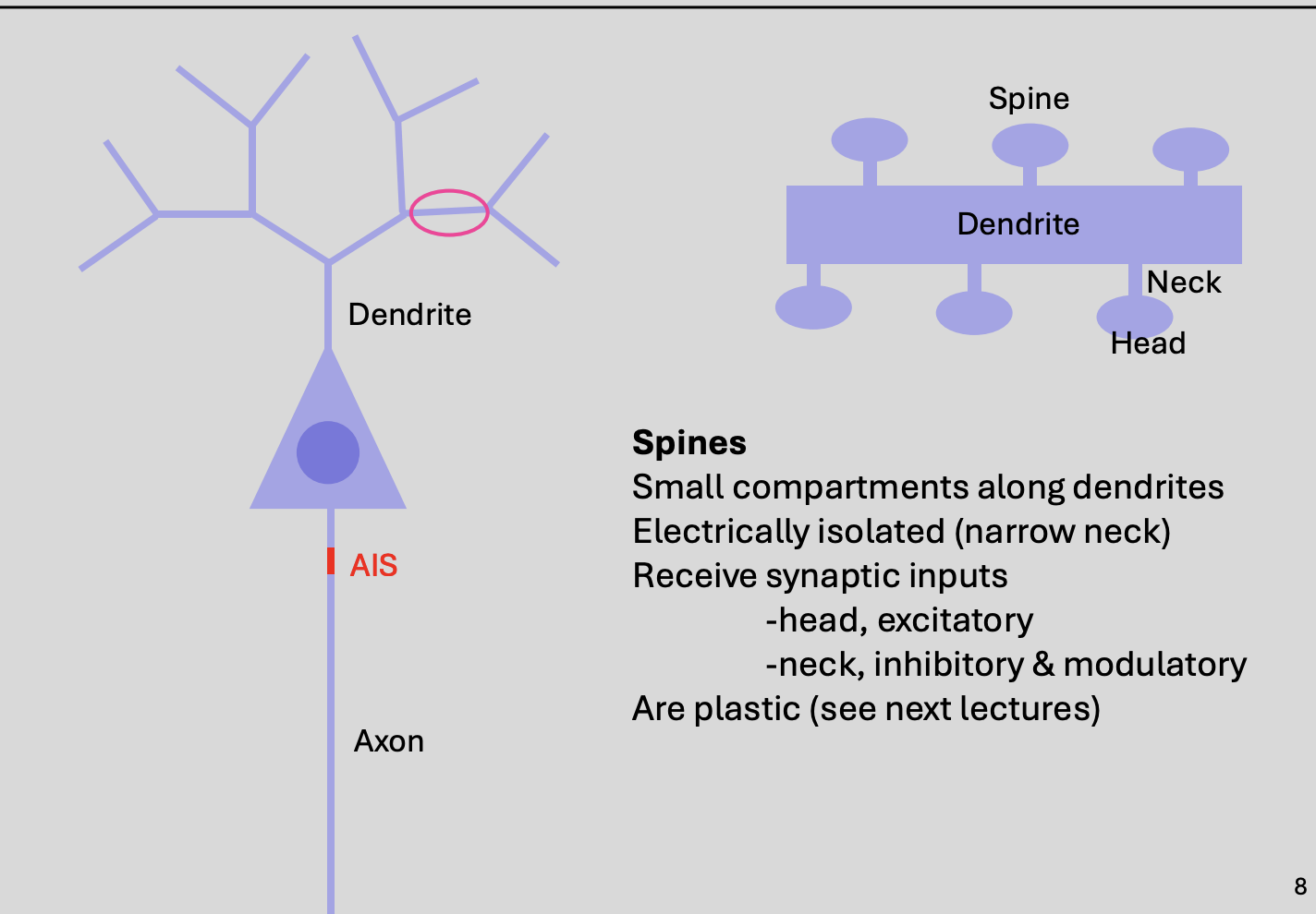
Spines on dendrites, features
WHAT small compartments along dendrites
to increase SA for synapses
electrically isolated→ narrow neck
WHERE receive synaptic inputs:
Head→ excitatory
Neck→ inhibitory
Plastic
most dendrites have spines
Another way dendrites can differ from eachother
distribution of spines
Dendritic potentials: dendritic signals are…
postsynaptic potentials
Dendritic potentials: what are dendritic signals mediated by in the mammalian CNS
ionotropic glutamate receptors
e.g AMPA receptors→ excitatory (EPSPs)
ionotropic GABA receptors
→ usually inhibitory (IPSPs)
this is due to their ionic permeability and the driving force on the permeant ions
Postsynaptic potentials are generated at…
chemical synapses
between presynaptic neuron and postsynaptic neuron
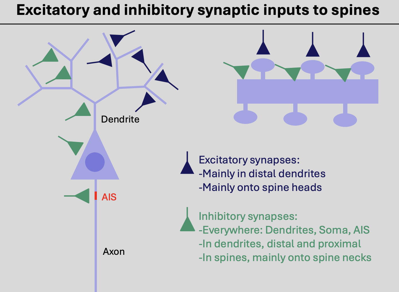
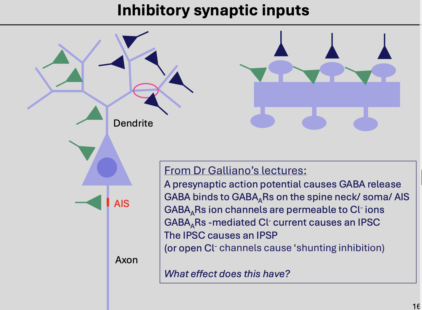
Excitatory and inhibitory synaptic inputs to spines, WHERE
Excitatory synapses form onto the
heads of dendritic spines
especially in the more distal dendrites
Inhibitory synapses form onto the
Necks of spines
WHERE: dendrites, soma, AIS → everywhere
distal AND proximal
note: that previous years of imagingand species looked at are limited→ so distribution may actually be different

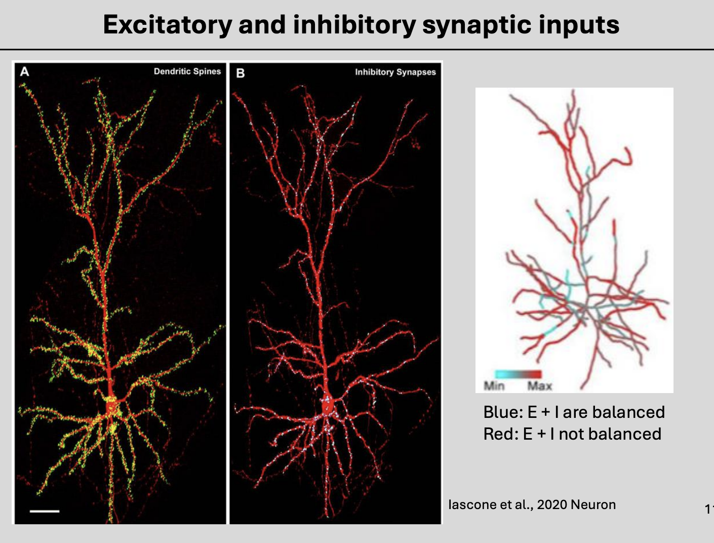
Relative number of inhibitory and excitatory synapses and location
will vary in different neurons
→ comparing distribution of the exand inhib synaptic inputs
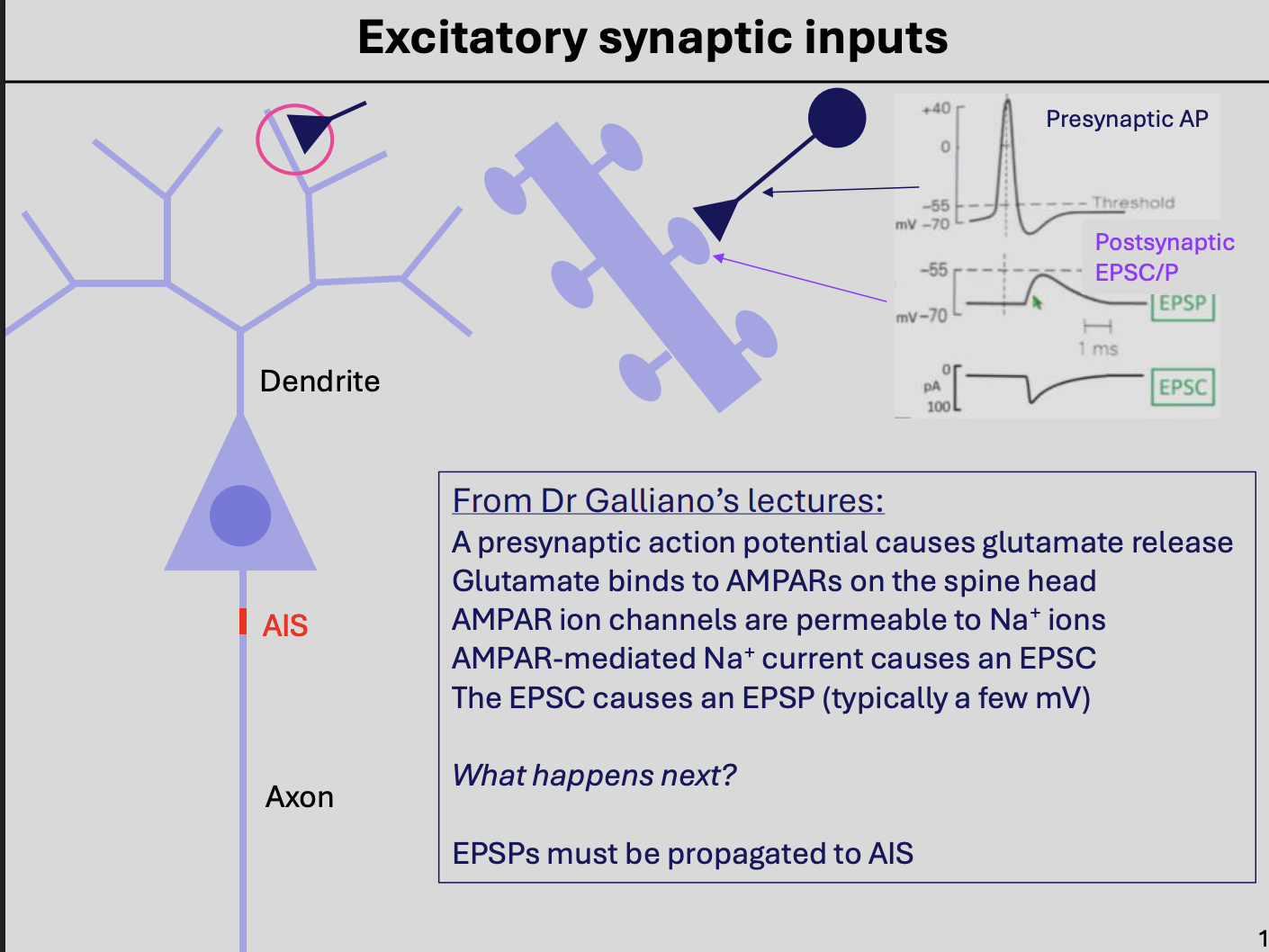
Excitatory synaptic inputs
presynpatic AP causes glutamte release
binds to AMPARs of the spine head
AMPAR ion channels→ permeable to Na+ ions
AMPAR-mediated Na+ current cuases EPSC
EPSC causes EPSP
But only a few mV
How does this propagate to AIS (even if so small?)
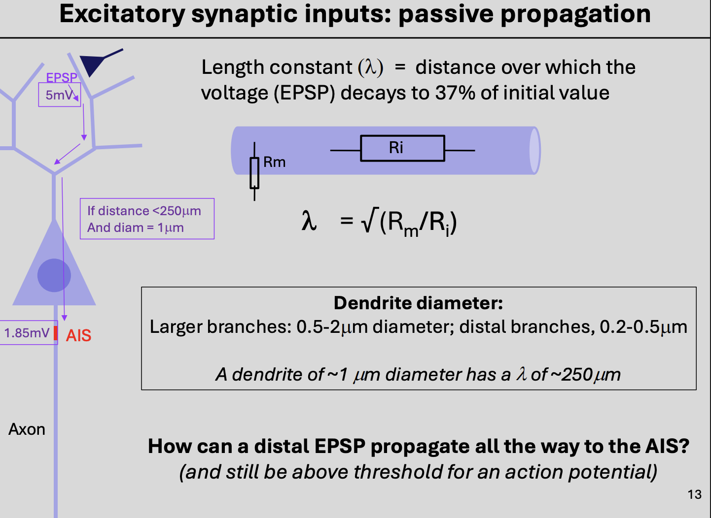
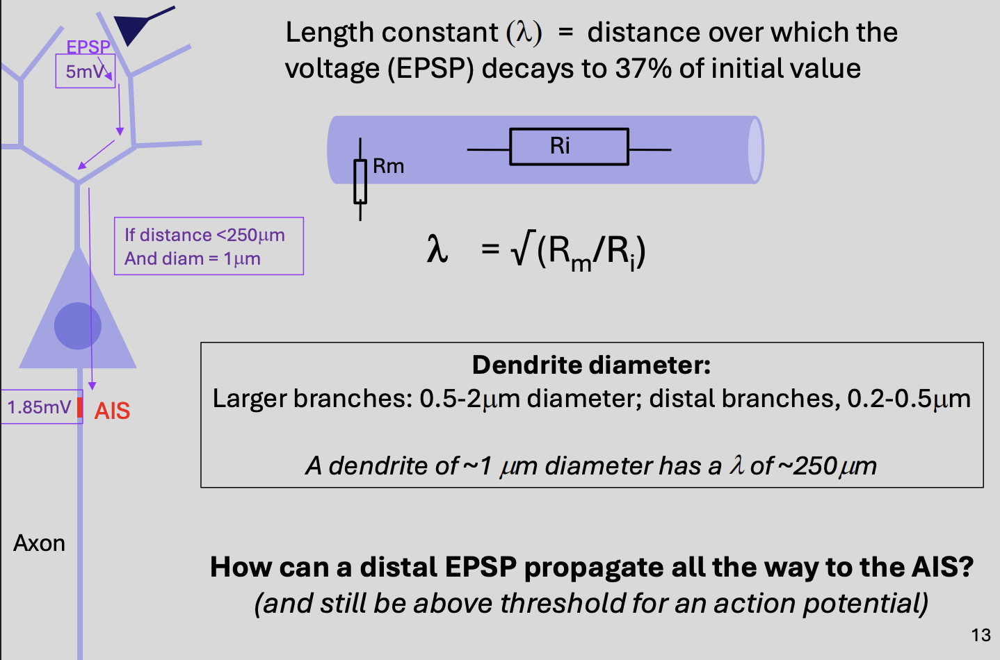
Length constant: what happens if an excitatory synpase is far out in the dendritic tree (distal dendrites)
EPSP must propagate along the dentries towards the soma
into axon to reach AIS
but…
Length constant: why is the passive propagation of a voltage signal along an axon or dendrite is not very effective?
Axons and dendrites are poor conductors of electricity
Due to high resistance to ionic movement within the cytoplasm and low resistance to ionic movement through ion channels in the membrane
i.e there is resistance:
low→ Causes ions to leak out (Rm)
high→ stopping ions moving through the neuron (Ri)
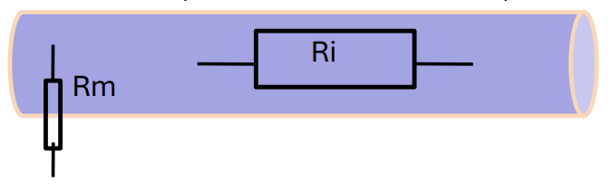
Length constant: what does the length constant describe
distance that a voltage signal will propagate bedore it decays to 37% of the initial value
For a large length constant we want
High Rm→ so stops ions leaking out
Low Ri→ so ions can move though axon quick

Length constant: if the resistance of the membrane is lower…
→ The length constant gets shorter
Why?:
More charged ions leak from the cytoplasm across the membrane
or
axon/dendrite diameter gets smaller (high resistance to current flow)
THEREFORE→ EPSPs will decay and be attenuated by the time they reach AIS

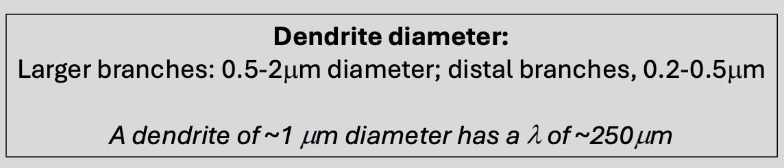
With an estimate of the denrite diamaer→ the length constant is
250 micro metres
How can a distal EPSP propagate ALL THE WAY to the AIS
However, because EPSPs are only a few mV in amplitude to start with
by the time they are passivley propagated to the AIS
→ they may depolarise the membrane there by less than 1-2mV
this is not enough to gate the Na channels at the AIS
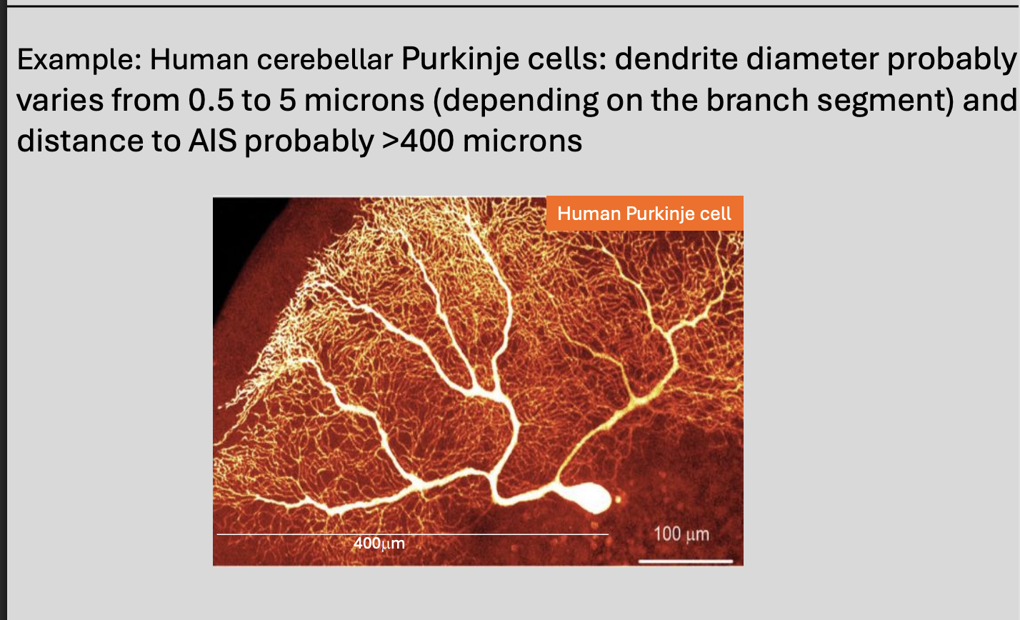
Example of human cell
Purkinje cells
length constant→ >400 microns
Then how does it travel so far???
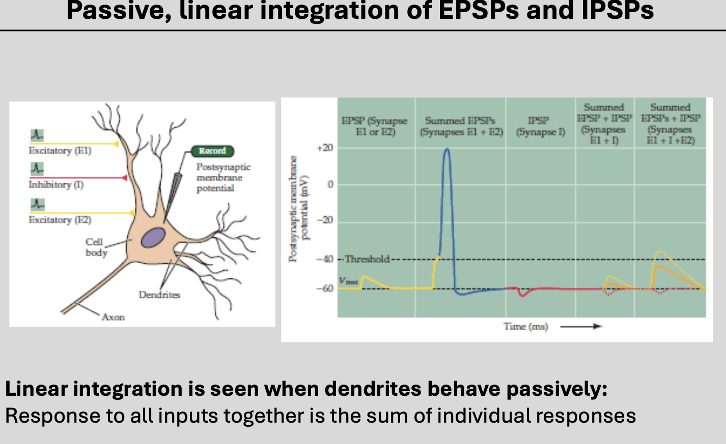
→ → SUMMATION (passive linear integration)
Passive integration properties of dendrites: EPSPs and IPSPs are graded signals, meaning…
if more neurotransmiter is released
will bind to and open more ligand-gated ion channels
cause more ions to move across the membrane of the dendrite
generate larger voltage changes
Passive integration properties of dendrites: what is the signal limited by?
Number of vesicles of neurotransmitter releasted
number of LGICs present at a synapse
how long they can be open for before neurotransmitter is inactiavted or removed
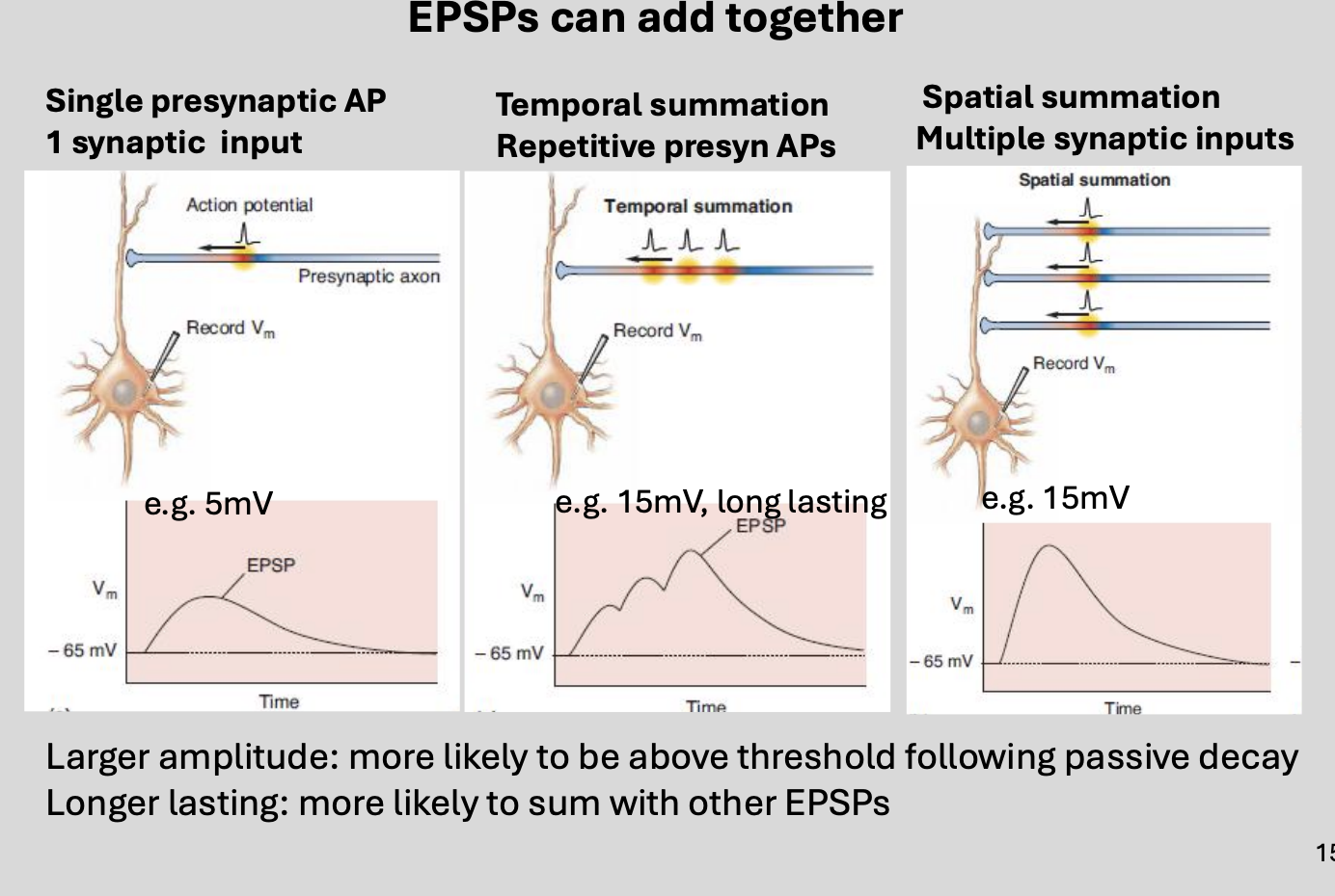
Passive integration properties of dendrites
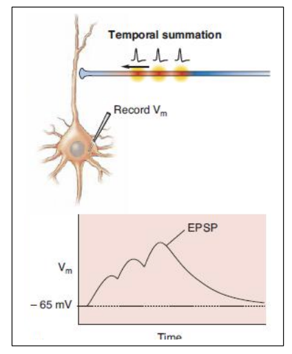
Temporal summation
Spatial Summation

1 Passive integration properties of dendrites: what happens at excitatory synapses
single presynaptic action potential releases glutamate
causing: EPSP

1 Passive integration properties of dendrites: what happens when a second presynaptic AP, before the first has decayed…
second EPSP might be added on top of the first
ALTHOUGH: depends on the number of releasable vesicle in the presynaptic active zone

1 Passive integration properties of dendrites: Therefore, a train of APs can lead to…
addition of EPSPs
→ SUMMATE TEMPORALLY to a larger amplitute

Passive integration properties of dendrites: spatial summation
typical CNS neuron have many different neuronal inuts
simulatenously activated
Spatial summation
Passive integration properties of dendrites: spatial summation why important?
can give EPSPs enough power to bring the membrane at AIS
→ to threshold voltage
Required to initiate an action potential

Different receptors have different likelihoods of summation…
AMPA
vs
NMDA
Because the AMPA receptor-mediated currents are transient…
a second synaptic stimulus will need to occur
within few milliseconds of the first one
→ in order to have an additive effect
however
NMDA receptors first have to be relieved of a voltage-dependent block of the channel by Mg2+ ions
things are complex (even without considering inhibition)
Because NMDA receptors mediate longer-lasting currents…
more likely to summate at lower frequencies of synaptic stimulation
But this summation of EPSPs must also work against…
IPSPs

Where do IPSPs typically occur at in the mammalian CNS
GABAergic synapses
or
glycinergic synapses

Features of these synapses
have integral anion channels
→ permeable to Cl-
usually mediate IPSPs or inhibition

How do inhibitory synaptic inputs occur
presynaptic AP causes GABA release
GABA binds to GABAARs on the spine/neck/AIS
GABAARs ion channels are permeable to Cl- ions
GABAARs→ mediated Cl- current cuases an IPSC
The IPSC causes an IPSP
or open Cl- channels cause ‘shunting inhibition’
What happens when the membrane permeability to Cl- increases?
Cl- ions will distribute themselves acorss the membrane
to maintain the Cl- equilibrium potential

why at rest or depolarised membrane potentials cause an inhibitory effect on membrane potential
Beause the movement of Cl- will tend to be from
outside→ into the cell

If ECl is near resting potential (as it often is), the effect of opening these channels…
increase the membrane permeability to Cl- ions
but
Without necessarily changing the membrane potential
→ ‘Shunting inhibition’
Why is this called shunting inhibition
positive charge from the EPSP is effectively shunted
(filtered or neutralised)
through raised conductance of the membrane
→ WITHOUT hyperpolarisation occurring
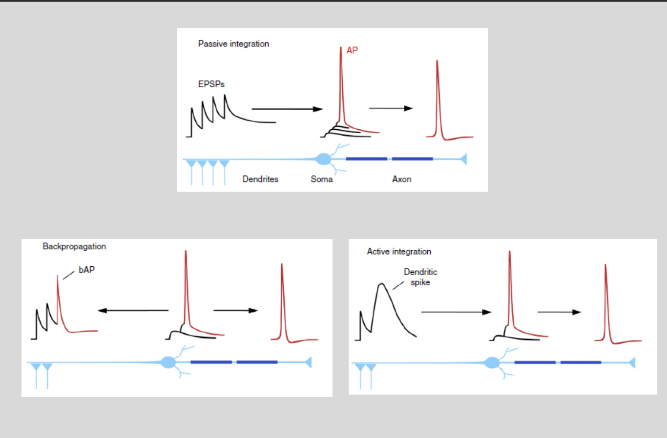
Active integration properties of dendrites: how do axons overcome their limitations as electrical conductors?
express voltage-gated ions channels
THEREFORE: they are active rather than passive conductors
Active integration properties of dendrites: are dendrites active or passive
most dendrites can passively conduct electricity less than 1mm
but there is some evidence to suggest dendrites are capable of active, non-linear transformations…
because they have VGICs
they do not show linear integration
e.g CNS dendrites can and do contain:
voltage-gated ion channels
including→ Na+, Ca2+ and K+ channels
=> leading to non-linear integration of inputs
Indications of this…
Dendrites of cerebellar Purkinje neurons→ report of active but poorly propagated signals in the
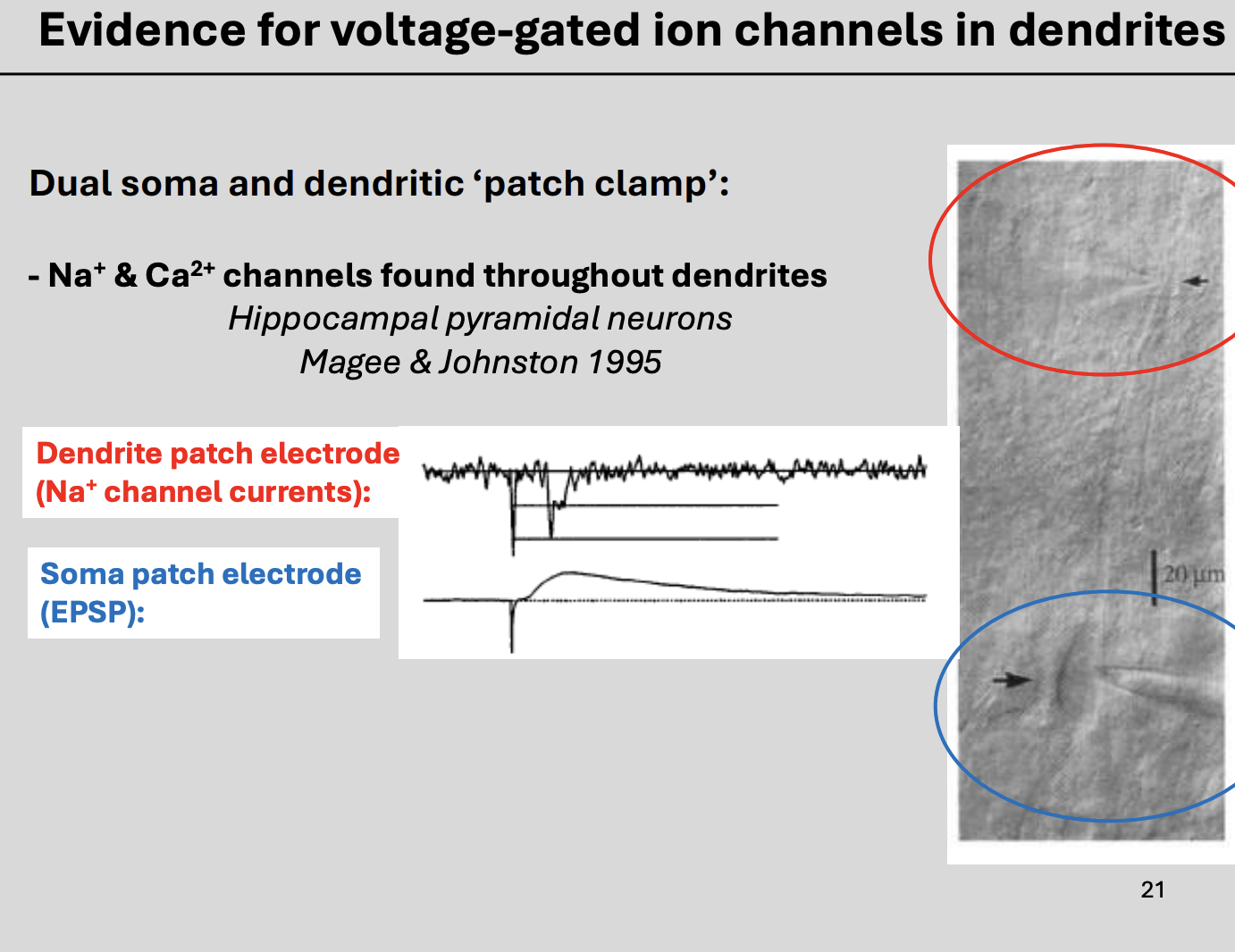
Mammalian CNS neurons→ direct measurements of voltage-gated ion channel currents→ see picture
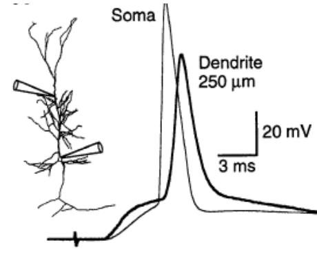
How was this second experimental reslt obtained?
better patch clamp recording method
development of higher resolution calcium imaging method
blue→ soma
red→ dendrite
Example of this new technology
simultaneous patch clamp recordings at the soma and in the dendrites of a pyramidal neuron showed:
Voltage-gated Na+ channels that resembled the action potential
seen in dendrite recordings
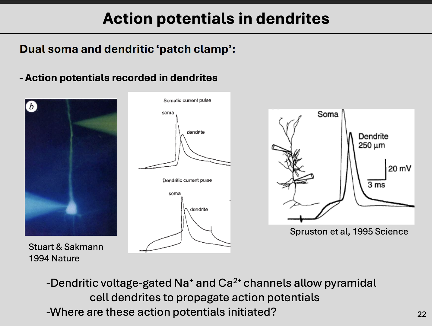
Example of this new technology: Because this occurred AFTER the AP at the soma
→ They are named ‘back-propagating action potentials (bAPs)’:
initiated at the AIS
then propagated simultaneously along the axon
back into the soma and dendrites
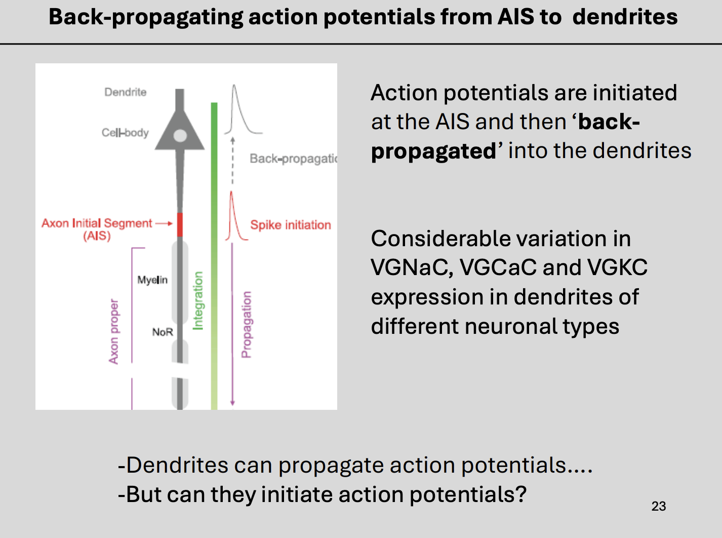
It is possible that these bAPS could contribute to…
depolarisation of the postynaptic membrane
at Excitatory synapses in spines
This enables…
relief of the magnesium block of the NMDARs
Facilitates NMDA receptor-dependent forms of synaptic plasticity
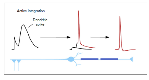
BUT could AP calso be initiated in the denrites?
in various neurons a high density of voltage-gated Ca2+ channels or voltage-gated Na+ channels in distal dendrites appear to support dendritic action potentials/spikes
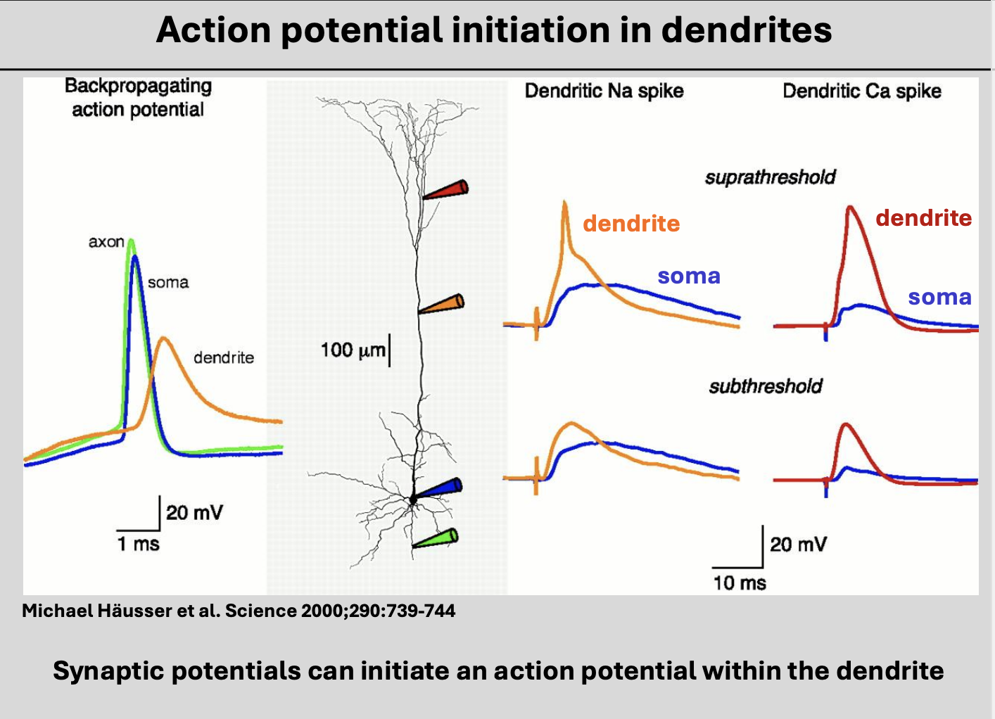
What does the initiation of Cav and Nav- mediated action potentials in distal dendrites likely to assist with?
→ forward-propagation of dendritic potentials from synapse to AIS
Overall types of dendritic integration
Top→ PASSIVE INTEGRATION
left→ BACKPROPAGATION (from AIS initiation)
right→ ACTIVE INTEGRATION (AP initiated in dendrites)
Review questions 1
1. What is a likely source of current that causes depolarization in the dendrites and activates VGICs?
2. What is meant by a ‘back-propagated action potential’?
3. Which ions/ ion channels can mediate action potentials in dendrites?
Review Questions 2
1. What is meant by ‘dendritic integration’?
2. Which intrinsic properties of dendrites influence integration.
3. What is a ‘spine’
4. What conductance is usually active during an IPSP?
5. Explain what is meant by ‘temporal summation’ and ‘spatial summation’.
6. Explain what is meant by ‘shunting inhibition’.