Cranial Nerves: Visual System - Visual Fields
1/24
There's no tags or description
Looks like no tags are added yet.
Name | Mastery | Learn | Test | Matching | Spaced | Call with Kai |
|---|
No analytics yet
Send a link to your students to track their progress
25 Terms
Sight — Visual Fields (VF) — CN II
visual field = what you see
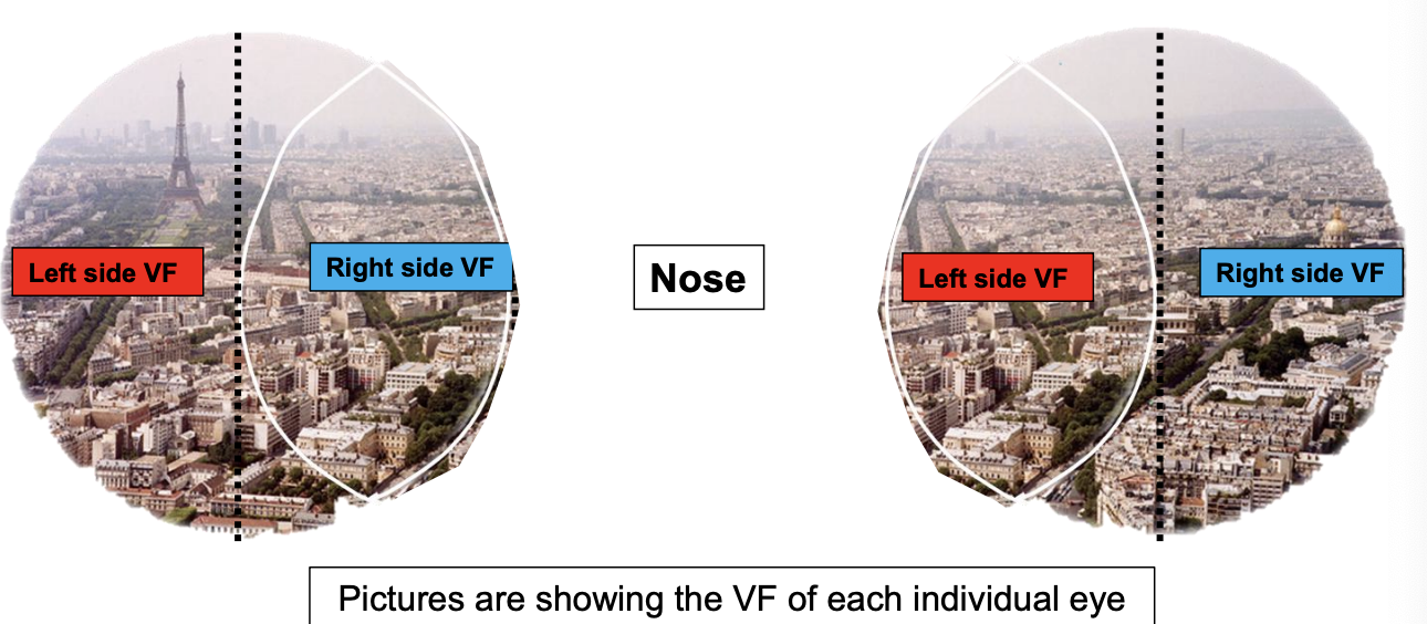
picture is showing the visual field of each eye when both eyes are opened
visual field is not the same as retina

Left vs Right VF
individually, each eye has a right VF and a left vf
Left vs Right Nasal VF
visual field for each individual eye can also be described as a temporal VF or a Nasal VF
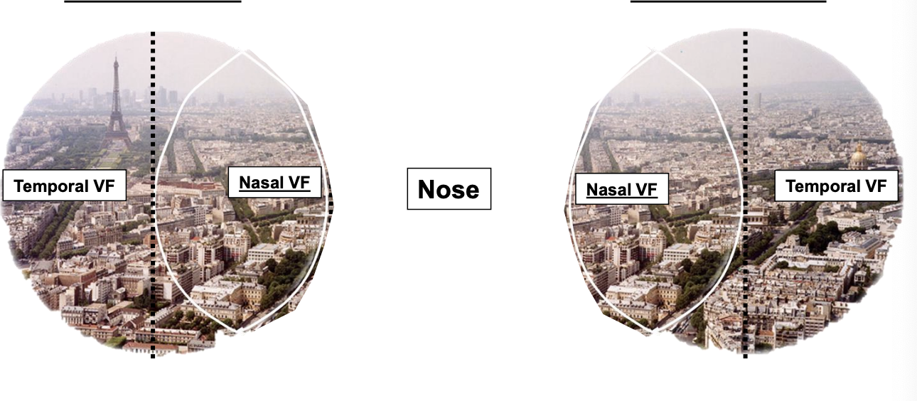
Temporal VF
the “outside” (lateral) image for each eye
Nasal VF
the “inside” (medial) image for each eye
Left/Right VF vs Nasal/Temporal VF
for the left eye:
its left side VF = temporal VF
right side VF = nasal VF
right eye:
left side VF = nasal VF
right side VF = temporal VF
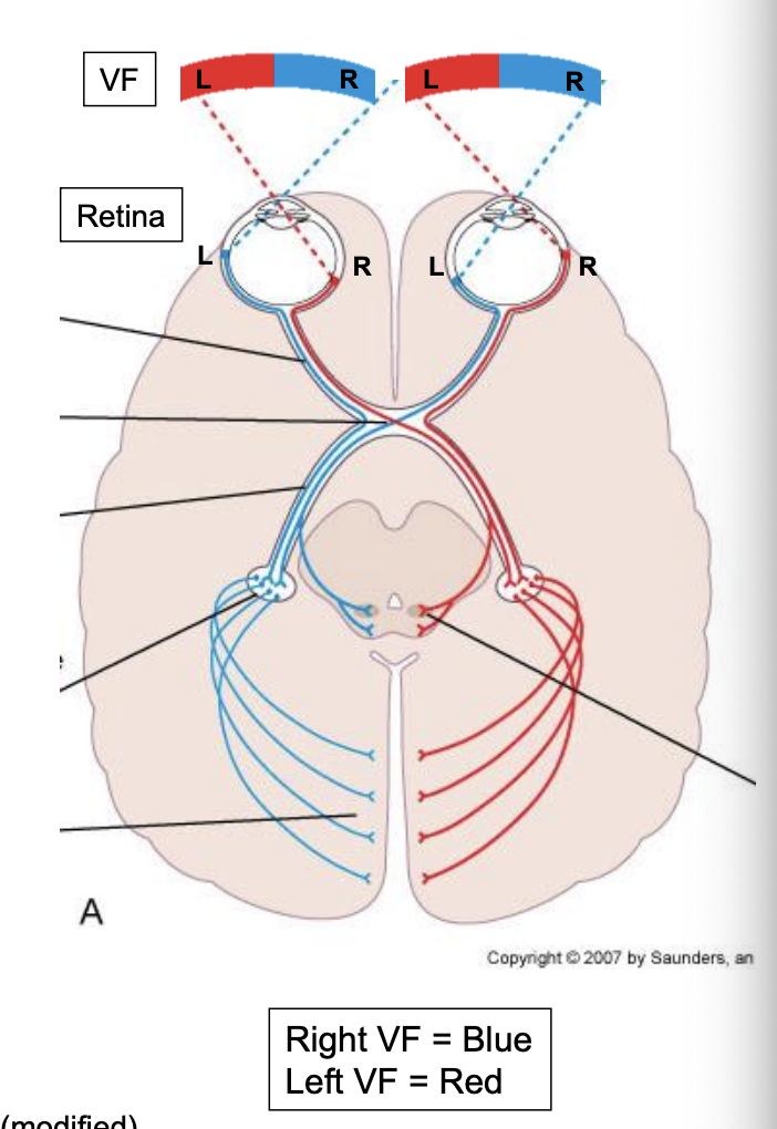
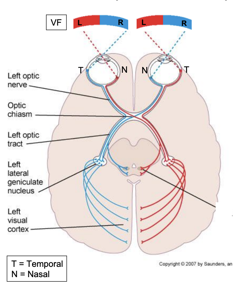
Visual Fields
information from the right visual field of each eye will end in the left primary visual cortex of the occipital lobe
information from the left visual field of each eye will end in the right primary visual cortex of the occipital lobe
uses 2 neurons:
1st neuron: from retina to the lateral geniculate nucleus (LGN) of the thalamus
2nd neuron": from LGN to primary visual cortex
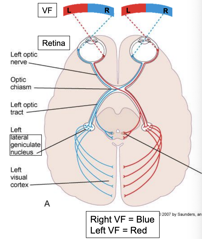
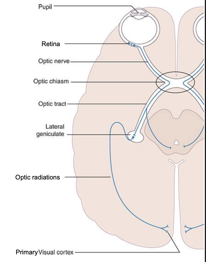
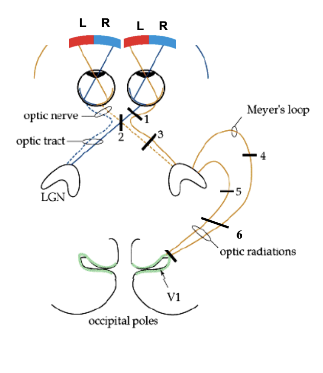
From Retina to Cortex
1st neuron axons in retina travel in CN II, through optic chiasm. optic tract then synapse in lateral geniculate nucleus (LGN) of thalamus
2nd neuron: axons travel form LGN through optic radiations to the primary visual cortex
this two neurons pathway is also called the retinogeniculocalcarine pathway
Primary visual cortex
processes conscious awareness of vision
registers basic information about size, shape, edges, texture of objects
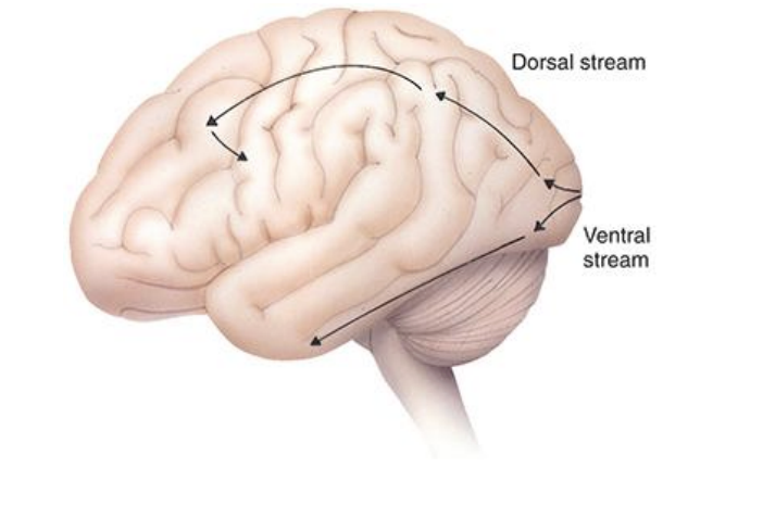
it does not provide information about color (secondary visual cortex) what the object is or how to move your eyes to follow the object. that requires input to other cortical areas
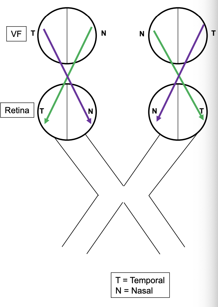
Retina — Overview
to reach the occipital lobe, the visual fields (light) need to first be processed by the retina
information from the right VF of each eye will end on the left side of the retina
information from the left VF of each eye will end on the right side of the retina
Remember
visual fields for each eye can be described as temporal VF or Nasal VF
the retina can also be described as having a nasal side and a temporal side
information from the nasal VF of either eye will end on the temporal side of the retina (Green)
information from the temporal VF of either eye will end on the nasal side of the retina (purple)
From optic to optic chiasm to LGN
info from temporal retina will stay ipsilateral on its way to primary visual cortex
info from nasal retina will cross over to the other side in the optic chias, and end in the contralateral primary visual cortex
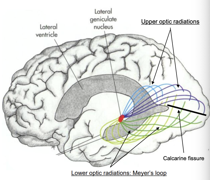
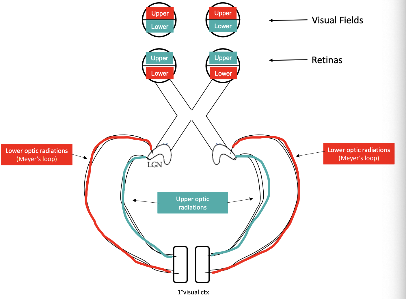
From LGN to Primary Visual Cortex
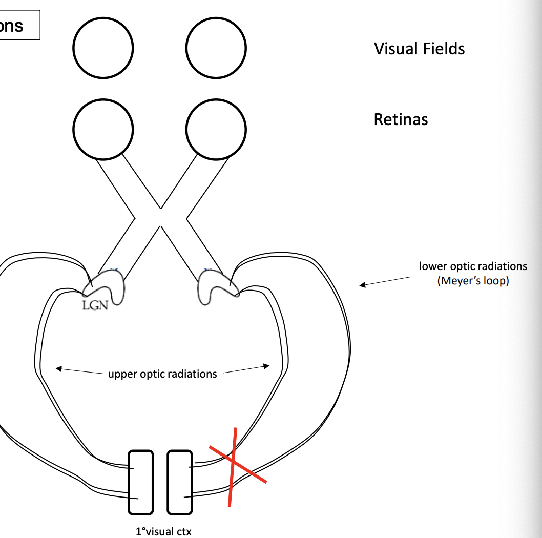
info from LGN reaches primary visual cortex via Optic radiations
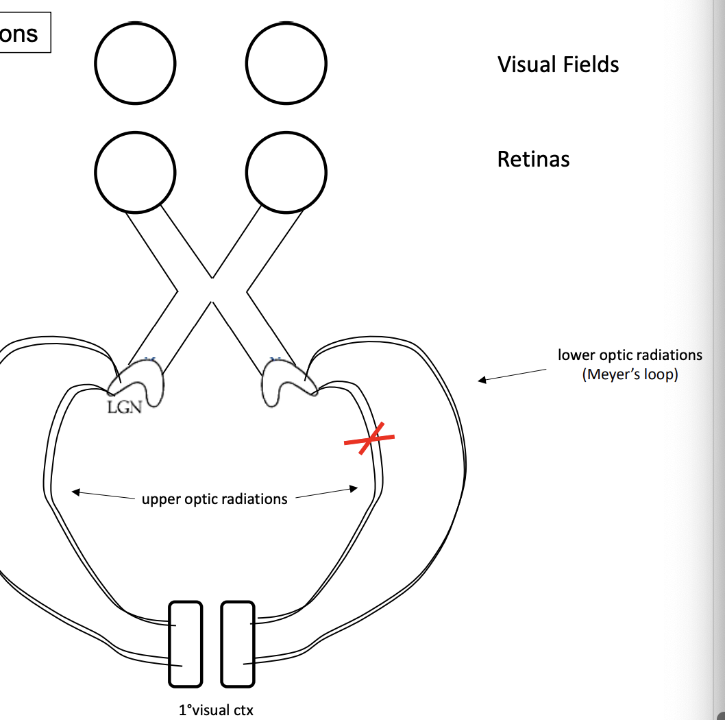
upper optic radiations
process lower visual field info from each eye
travel through parietal lobe then synapse in cuneus gyrus of primary visual cortex (occipital lobe)
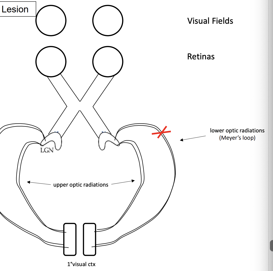
lower optic radiations (Meyer’s loop)
process upper visual field info from each eye
travel through temporal lobe then synapses in lingual gyrus of the primary visual cortex (occipital lobe)
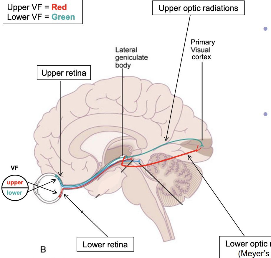
Upper/Lower Visual field and upper/lower retina
info from lower visual field goes to upper retina
once in LGN, axons use upper optic radiations (parietal lobe) to synapse in cuneate gyrus
info from upper visual field goes to lower retina
once in LGN, axons use lower optic radiations (temporal lobe) to synapse in lingual gyrus
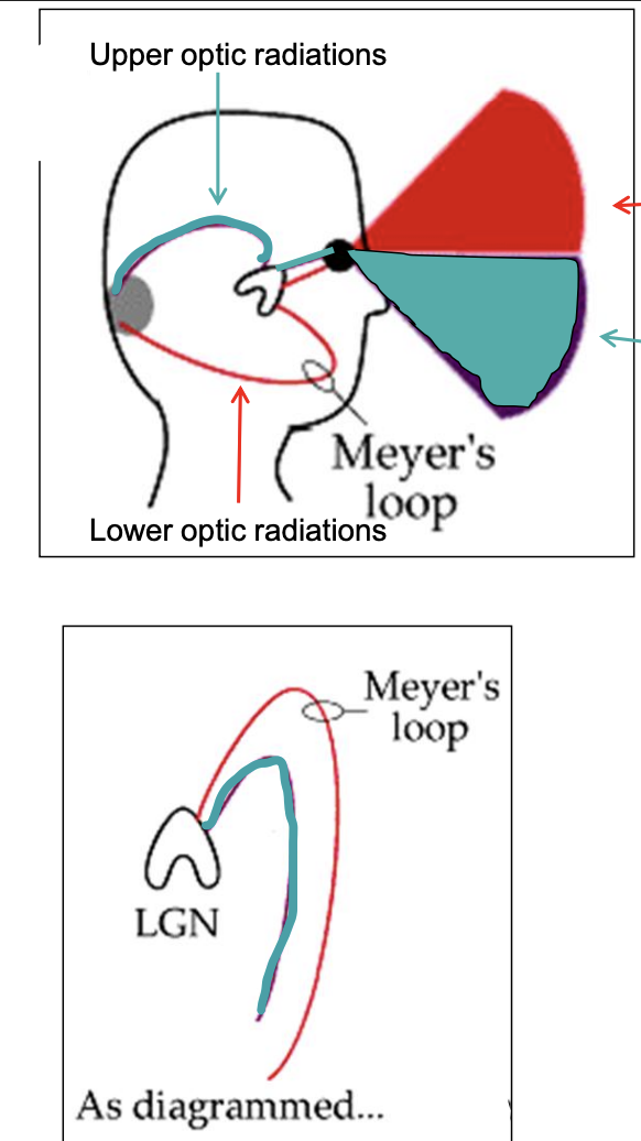
Upper/Lower Optic Radiation
upper optic radiation = green
represent lower visual field
travels in parietal lobe
Meyer’s loop (lower optic radiation) = red
represents upper visual field
travels in temporal lobe
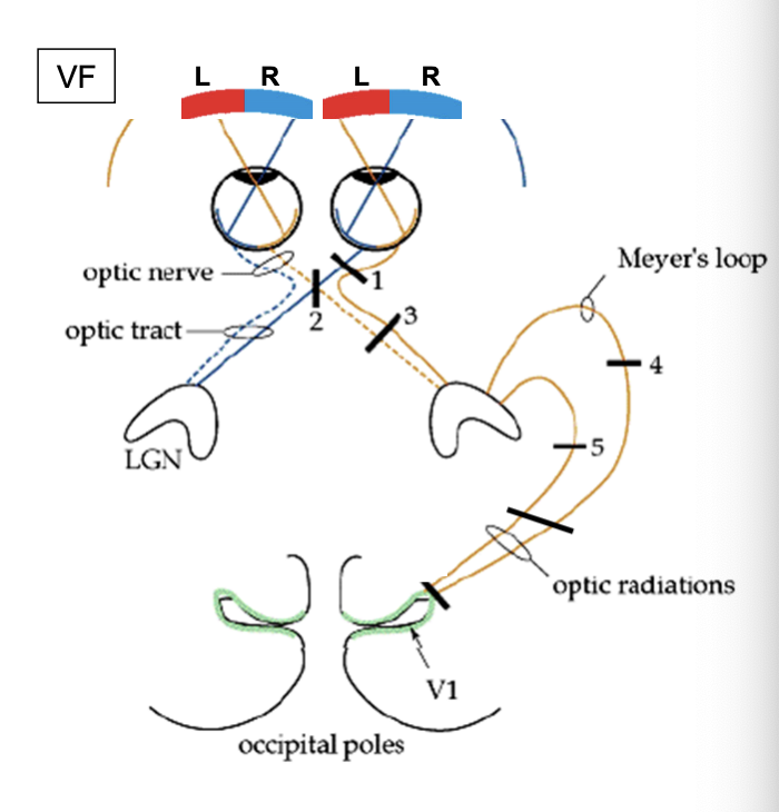
Visual Tract
CN II — Visual Field Deficits
what happens to someone’s visual field if there are lesions/defects along the visual pathway?
Circle of Willis
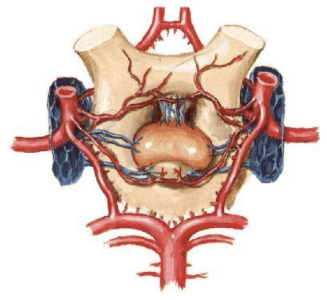
different lesions along the visual pathway
R. Optic nerve
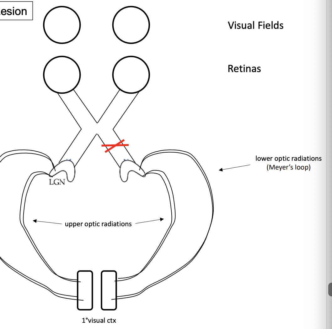
optic chiasm
R. Optic tract
R. Meyer’s loop (lower optic radiations)
R. upper optic radiations
R. optic radiations
R. Optic nerve: R. eye blindness
can see out of left eye
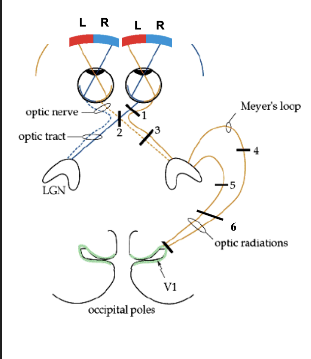
Optic Chiasm: Bitemporal hemianopsia
tunnel vision (can see medial half)
R. optic tract: Left homonymous hemianopsia
can see through right visual field
R. Meyer’s loop: left homonymous superior quadrantic anopsia (quadranopsia)
pie shape, can see everything expect top left quarter of visual field
R. Upper Radiations: Left homonymous inferior quadrantic anopsia (quadranopsia)
pie shape, can see everything expect bottom left quarter of visual field
R. Optic Radiations: Left homonymous hemianopsia (same as lesion 3)
cannot see out of left visual field