HW9: Pericarditis Quiz
1/19
Earn XP
Description and Tags
21/23
Name | Mastery | Learn | Test | Matching | Spaced | Call with Kai |
|---|
No analytics yet
Send a link to your students to track their progress
20 Terms
The following are the layers of the pericardium, EXCEPT:
Fibrous
Serous visceral
Endocardium
Serous parietal
Endocardium
The causes of Pericarditis include all the following, EXCEPT:
Pericardial effusion
Following a heart attack
Bacterial infection; Tuberculosis
Viral infection
Trauma
Pericardial effusion
What murmur might you hear with Pericarditis?
"Pericardial Rub"
Mid systolic click
Harsh systolic ejection murmur
Loud long systolic blowing
Diastolic rumble with an opening snap
"Pericardial Rub"
Pericarditis can be an acute or chronic condition.
True
False
True
The role of the pericardium is to protect the heart, to provide lubrication between the pericardium and the heart muscle in order to prevent friction and to provide nutrients to the heart muscle.
True
False
False
Pericarditis is an infiltration or inflammatory process of the ________________ of the pericardium.
endocardial and visceral layers
parietal and myocardial layers
parietal and visceral layers
fibrous and serous layers
parietal and visceral layers
All the following are complications of Constrictive Pericarditis, EXCEPT:
Restrictive diastolic filling
Pericardial effusion
Pressure equalization of all four chambers
Decreased cardiac output
Decreased pressures in all four chambers
Decreased pressures in all four chambers
The 2-D echo findings for Constrictive pericarditis include all the following EXCEPT:
IVS bounce/shuttering
Bright-dense, thick pericardium
Reduced EF with LVE
Dilation of the IVC and hepatic veins
Pericardial effusion
Reduced EF with LVE
The M-Mode findings for constrictive pericarditis include all the following, EXCEPT:
Flat inferior-lateral wall in diastole
Paradoxical septal motion/notching of the septum
Left atrial enlargement
Bright dense pericardium
Left ventricular enlargement
Left ventricular enlargement
Doppler findings for Constrictive pericarditis will show respiratory variations on the PW spectral doppler. The mitral valve velocity will increase with inspiration and decrease with expiration, while the tricuspid valve velocity will decrease with inspiration and increase with expiration.
True
False
False
Dressler's Syndrome, also known as "post myocardial infarction syndrome", includes of all the following findings, EXCEPT:
Pleural effusion
Tachycardia
Fever, CP, and joint pain
Pleurisy
Pericardial effusion
Tachycardia
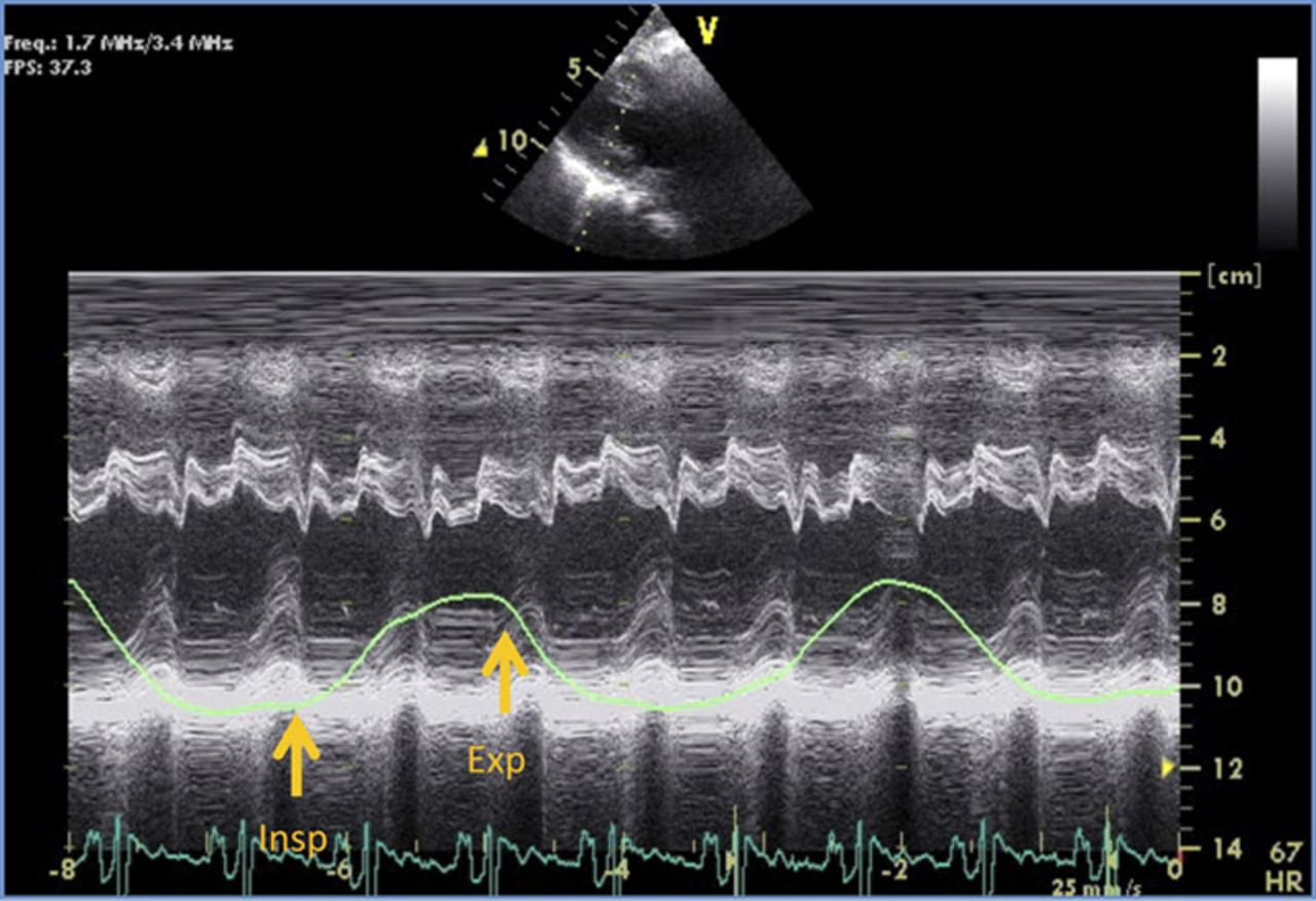
The M-Mode demonstrates and is an example of what cardiac pathology?
Constrictive Pericarditis
Acute pericarditis
Pulmonary hypertension
Dressler's syndrome
RVVO
Constrictive Pericarditis
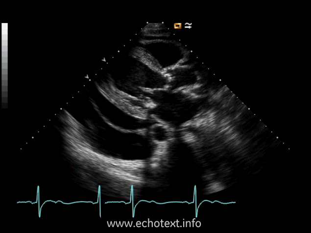
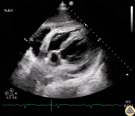
The images are examples of what cardiac pathology?
Constrictive Pericarditis
Constrictive Pericarditis with Pericardial effusion
Acute Pericarditis
RVVO
Constrictive Pericarditis
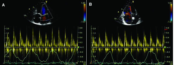
The Doppler images are suggestive of what cardiac pathology or condition?
Constrictive Pericarditis
Pericardial effusion
Dressler's syndrome
Acute pericarditis
Restrictive Cardiomyopathy
Constrictive Pericarditis
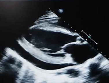
The image below is an example of what cardiac pathology:
Pericardial effusion
Constrictive pericarditis
Constrictive Pericarditis with pericardial effusion
Acute pericardial effusion
Constrictive pericarditis
The clip below is sometimes visualized after a cardiac incident, infarction or surgery, followed by acute pericarditis. What does this image clip represent?
Dressler's syndrome
Pleural effusion
Constrictive pericarditis
Pericardial effusion
Dressler's syndrome
The image below is an example of what cardiac pathology:
Pericardial effusion
Constrictive pericarditis with pericardial effusion
Constrictive pericarditis with pericardial effusion
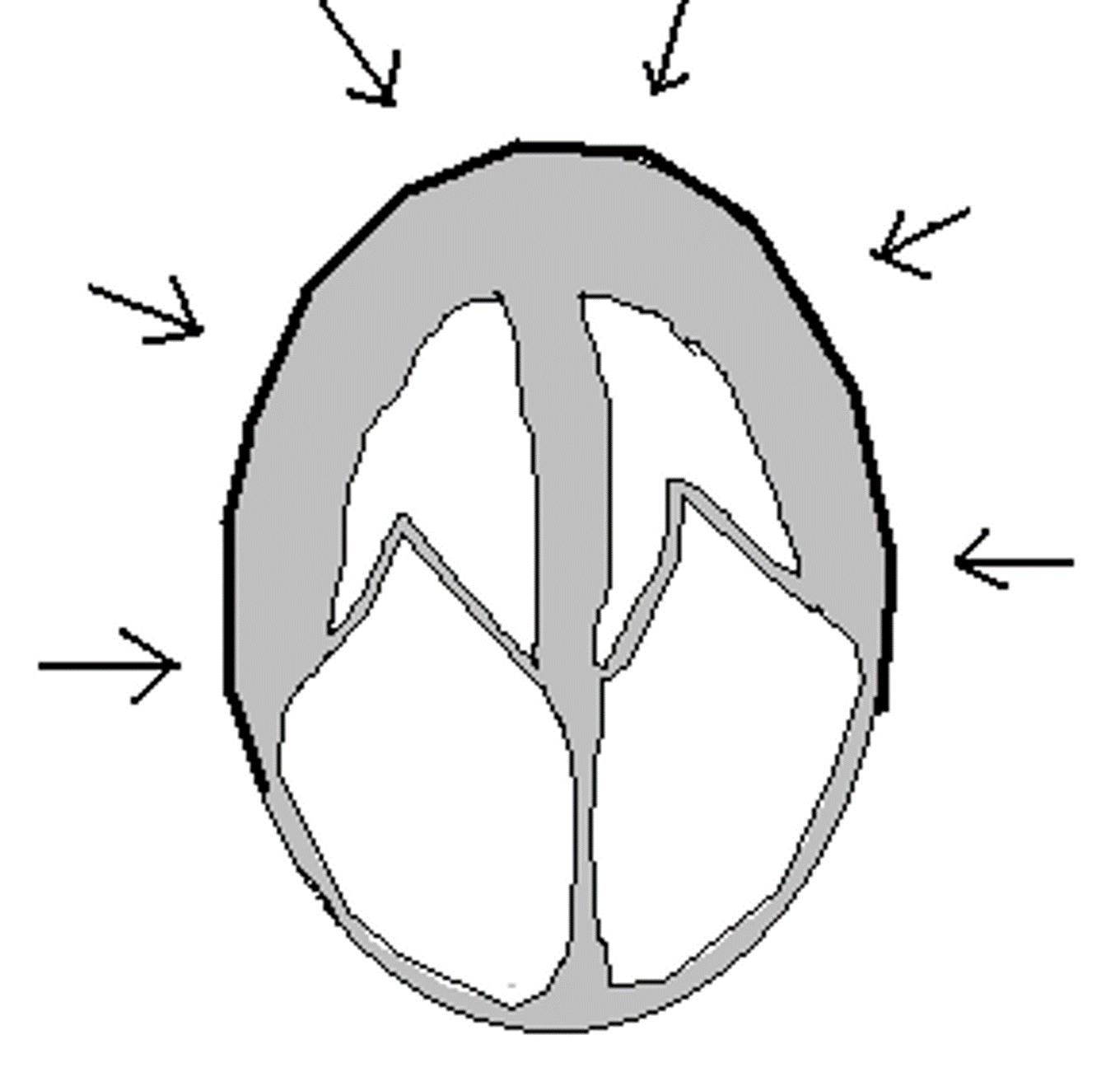
Pericarditis versus Restrictive Cardiomyopathy: Select all the following answers (multiple answers) that apply to the image:
Surrounds entire heart, impedes diastolic filling
Restrictive/ Infiltrated Cardiomyopathy
Constrictive pericarditis
Large MV E, small A, with respiratory changes
Involves infiltrated myocardium, stiff ventricular walls, impedes diastolic filling
Large MV E, small A, without respiratory changes
Restrictive/ Infiltrated Cardiomyopathy
Involves infiltrated myocardium, stiff ventricular walls, impedes diastolic filling
Large MV E, small A, without respiratory changes
Pericarditis verses Restrictive Cardiomyopathy: Select all the following answers (multiple answers) that apply to the image:
Large MV E, small A, without respiratory changes
Large MV E, small A, with respiratory changes
Constrictive pericarditis
Restrictive/ Infiltrated Cardiomyopathy
Involves infiltrated myocardium, stiff ventricular walls, impedes diastolic filling
Surrounds entire heart, impedes diastolic filling
Surrounds entire heart, impedes diastolic filling
Large MV E, small A, with respiratory changes
Constrictive pericarditis
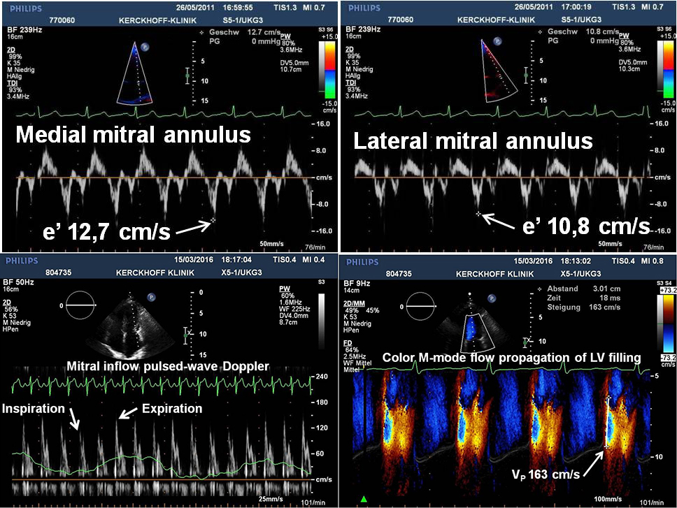
Explain 2 findings in these images that support the diagnosis of constrictive pericarditis.