Topic 2B- Basic Biology for Biomechanics
1/52
Earn XP
Description and Tags
We will learn about the different types of cells which form the most important biological tissues
Name | Mastery | Learn | Test | Matching | Spaced | Call with Kai |
|---|
No analytics yet
Send a link to your students to track their progress
53 Terms
What is a cell
The cell is the basic functional unit in the body
There are more than 200 different types of cells
all share the same genetic information
differ in terms of shape, size and constituent molecules
liver cells have abundant enzymes
red blood cells have haemoglobin
Behaviour is cell-dependent
Most cells have got similar structure
What is the cell membrane
The cell membrane is a thin, flexible barrier that surrounds the cell and regulates what enters and leaves the cell.
What is the cytoplasm
The cytoplasm is the jelly-like substance that fills the cell. It contains all of the cell’s organelles and provides a supporting environment for them to carry out their functions.
What is the nucleus
The nucleus is the control center of the cell
What are the organelles
Organelles are tiny structures within the cell that have specific functions
What are the mitochondria
They generate energy from food molecules in the form of ATP
What are the ribosomes
Ribosomes are the protein factories of the cell. They synthesize proteins from amino acids. They are bound to the endoplasmic reticulum
What are the endoplasmic reticulum
The endoplasmic reticulum is a network of tubes that transport materials throughout the cell. t is also involved in protein synthesis and lipid production; it envelopes the mitochondria
What are the Golgi apparatus
The Golgi apparatus is a packaging and shipping center for the cell. It modifies and packages proteins and other molecules for transport to other parts of the cell or outside of the cell
What is the cytoskeleton
The cytoskeleton is an internal skeleton that gives rise to the cell shape and coordinates cell division and cell movement. It is composed by microfilaments (actin filaments), microtubules and intermediate filaments
What does the cytoskeleton do
Establishes and maintains the shape of the cell
Allows the cell to move (the process of locomotion)
Provides mechanical strength and integrity to the cell
Is central to the intracellular transport of organelles, especially in large cells
Is essential during cell division, where it plays a key role in many processes including chromosome separation in mitosis and meiosis
What needs to happen for a cell to function properly
To function properly, cells must stay attached to their substrates and to their neighbours
Vascular endothelial cell in a large artery lives on a continuously deforming substrate to which it must adhere while maintaining close contact with neighbouring cells
If necessary, it has to crowl and move
local injury
allow macrophages to enter/exit the blood stream
What happens when cells do diffusion
Facilitated transport
Ion channels
Active transport
Receptor mediated signalling
What happens when living cells apply a mechanical stimulus
In living cells, application of a mechanical stimulus causes not only a mechanical response, but also a biological response: using complex networks of sensors, transducers and actuating mechanisms, cells are able to respond and adapt to their mechanical environment
What are integrins
Integrins are transmembrane proteins that link the ECM to the cytoskeleton
What can transmission of mechanical signals via integrins can lead to
Deformation of the cytoskeleton, which in turn can affect the biochemical state of the cell. This means a perturbation applied locally to an integrin can lead to movement of organelles and distortion of the nucleus, possible influencing gene expression
decentralization mechanism
A locally applied stimulus results in mechanotransduction at multiple, mechanically coupled sites, allows for greater diversity in the cellular response
What is an example of integrin activation
Integrin activation is the process of leukocyte extravasation (the movement of white blood cells from the bloodstream into the surrounding tissue) in response to an inflammatory signal
What do activated integrins do to their ligands
Activated integrins bind tightly to their ligands on the endothelial cells, facilitating firm adhesion of the leukocyte to the endothelium
What does the stretch activated ion channels do
Stretch activated ion channels connect the cytoplasm to the cell exterior
Physical deformation of the plasma membrane causes conformational changes in the embedded channel proteins leading to its activation
Usually, they are ionic channels for Na,K,Ca,CI which are involved in a multitude of cellular activities and signaling
Some are activated by stretch while others are inactivated
What are the 4 types of tissues (general)
muscle, nervous, epithelial, connective
muscle: specialised for movement
nervous: initiation and transmission of signal
epithelial: separates body from environment is organised into sheets
connective: is rich in extracellular material that provides mechanical strength and anchors adjacent tissues
Types of connective tissues
connective tissue proper
loose connective tissue → hold organs together
dense connective tissue → high fibre density
cartilage
osseous tissue
blood
What are connective tissues
Connective tissues are made up of many types of specialized (resident or migrant) cells together with a large amount of non-living material referred to as the ECM, composed of ground substance and fibres. Typically this matter is synthesized and secreted by specific connective tissue cells
What is ECM
ECM is a gel-like substance (ground substance) and the properties are determined by the types of fibers present
Structurally, all connective tissues contain cells embedded in an ECM stabilized by proteins. the chemical nature and physical layout of the ECM and proteins vary enormously among tissues, thus reflecting the variety of functions that connective tissue fulfils in the body
what are osteoblasts
Responsible for the initial synthesis and deposition of bone matrix. Osteoblasts deposit in the form of hydroxyaptite. Upon completion of bone matrix formation, some mature osteoblasts remain entrapped in bone as osteocytes
what are osteocytes
Osteocytes are the primary cells responsible for the adaptation of bone to mechanical forces through signalling pathways that regulate bone formation and resorption
What are chondroblasts
Chondroblasts are specialized cells that produce the extracellular matrix of cartilage; they actively synthesize and secrete the components of the cartilage matrix, including collagen (mainly type II collagen), proteoglycans, and other structural molecules; as they produce the matrix, chondroblasts become embedded within it and transform into chondrocytes
What are chondrocytes
Chondrocytes are mature cartilage cells that reside within the lacunae, which are small spaces within the cartilage matrix, they are responsible for the homeostasis of cartilage, regulating the turnover between matrix synthesis and degradation
What are fibroblasts
Fibroblasts responsible for production of both the ground substance and fibres. They play a crucial role in collagen production and deposition in other tissues of the body, such as tendons, ligaments, and various types of connective tissues outside the bone.
These cells are responsible for synthesizing collagen and other extracellular matrix components that provide structural support and integrity to these tissues
What is coloured in the Hematoxylin and Eosin Stain (H&E Stain) (what colour is cell nuclei, ECM and cytoplasm)
Hematoxylin is a basic dye and complexes with nucleic acids (DNA and RNA in the nucleus)
Eosin is an acidic dye which stains membranes and most proteins
Cell Nuclei→ Blue
ECM→ Pink
Cytoplasm→ Pink
What is Trichrome staining (what colour is collagen fibers, nuclei and cytoplasm?)
Trichrome staining is used to visualise connective tissues, particularly collagen, in tissue sections.
Collagen fibers → Blue
Nuclei → Dark Red
Cytoplasm → Pink
What is Verhoeff-van Gieson Stain (what colour is collagen, elastin and nuclei?)
Verhoeff-van Gieson stain is a histological staining procedure one of the most commonly used stains to
What does Alcian blue stain
Alcian blue binds strongly to GAGs and glycoproteins. Because cartilage contains higher concentrations of GAGs than any other tissue, it binds more Alcian blue
What does Alizarin red stain
Alizarin Red strains calcium rich tissues becuase 99% of the calcium in the body is localized in bone, Alizarin red is highly specific for bone
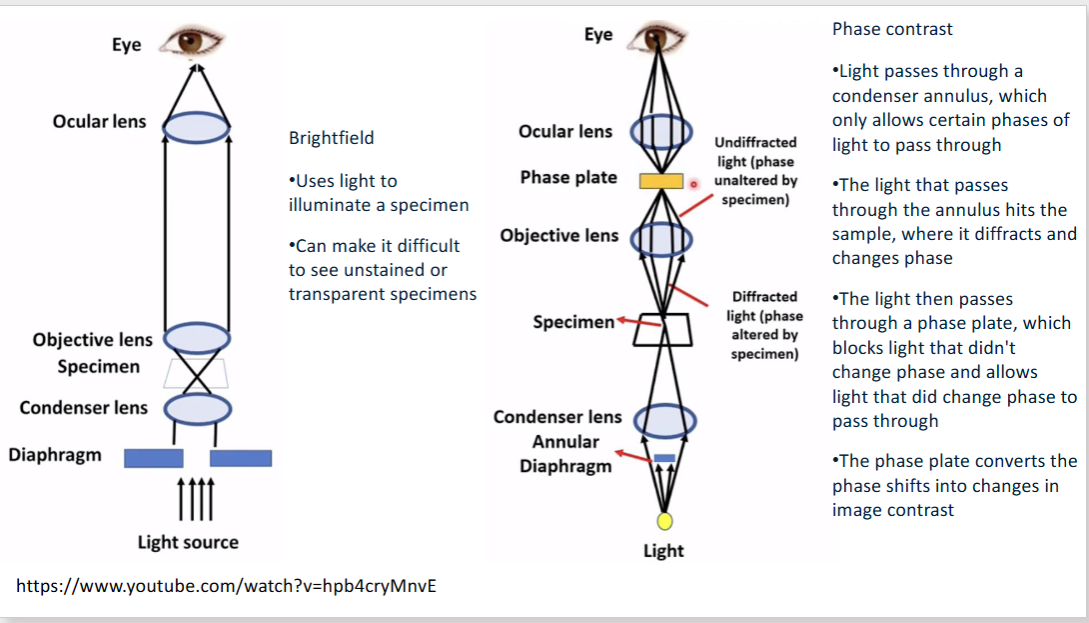
What is Brightfield Microscopy
illumination light positioned below or above the sample and it is transmitted through the sample and the contrast is generated by the absorption of light in dense areas of the specimen
What is Phase-Contrast Microscopy
Quantitative phase imaging (QPI) refers to a subset of label-free microscopy techniques where contrast in the image is generated by the variation of optical path length across the sample. The resulting image is a phase map that is a quantitative measure of the product of the difference between the refractive index of the tissue and that of its surrounding medium
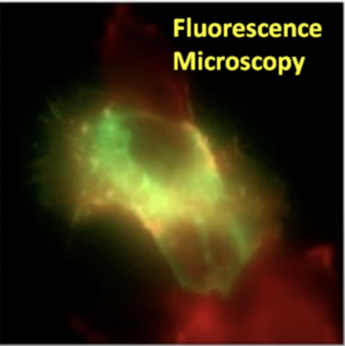
What is Fluorescence Microscopy
Utilises fluorescent dyes, proteins. or antibodies to label specific cellular components or molecules. It enables visualisation of specific proteins, organelles, or structures within cells
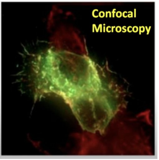
What is Confocal Microscopy
Similar to fluorescence microscopy, confocal microscopy uses fluorescent labels. confocal microscopy provides a means of rejecting the out-of-focus light from the detector such that it does not contribute blue to the images being collected. It generates 3D images by stacking serial optical sections
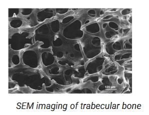
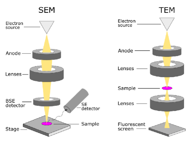
What is Scanning Electron Microscopy (SEM)
In an SEM, an electron beam is directed at the surface of a sample. This beam excites atoms of the sample, emitting secondary photons which are received by the detector to form an image. It can produce 3D images
What is Transmission Electron Microscopy (TEM)
Uses electrons to visualise ultrastructural details within cells at high resolution, allowing for detailed imaging of cell organelles and membranes, the primary electron beam is transmitted through a thin sample, and then detected on the other side. The electrons are interacting (being absorbed and emitted) with structures inside the sample and are therefore able to produce a higher resolution image than through the collection of scattered secondary electrons. It can only produce 2D images of a small area
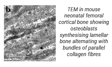
What are the resolutions of SEM and TEM
SEM has a resolution is limited to 0.5nm, while TEM has a resolution of 0.5 angstroms
What are the magnification levels of SEM and TEM
SEM has a maximum magnification level of 2 million, while TEM has up to a 50 million magnification level
What does TEM require in comparison to SEM
TEM requires thin layers of samples (usually <150nm) for sufficient amounts of electrons to be transmitted , while SEM can be used for full reconstructions
How does one measure mechanical properties for a cell or molecule?
It is not trivial
Consider, for instance the magnitudes we are dealing with at the cellular level
Measurement of a single-cell mechanical properties has attracted great interest from both academia and industry, due to its importance in a variety of applications, such as cell separation, disease diagnostics, immune status analysis and drug screening
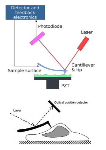
What is Atomic Force Microscopy
The key element of the microscope is a tapered probe, typically made from silicon or silicon nitride, which is attached to a cantilever arm. When the probe tip interacts with a sample, the arm is deflected. This deflection can be measured by sensing the position of a laser beam that reflects off the cantilever arm.
By suitable placement of the laser and detector to take advantage of the “optical level arm” effect, displacements of less than 1 nm can be measured
The AFM is typically used in conjunction with an optical microscope that can be used for real-time visualization for example, to help to localize the AFM probe over a target cell. One of the great advantages of the AFM is that imaging can be carried out on living cells or intact molecules in an aqueous environment
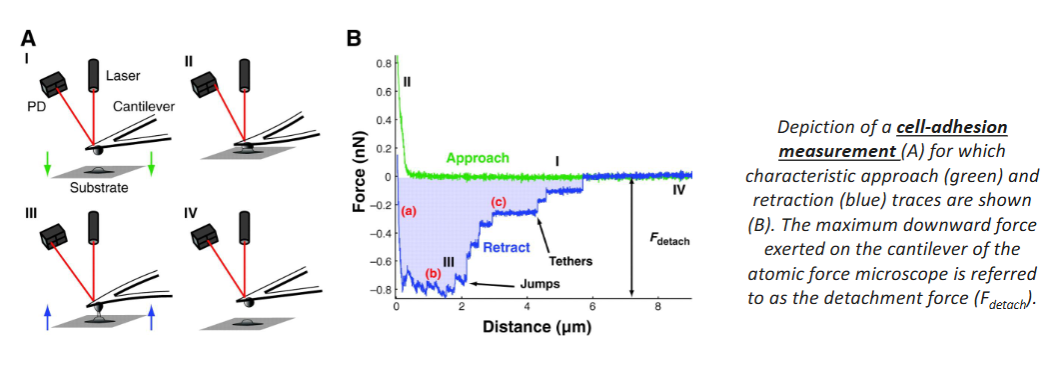
How does Cell-Adhesion Measurement (CAM) work
The vertical location of cantilever z and the cantilever deflection signal d is recorded during the process
The cantilever starts from point a, a few micrometres above the cell. While approaching the cell, the sample indentation remains zero until it reaches point b, where the tip comes into contact with cell
The coordinates of point b in the plot are critical values for data analysis, denoted (z0,d0)
Once the deflection signal reaches the preset maximum value, the cantilever is then withdrawn from the cell
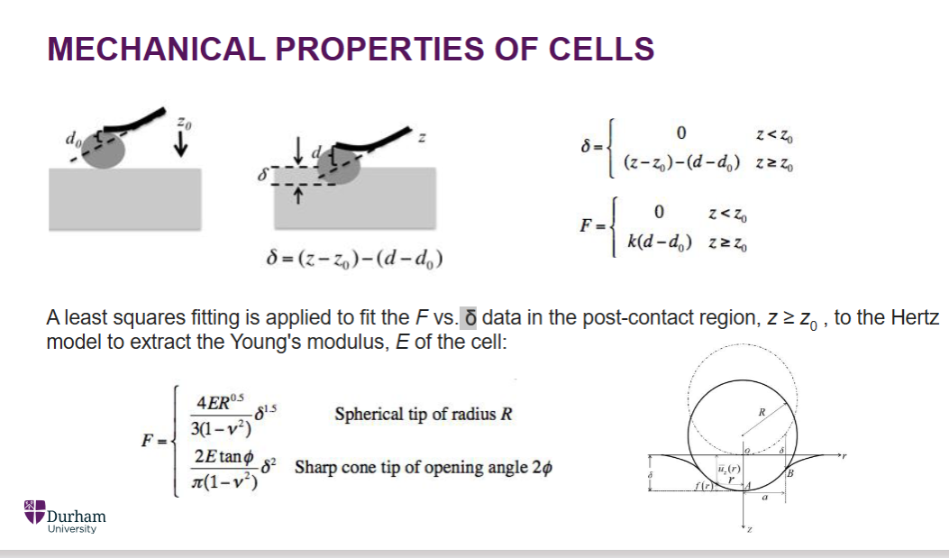
In CAM what can be applied
A least squares fitting is applied to fit the F vs δ data in the post-contact z>= z0, to the Hertz model to extract the Young’s modulus , E of the cell e.g. turn in a spherical shape to find the Force
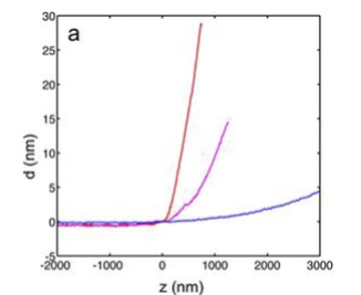
What does the force curve data and the analysed force indentation curve show
Force curve data and the analysed force-indentation curve- a set of 3 representative force curve data acquired for 3T3 fibroblasts culture on glass, 17kPa polyacrylamide gel (purple), and 3kPa polyacrylamide gel (blue)
What does the fluorescence image of a 3T3 fibroblast
Only a part of the cell is shown in the image. Scale bar represents 20 μm.
A 32 × 32 pixel stiffness map of the same area. Each pixel represents 2.5 μm
What is micropipette aspiration
Micropipette aspiration is one of the oldest techniques for measuring cellular (and subcellular) biomechanical properties.
This technique has been applied in numerous ways from examining passive material properties and mechanical responses in lipid vesicles and cells, to measuring molecular adhesion and active cell contractility against mechanical load
What are the steps for micropipette aspiration
A very fine glass micropipette, typically having internal diameter of 1-10 μm and with a tip that can be moved about by a micromanipulator
A negative pressure is applied to aspirate the cell into the micropipette. Cell deformation is recorded through imaging on an optical microscope at varying known suction pressures
If the applied suction pressure is low enough to avoid red cell rupture, an equilibrium is established in which the applied suction pressure is balanced by mechanical stresses within the cell (membrane and cytoskeleton)
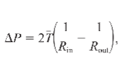
What is the simplest model for analysing micropipette aspiration result
Young-Laplace equation
this model basically assume a homogenous elastic membrane holding a drop of homogenous Newtonian liquid inside. When the cell is aspirated into the micropipette forming a perfect semi-hemisphere, the membrane tension T of the cell can be calculated under the force balance with the liquid’s internal pressure
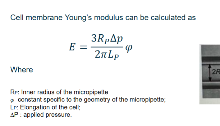
What is the equation of the cell membrane Young’s modulus
E=(2Rpp/2piLp)w