Neurophysiology 1 and 2.
1/75
Earn XP
Description and Tags
Transmission and integration of neuronal signals
Name | Mastery | Learn | Test | Matching | Spaced |
|---|
No study sessions yet.
76 Terms
Electrical signals
Are the vocal of the nervous system.
Neurophysiology
Is the study of life processes within neurons that use electrical and chemical signals.
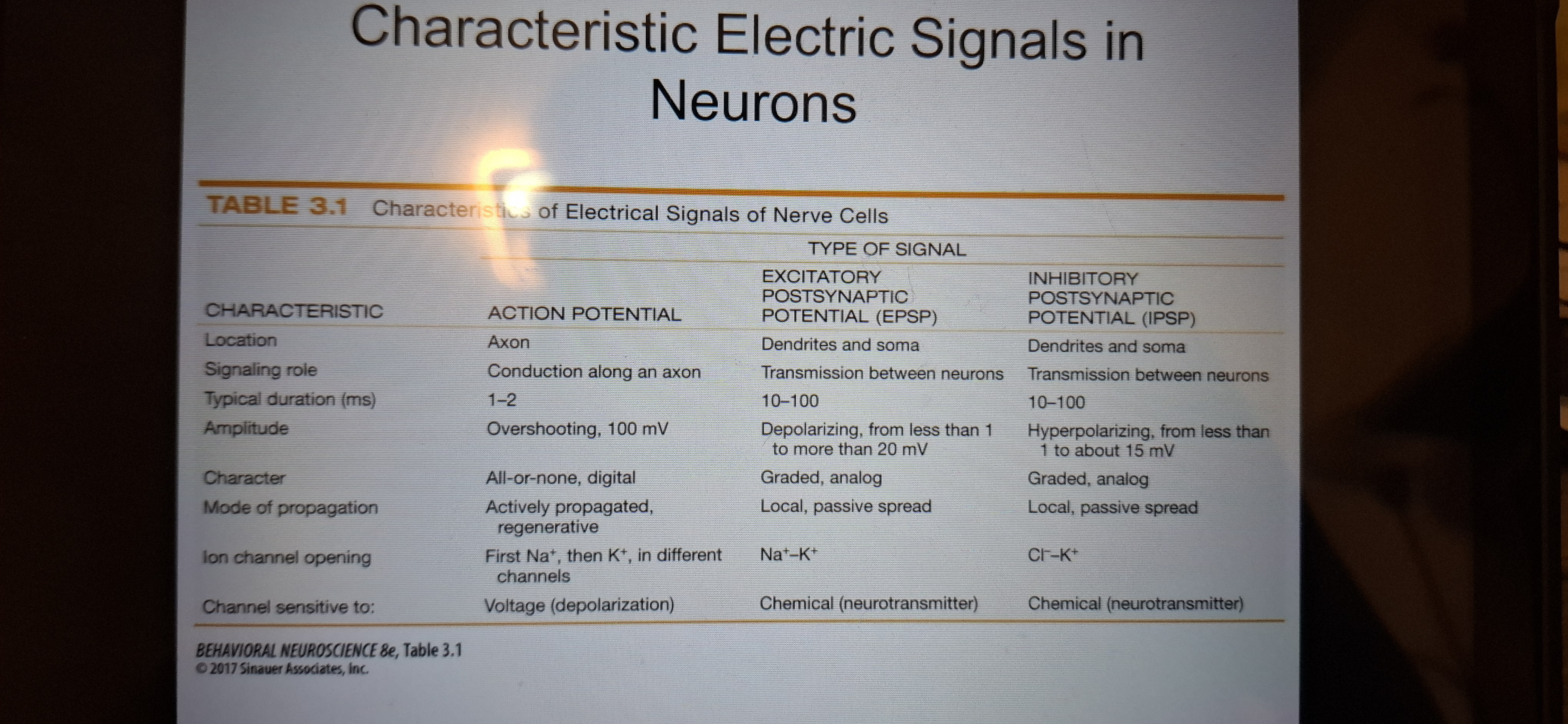
Action potential
Is a rapid electrical signal that travels along axons of a neuron.
Brief but large change in membrane potential.
Neurotransmitters
A chemical messenger between neurons.
Neurons at rest
A balance of electrochemical forces.
Ions
Electrical charged particles.
Anions: negatively charged
Cations:positively charged
Ions
Dissolved in intracelkular fluid, separated from the extracellular fluid by the cell membrane(lipid bilayer)
Resting membrane potential
-50millivolts to -80millivolts
Cell membrane
A Lipid bilayer, with 2 layers of lipid molecules.
Ion channels
Are proteins that span the membrane and allow ions to pass.
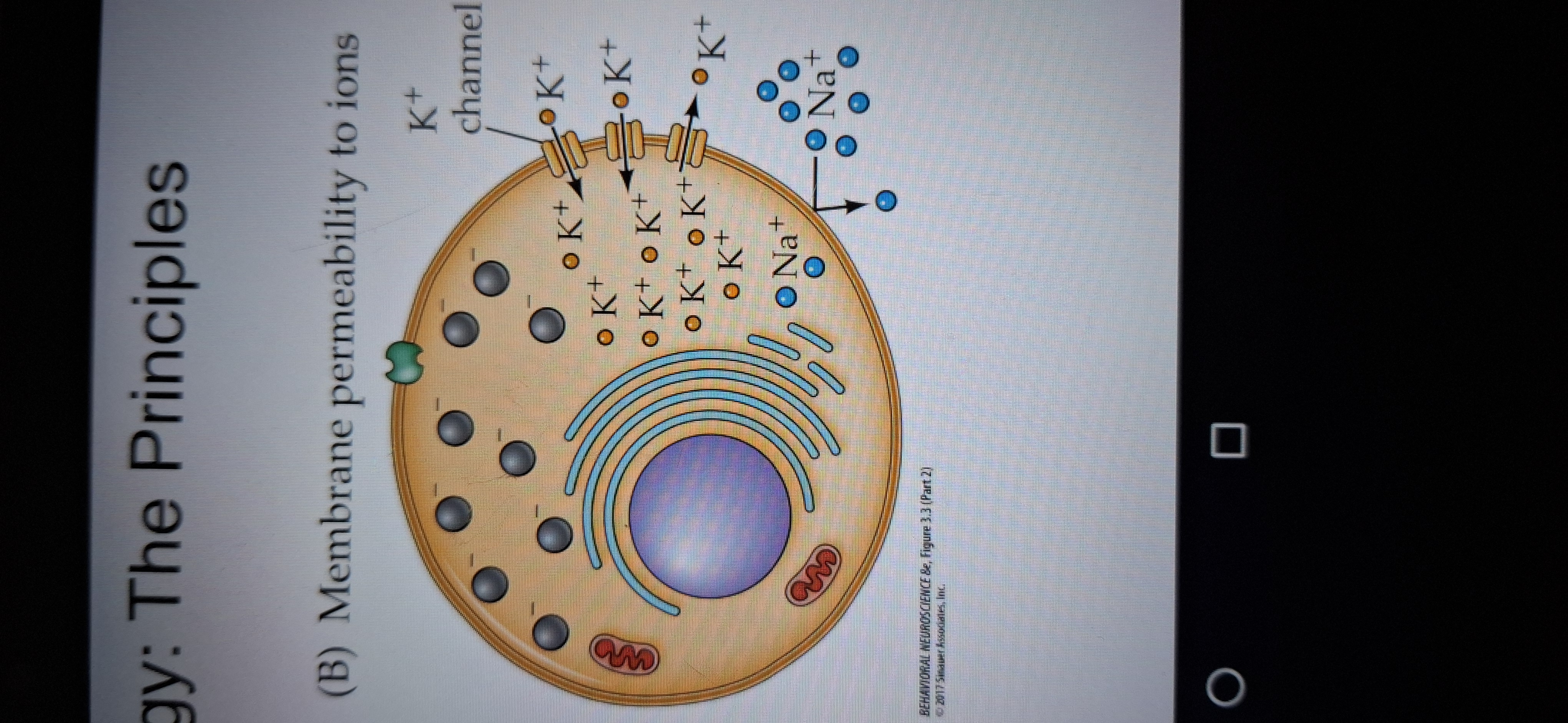
Why do ion channels open and close?
In response to voltage changes,chemicals and mechanical actions.
Selective permeability
It allows k+ to enter or leave the cell freely, but restricts the flow of other ions.
Forces that drive ion movement
1.diffusion
2.electrostatic forces and pressure
Diffusion
Causes ions to flow from areas of high to low concentration, along their concentration gradient.
Electrostatic forces and pressures.
Causes ions to flow towards oppositely charged areas.
Sodium-potassium pump
-used to maintain resting potential
-it pumps 3 Na+ions out fr every 2 k+ions pumped in.
-cause turnover of 70kgs of ATP per day.
Equilibrium
When the movement out is balanced by the movement in.
Resting potential value
-65mV
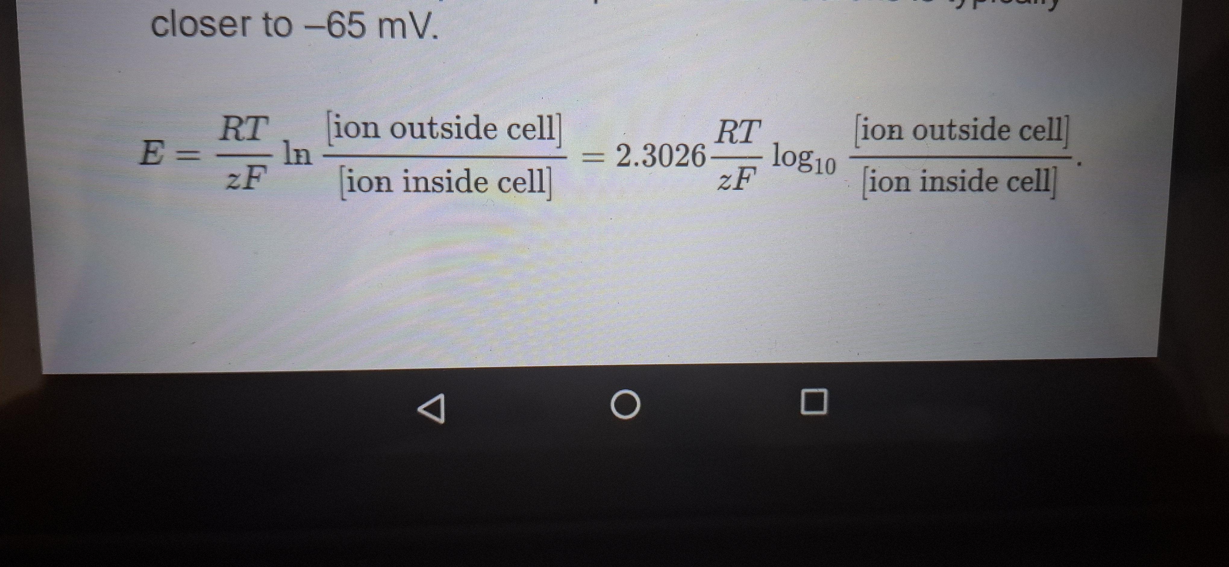
Nernst equation
Predicts the voltage needed to counterbalance the diffusion force pushing ions across a membrane.
Predicts the equilibrium potential of an ion. K+= -80mV.
Nernst equation formula
Find equation below
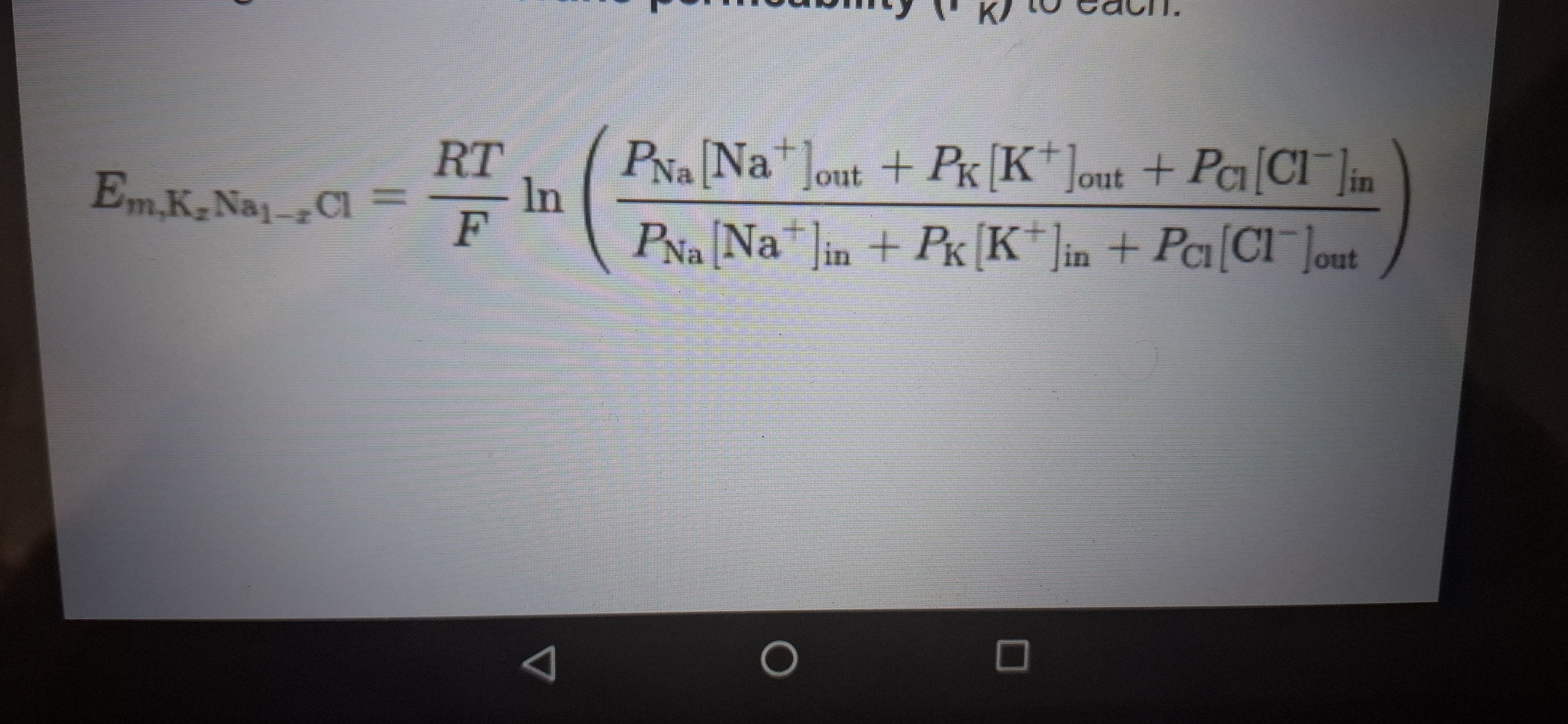
Goldman-hudginkin-katz (GHK)Equation
Predicts voltage potentials that are quite close to Observed resting potentials.
Takes into account intracellular and extracellular concentrations of several ions and the degree of membrane permeability to each.
Flow of action potentials
Originate in the axon hillock, and move along the axon. Patterns of axon potentials carry information to the presynaptic targets.
Hyperpolarization
Is an increase in membrane potential the interior of the membrane becomes even more negative, relative to the outside.
Depolarization
Is a decrease in membrane potential the interior of the cell becomes less negative.
Hyperpolarizing.
stimulus produces an immediate response that passively follows the stimulus.
Graded response
Change in potential.
Local potential
An electrical potential that spreads passively across the membrane ,diminishing as it moves away from the point of Stimulation.
Potential threshold
-40mV, triggers action potential.
All or none property of action potential
•neuron fires at full amplitude or not at all.
•does not reflect increased stimuli strength.
Frequency of action potential
Increased frequency =Increased stimulus strength.
Afterpotential
Are changes in membrane potential after action potentials.
When are action potentials produced?
When Na+ions move into the cell.
Peak of action potential
When the concentration gradient pushing Na+ions into the cell equals the positive charge driving them out.
Voltage-gated Na+channels
Opens in response to the initial depolorisation
Na+ equilibrium potential
+40mV
Refractory period
Time when only some stimuli can produce an action potential
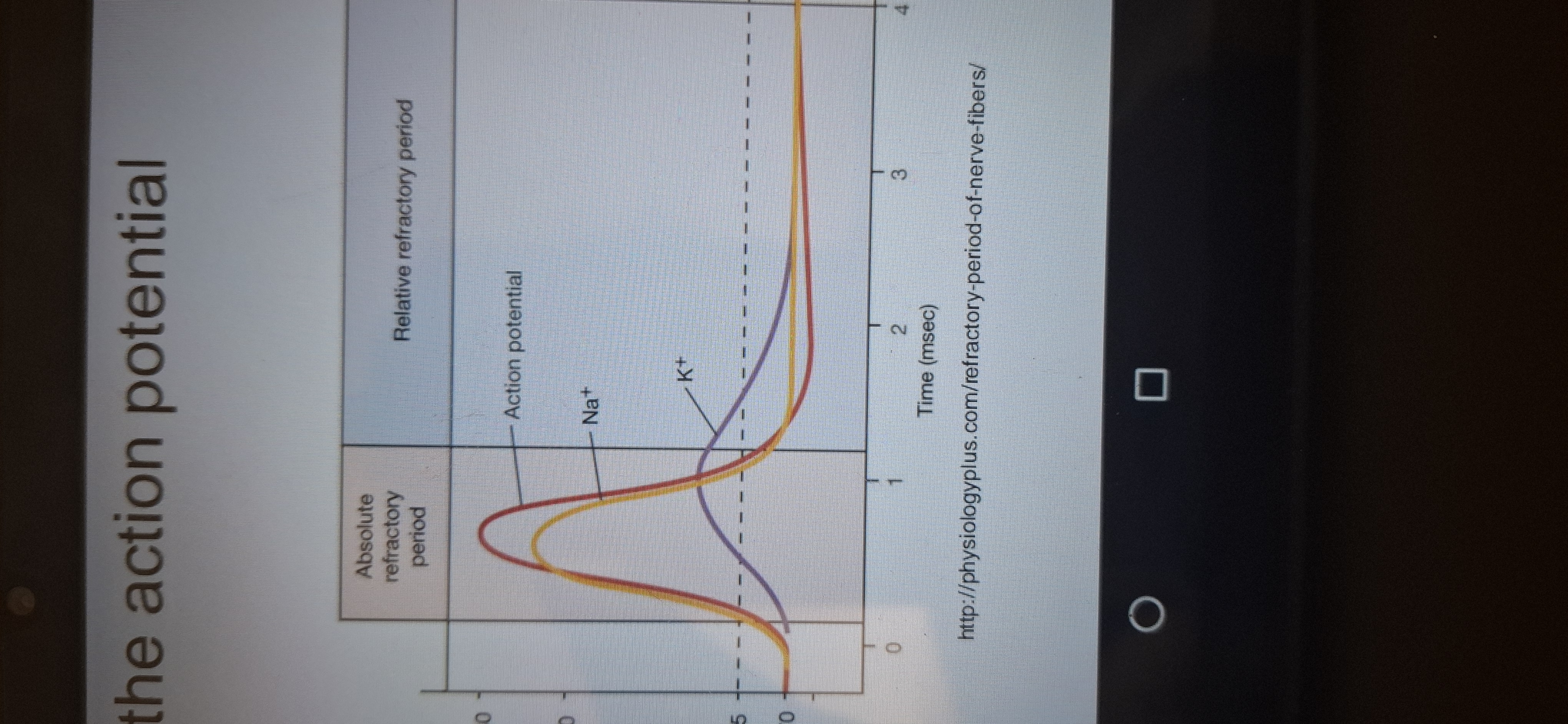
Absolute refractory period
Time when no action potential are produced.
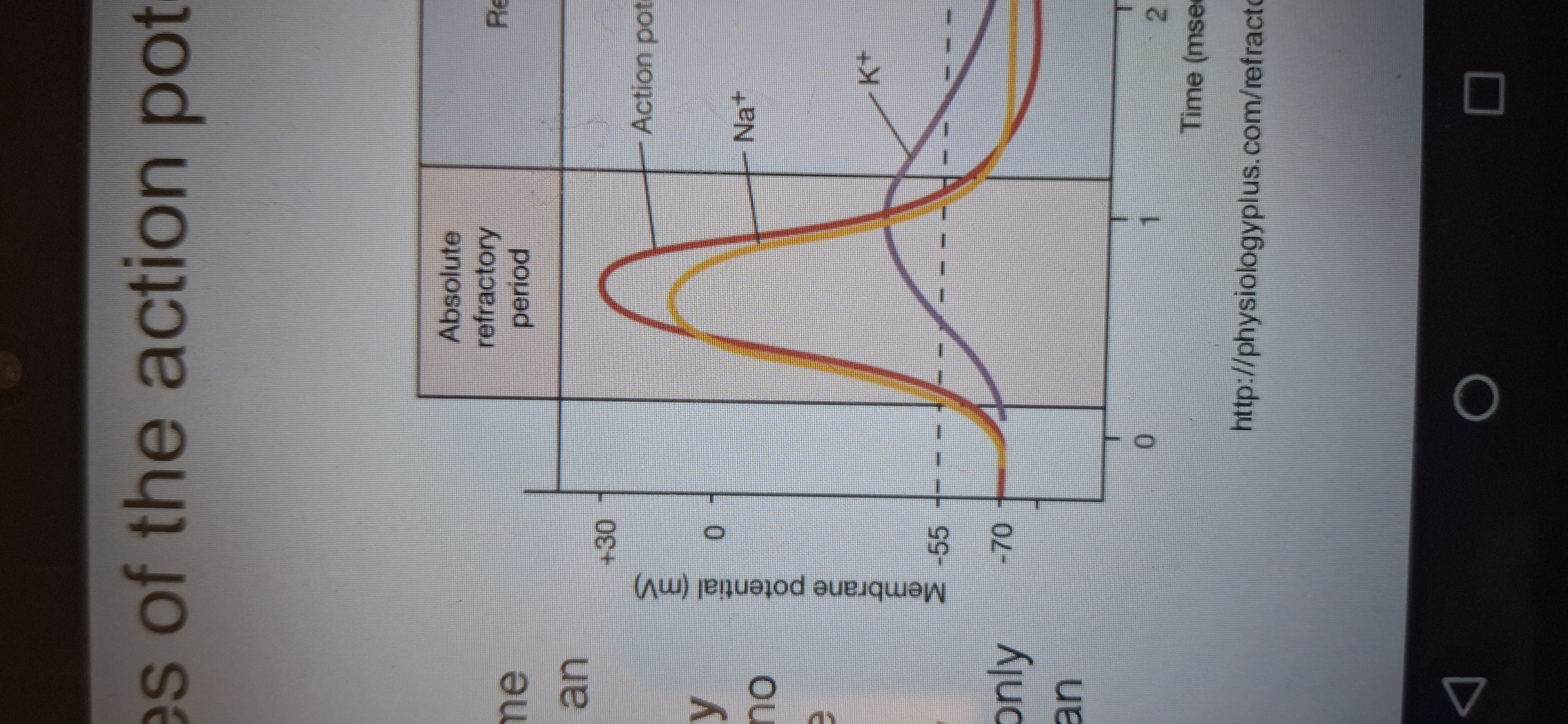
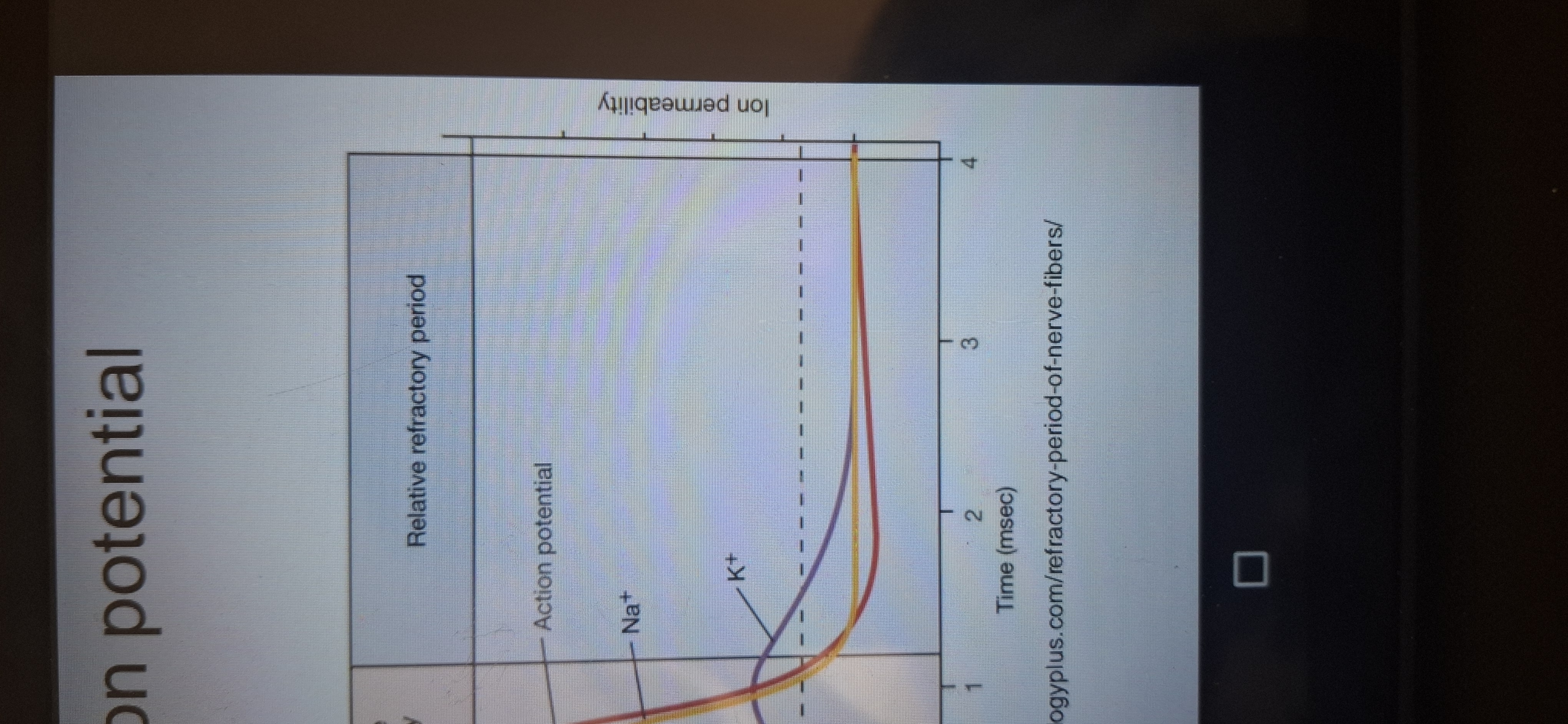
Relative refractory period
Time when only strong Stimulation can produce an action potential.
Action potential generation
Each adjacent section is depolarized and a new action potential occurs.
Why action potential travel in one direction?
Because of the refractory state of the membrane after a depolarization.
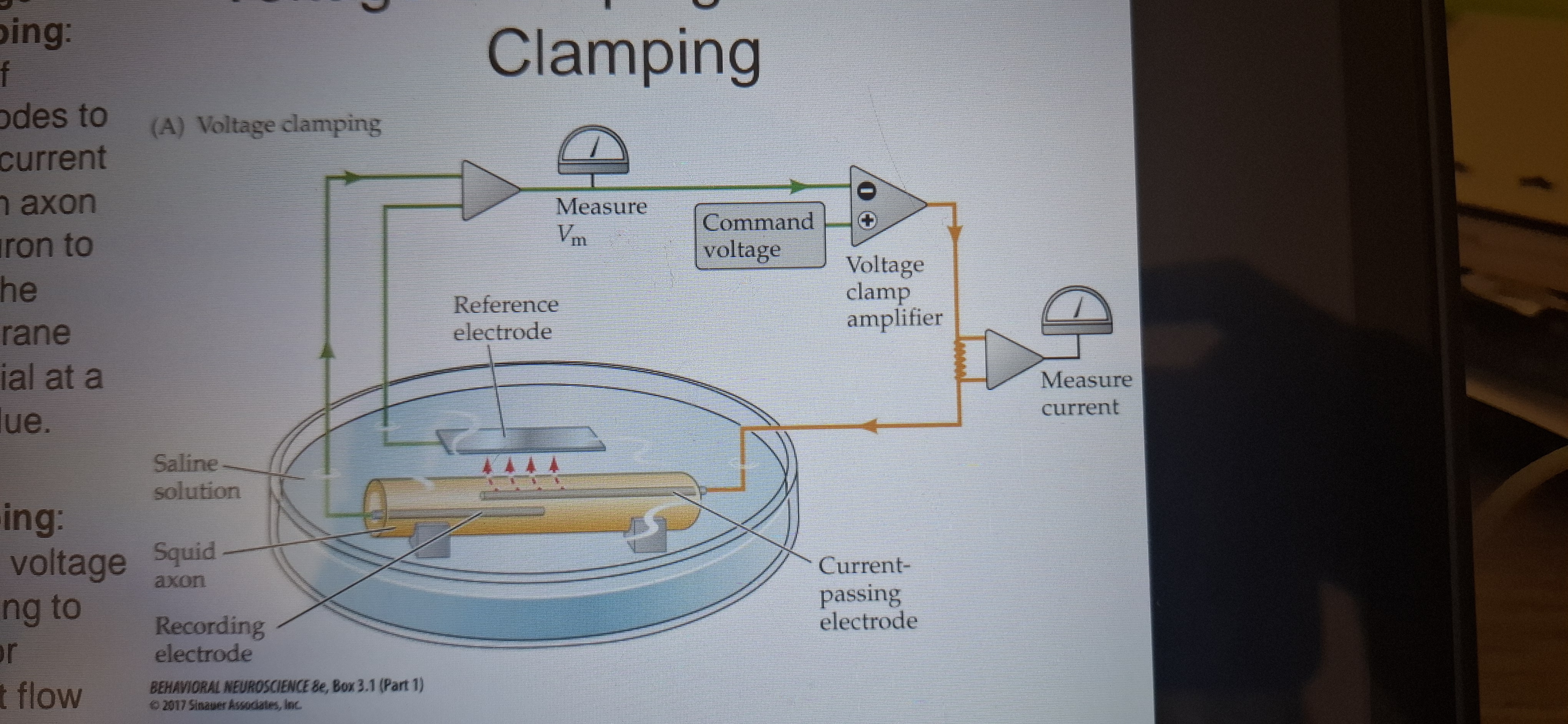
Voltage clamping
Use of electrodes to inject current into an axon or neuron to keep the membrane potential at set value.
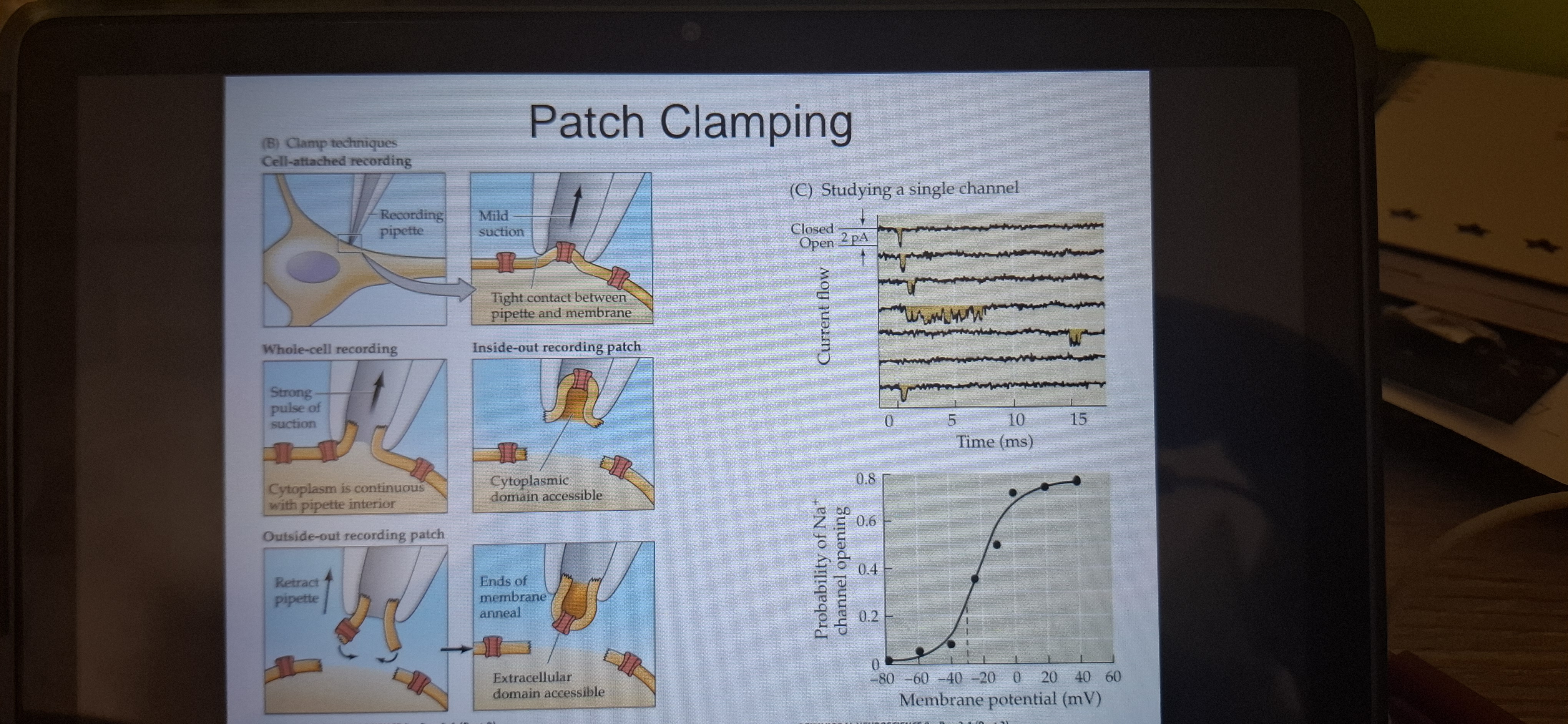
Patch clamping
Use of voltage clamping to monitor current flow across a tiny patch of neuronal membrane.
Conduction velocity
The speed of propagation of action potentials, varies with diameter.
Nodes of Ranvier
Small gaps in the insulating myelin sheath.
Saltatory conduction
The axon potential travels inside the axon and jumps from node to node.
How k+ channels are specific to their function
●the channels are lined with oxygen atoms that mimics water molecules
●k+ions pass through this selectivity filter, Na+does not.
Channelopathy
Genetic abnormality of ion channels.
Tetrodotoxin(TTX) AND Saxitoxin(STX)
Block voltage gated Na+ channels.
Batrachotoxin
Forces Na+channels to stay open.
Postsynaptic potentials.
Are brief changes in the resting potential.
Excitatory postsynaptic potential. (EPSP)
Produces a small local depolarization, pushing the cell closer to threshold.
Synaptic delay
The delay between an action potential reaching the axon terminal and creating a postsynaptic potential.
Inhibitory postsynaptic potential
A small localised hyperpolarization which pushes the cell to move away from the threshold.
Caused by chloride (Cl-)ions making inside the cell negative.
Neurons
Integrate synaptic inputs.
Spatial summation
Is the summing of potentials that come from different parts of the cell.
Temporal summation
Is the summing of potentials that arrive st the axon hillock at different times. The closer the time of arriving together to more likely for an action potential.
Comparison
Characteristics.
Gap junctions/electrical sypnases
Regions where action potential can jump directly to the postsynaptic region without fiest being transformed into a chemical signal.
Connexons
Large channels aiding in gap unconscious, reducing time delay.
Chemical synapse
Use of neurotransmitters for action potential transportation.
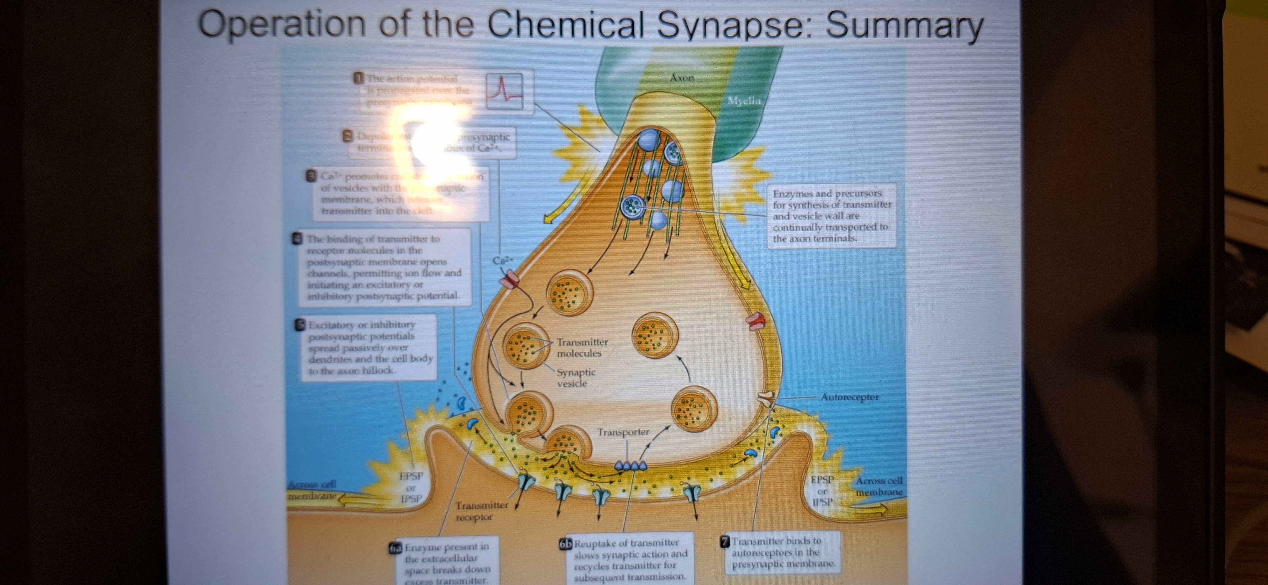
Sequence of chemical synapses
1.action potential travels down the axon to the axon terminals.
2.voltage-gated calcium channels open and ca2+enter.
3.synaptic vesicles fuse wirh membrane and release transmitter into the cleft.
4.transmitterscross the cleft and bind to postsynaptic receptors and cause an EPSP or IPSP.
5.EPSP or IPSP spread towards thee postsynaptic axon hillock.
6.transmitter is inactivated by enzymatic degradation or removed by transporters for reuptake and recycling.
7.transmitter may activate presynaptic autoreceptors, decreasing release.
SNAREs
-serve ad tethers
V-snares attach to vesicles
T-snares attach to the presynaptic membrane.
Synaptotagmin
A protein attached to the vesicle. is activated by Ca2+,triggering fusion of the vesicle to the presynaptic membrane,thus releasing the neurotransmitters.
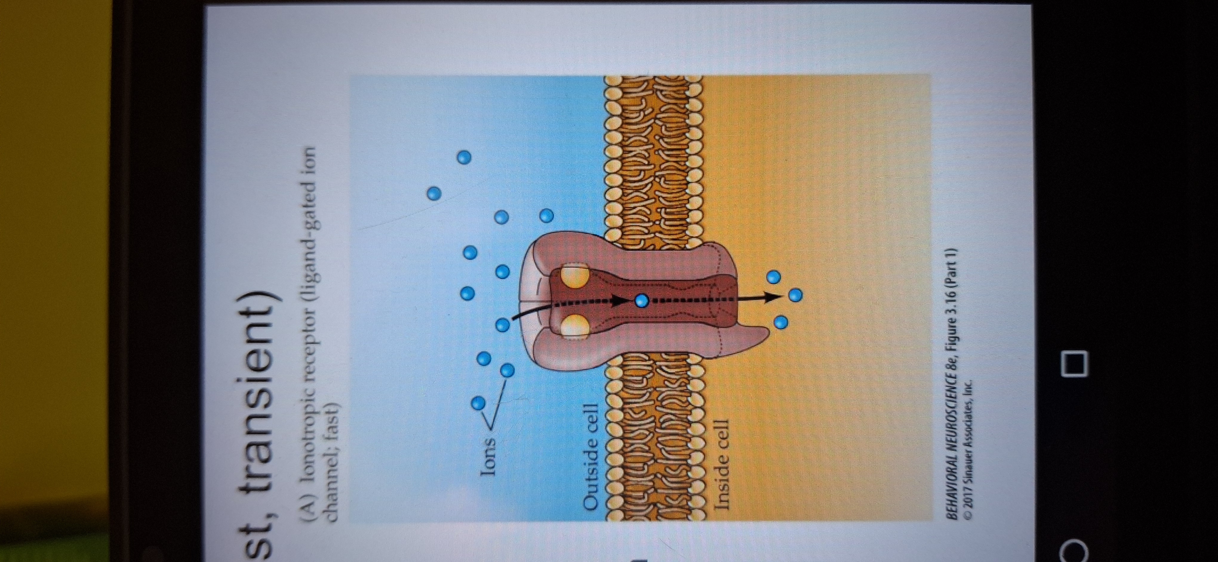
Ionotropic receptors
Fast. Transmitter binds to channel protein. Channel opens, and ions go in.
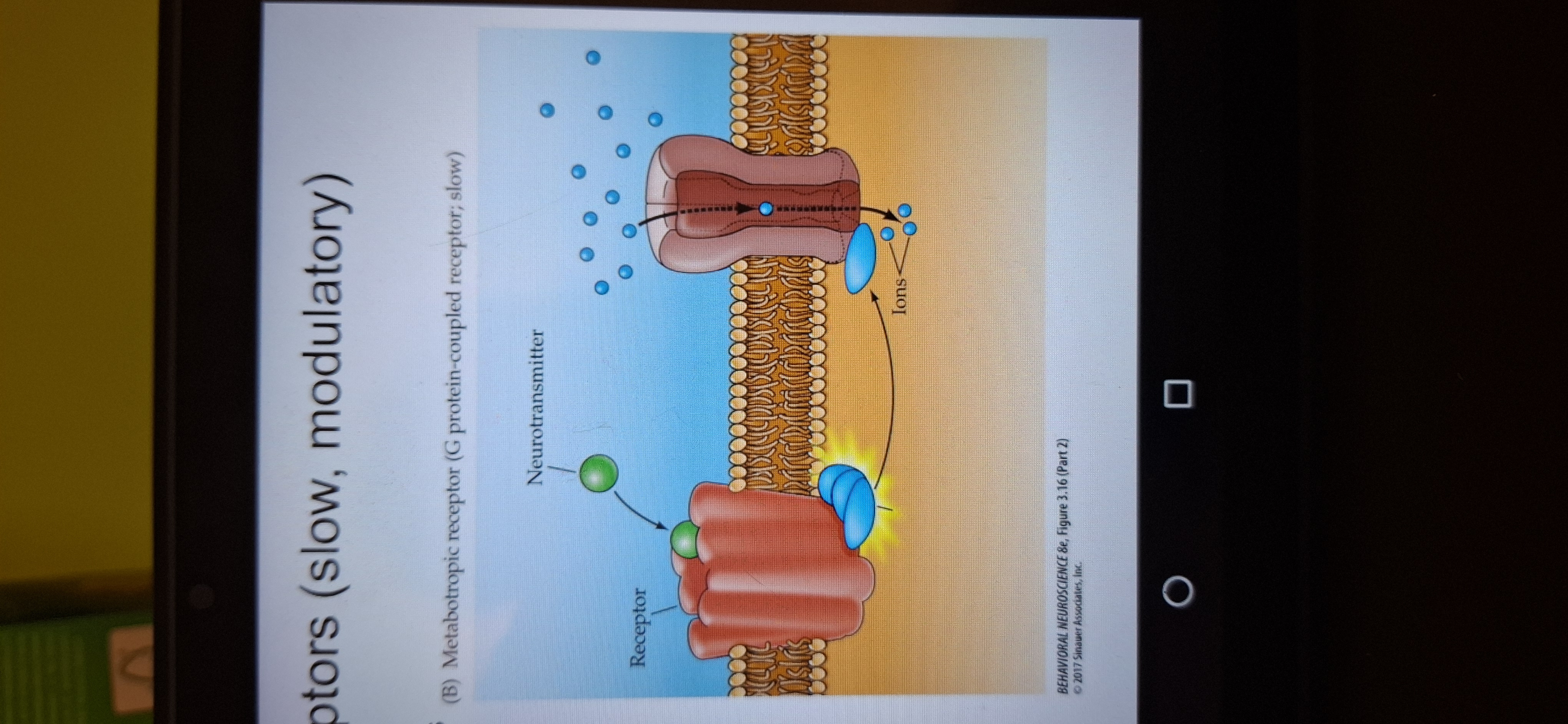
Metabotropic receptors.
Transmitter bind to Gprotrin. Protein is released. Binds to channel prot3in and channel opens. This is slow and has delay.
Ligands
Fit receptors exactly and activate or block them.
Endogenous ligands
Neurotransmitters and hormones
Exogenous ligands
Drugs and toxins from outside the body.
Antagonists
Block ACh receptors
Agonists
Mimic ACh stimulating the receptor.
Nicotine ACh receptors
Found in the brain and at synapses on muscles and in autonomic ganglia.blockage of these receptors are responsible for paralysis caused by curate and bungarotoxin.
Muscarinic Ach Receptor mACnR
Found in the brain. Found also in organs innervation by the parasympathetic division of the autonomic system.
Played a role in discovery of neurotransmitters. By ottoman loewi.
Types of synapses
Axo-dendritic-axosomatic-axo-axonic,dendro-dendritic.
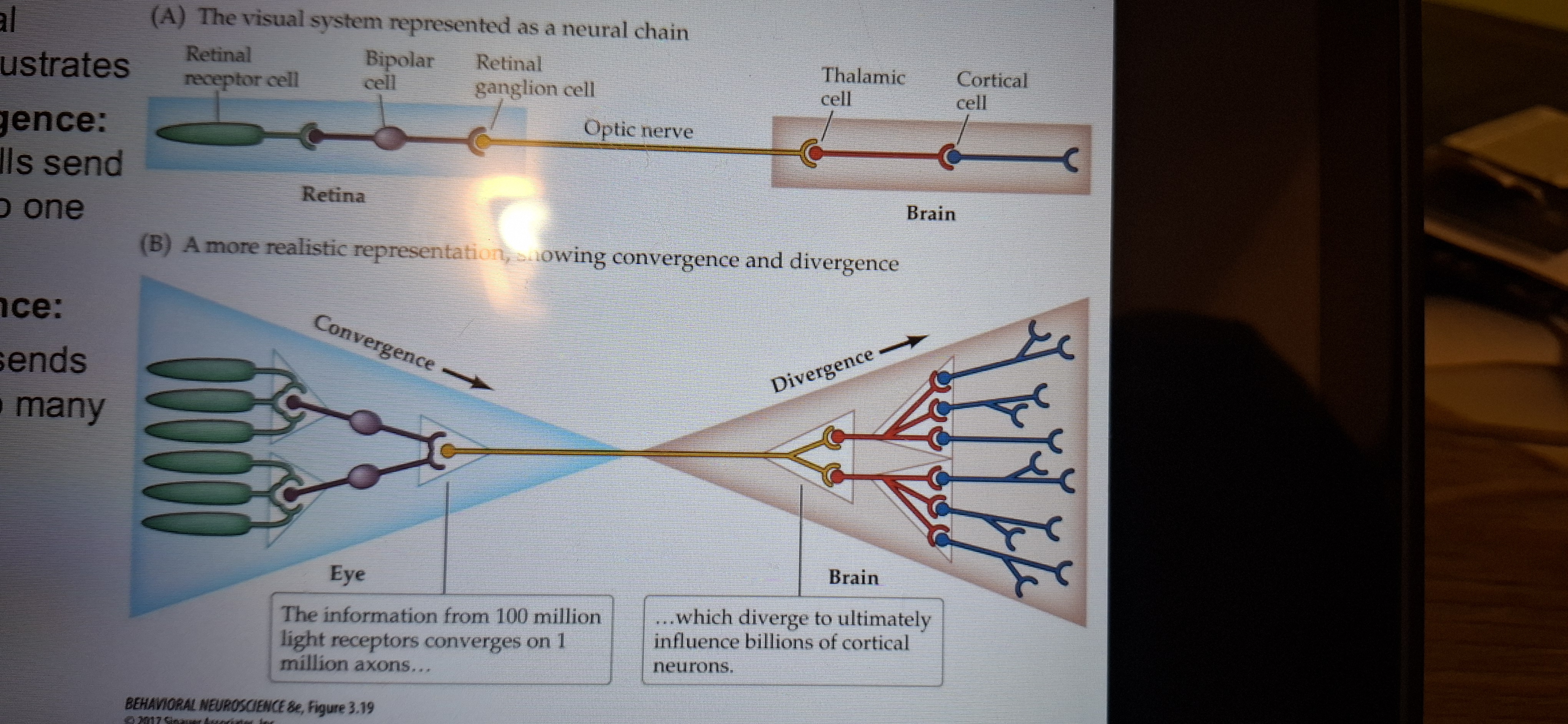
Visual pathway
Convergence- many cells send signals to one cell
Divergence-one cell signals to many cells.
EEG(electroenceplalogram)
Is a recording of brain potentials, or brain waves. Brain potentials indicate sleep states andprovide data in seizure disorders.