Key Concepts in Human Anatomy and Physiology
1/323
There's no tags or description
Looks like no tags are added yet.
Name | Mastery | Learn | Test | Matching | Spaced |
|---|
No study sessions yet.
324 Terms
Sagittal Plane
Divides the body into left and right sections.
Frontal (Coronal) Plane
Divides the body into anterior (front) and posterior (back) sections.
Transverse (Horizontal) Plane
Divides the body into superior (upper) and inferior (lower) sections.
Cellular Level
Basic unit of life, including various cell types.
Tissue Level
Groups of similar cells performing a specific function (e.g., epithelial, connective, muscle, nervous).
Organ Level
Structures composed of two or more tissue types working together (e.g., heart, lungs).
Organ System Level
Groups of organs that work together to perform complex functions (e.g., cardiovascular system).
Dorsal Body Cavity
Contains the cranial cavity (housing the brain) and the spinal cavity (housing the spinal cord).
Ventral Body Cavity
Contains the thoracic cavity (housing the lungs and heart) and the abdominopelvic cavity (housing digestive organs, reproductive organs, and urinary organs).
Anatomical Position
Refers to the body standing erect with the face forward, feet together, and arms hanging at the sides with palms facing forward.
Anatomy
The study of the body's structure.
Physiology
The study of how the body functions.
Midsagittal Plane
A vertical plane that divides the body into equal left and right halves.
Sagittal Plane
Any vertical plane parallel to the midline that divides the body into unequal left and right portions.
Horizontal Plane
Also known as the transverse plane, divides the body into superior (upper) and inferior (lower) portions.
Frontal Plane
A vertical plane at right angles to the midsagittal plane that divides the body into anterior (front) and posterior (back) portions.
Basic Components of a Cell
Include the cell membrane, nucleus, cytoplasm, and chromosomes.
Role of the Nucleus
Contains genetic codes that are essential for the functioning and reproduction of the cell.
Role of the Cytoplasm
Comprises all the substance of a cell except the nucleus.
Chromosomes
Found in the nucleus and contain DNA.
Homeostasis
The harmony maintained by cells, tissues, organs, and systems working together in the body.
Differentiation
The term for the specialized function of cells.
Stem Cells
Immature, unspecialized cells in the body that can be induced to become other types of cells.
Embryonic Stem Cells
Come from embryos developed from fertilized eggs used for research.
Adult Stem Cells
Found in the tissues of both adults and children.
Formation of Tissues
Tissues are formed when many millions of the same type of cell join together to perform a specific function for the body.
Main Types of Tissues
The four main types of tissues in the human body are epithelial, connective, muscle, and nerve tissue.
Epithelial tissue
The primary functions of epithelial tissue are to provide protection, produce secretions, and regulate the passage of materials across them.
Connective tissue
Connective tissue serves as the major support material of the body, providing support and connecting organs and tissues.
Muscle tissue
Muscle tissue has the ability to lengthen and shorten, allowing for the movement of body parts. Skeletal muscles can be classified as either voluntary or involuntary.
Nerve tissue
Nerve tissue is found in the brain, spinal cord, and nerves. Its primary function is to coordinate and control many body activities.
Organ
An organ is defined as a structure formed when several types of tissue group together to perform a single function, such as the stomach, which contains all four tissue types and performs digestive functions.
Body system
A body system is composed of a group of organs that work together to perform a major function to keep the body healthy and functional.
Dorsal Cavity
The dorsal cavity contains the spinal canal, which contains the spinal cord, and the cranial cavity, which contains the brain.
Thoracic Cavity
The thoracic cavity contains the lungs, heart, and accessory parts for their functioning.
Abdominal Cavity
The abdominal cavity includes most of the digestive tract and supporting organs for digestion.
Pelvic Cavity
The pelvic cavity contains the urinary bladder, rectum, and reproductive system.
Body systems
Body systems consist of specific organs and serve specific purposes essential for maintaining homeostasis and overall health.
Axial skeleton
The axial skeleton consists of 80 bones and its primary function is to protect the major organs of the nervous, respiratory, and circulatory systems.
Appendicular skeleton
The appendicular skeleton consists of 126 bones and includes the upper extremities (shoulders, arms, hands) and lower extremities (hips, legs, feet).
Bone tissue
Bone tissue, or osseous tissue, is composed of connective tissue that includes an organic component (cells and matrix) and an inorganic component (minerals).
Minerals in bone structure
Minerals, primarily calcium and phosphate, give rigidity to bone and act as reservoirs to maintain essential blood mineral concentrations when the body's supply is inadequate.
Skull
The skull consists of 28 bones.
Main components of the axial skeleton
The main components of the axial skeleton are the skull, spinal column, ribs, and sternum.
Function of appendicular skeleton
The function of the appendicular skeleton is to protect the organs of digestion and reproduction.
Three layers of bone
Periosteum – the dense, tough outer shell that contains blood vessels and nerves. Compact or dense tissue – the hard, smooth layer that protects the tissue within. Spongy or cancellous tissue – the porous, honeycombed material found inside most bones, which allows the bone to be strong yet lightweight.
Appendicular Skeleton
Consists of the bones of the limbs and the girdles that attach them to the axial skeleton.
Periosteum
A thin layer of connective tissue containing nerves and blood vessels, with an inner layer of loose connective tissue that has osteoblasts.
Compact Bone
The strong and hard section of the bone, dense and forming the main shaft of long bones and the outer layer of other bones.
Cancellous Bone and Marrow
Found inside the bone, lighter in weight and not as strong as compact bone, with marrow that produces blood cells.
Function of Periosteum
Serves as a protective layer and contains nerves and blood vessels; has an inner layer of loose connective tissue that contains osteoblasts, essential for bone growth and repair.
Sharpey's Fibers
Fibers that anchor the periosteum to the bone.
Difference between Compact and Cancellous Bone
Compact bone is dense, strong, and forms the main shaft of long bones and the outer layer of other bones, while cancellous bone is lighter in weight, found inside the bone, and is not as strong.
Role of Bone Marrow
Produces white blood cells (fight infection), red blood cells (carry oxygen), and platelets (help stop bleeding).
Fibrous Joints
Joints that do not move, such as the sutures of the skull.
Cartilaginous Joints
Joints made of connective tissue and cartilage that move only slightly, like the joints between the vertebrae.
Synovial Joints
Movable joints that account for most joints in the body and may be lined with a bursa filled with synovial fluid for cushioning.
Function of Bursa
Acts as a cushion to ease movement by reducing friction between the moving parts of the joint.
Difference between Fibrous and Cartilaginous Joints
Fibrous joints do not move at all, while cartilaginous joints allow for very slight movement.
Ball-and-Socket Joints
A type of synovial joint that allows for a wide range of movements, including flexion, extension, and rotational movements.
Hinge Joints
Allow movement in only one direction, similar to the hinge on a door.
Gliding Joints
Allow the bones to slide over one another, facilitating a range of movements.
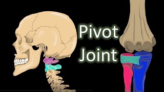
Pivot Joint
Found at the base of the skull and allows the head to rotate.
Saddle Joint
Allows movements such as touching the thumb to the fingers.
Gomphoses
Specialized joints that allow only very slight movement, such as attaching a tooth to the socket.
Percentage of Body Weight by Muscular System
The muscular system makes up 30 to 40 percent of total body weight.
Types of Muscles
The three types of muscles identified are striated, smooth, and cardiac.
Function of Striated Muscles
Provide for external body movement, such as facial expressions and activities like bike riding.
Voluntary Muscles
Striated muscles that are under conscious control.
Characteristics of Smooth Muscle Tissue
Non-striated tissue that is involuntary, controlled by the autonomic nervous system and not consciously controlled.
Locations of Smooth Muscle
Found in internal organs (except the heart), blood vessels, skin, and ducts from glands.
Smooth muscle
Characterized as nonstriated tissue and is involuntary, meaning it is controlled by the autonomic nervous system and not consciously controlled.
Cardiac muscle
Has a striated appearance like skeletal muscle but is involuntary in action and is only found in the heart.
Muscle contraction
The tightening of a muscle, making it shorter and thicker.
Muscle relaxation
The return to its original form or shape.
Muscle origin
Refers to the place where the muscle begins, which is the more fixed attachment toward the midline of the body.
Muscle insertion
The place where the muscle ends, referring to the more movable attachment that is away from the midline of the body.
Cardiovascular system
Consists of the circulatory system, heart, and lymphatic system.
Primary function of the cardiovascular system
To provide life-sustaining functions for the survival of bodily cells and tissues.
Disorders of the cardiovascular system
Include those affecting the heart and lymphatic system, which have specific signs and symptoms.
Functions of the circulatory system
1. Transports oxygen and nutrients to body cells, carbon dioxide and waste products from body cells, hormones and antibodies throughout the body. 2. Regulates body temperature and maintains chemical stability.
Function of the heart in the circulatory system
Acts as a pump that circulates blood throughout the body.
Layers covering the heart
1. Pericardium: Outer layer composed of a double-walled sac. 2. Myocardium: Tough muscular wall.
Function of the heart's right side
Pumps blood to the lungs.
Types of chambers in the heart
The atria, which receive blood, and the ventricles, which pump blood.
Role of one-way valves in the heart
Prevent the backflow of blood and separate the chambers of the heart by opening and closing with each heartbeat.
Tricuspid valve
Located between the right atrium and the right ventricle and has three cusps.
Mitral valve
Lies between the left atrium and the left ventricle and has two cusps.
Functions of the pulmonary and aortic semilunar valves
The pulmonary semilunar valve allows blood to flow from the right ventricle into the pulmonary artery, while the aortic semilunar valve allows blood to flow from the left ventricle into the aorta.
Blood flow through the heart
1. Blood enters the right atrium from the superior and inferior venae cavae. 2. It flows into the right ventricle. 3. The right ventricle pumps blood into the pulmonary artery to the lungs. 4. Oxygenated blood returns to the left atrium via the four pulmonary veins. 5. Blood flows from the left atrium into the left ventricle. 6. The left ventricle pumps blood into the aorta, distributing it to the body (except the lungs).
Major types of blood vessels
1. Arteries 2. Veins 3. Capillaries.
Functions of blood
1. Transportation of nutrients, gases, waste products, and hormones.
Regulation
The process of maintaining body fluids, pH balance, and body temperature.
Protection
The mechanism that defends against pathogens and prevents blood loss after injury through clotting.
Erythrocytes
Red blood cells that contain hemoglobin, which allows them to carry oxygen.
Leukocytes
White blood cells that protect the body from infection and disease.
Plasma
A straw-colored fluid that transports nutrients, hormones, and waste products; it is 91% water and 9% plasma proteins, including albumin and globulin.
Arteries
Large blood vessels that carry blood away from the heart to all regions of the body, having thicker, more elastic walls to withstand high pressure.
Veins
Blood vessels that form a low-pressure collecting system that returns waste-filled blood to the heart, having thinner walls compared to arteries and being less elastic.
Capillaries
Microscopic vessels that connect the arterial and venous systems, allowing for the exchange of nutrients and waste between blood and tissues.