Study Module 2, NHA CET Exam Preparation and EKG Fundamentals
1/127
There's no tags or description
Looks like no tags are added yet.
Name | Mastery | Learn | Test | Matching | Spaced | Call with Kai |
|---|
No analytics yet
Send a link to your students to track their progress
128 Terms
Epicardium
connective tissue that forms a sac around the heart
Myocardium
consists of involuntary striated muscle tissue and is responsible for physical contraction of the heart muscle
Endocardium
lines the chambers of the heart and forms the surface of the valves
4 chambers of the heart
right atrium, right ventricle, left atrium, left ventricle
Heart location
center of thoracic cavity, behind the sternum
Interatrial septum
wall dividing the atria
Interventricular septum
wall dividing the ventricles
Valves
control the direction of blood flow through the heart
Chordae Tendineae
support floppy valve leaflets and prevents regurgitation of blood into other chambers of the heart
4 valves of the heart
tricuspid valve, bicuspid valve, pulmonary valve, aortic valve
Atrioventricular valves
tricuspid and bicuspid valves
Semilunar valves
pulmonary and aortic valves
Tricuspid valve
separates right atrium from right ventricle
Bicuspid valve
separates the left atrium from the left ventricle
Pulmonary valve
between the right ventricle and pulmonary arteries
Aortic valve
between the left ventricle and the aorta
Right atrium receives blood from
inferior and superior vena cavas
Right atrium pumps blood through the _____ valve and into the ____ _____
tricuspid valve, right ventricle
Right ventricle pumps blood through the pulmonary ______ to the pulmonary arteries and then to the _______
veins, lungs
Blood becomes oxygenation through the ____ ____ of the lungs
capillary beds
Oxygenation blood returns to the _____ _____ through the pulmonary veins
left atrium
Left atrium pumps blood through the _____ valve to the ____ _____
bicuspid valve, left ventricle
Left ventricle pumps blood through the ______ valve into the aorta
aortic
The _____ delivers oxygenated blood to the heart muscle and the rest of the body
aorta
Right Coronary Artery (RCA)
carry oxygenated blood to the right atrium and ventricle, part of the left atrium, and the inferior wall of the left ventricle
SA node is a natural
pacemaker
AV junction acts as a
pacemaker if SA node fails
PR interval
time needed for an electrical impulse to travel from the SA node through the AV node to the ventricles
PR interval starts
P wave begins and ends at the beginning of Q wave
PR interval expected interval is
0.12-0.20 seconds
Ectopic beats
cause premature atrial, junctional, or ventricular complexes
Bundle of His
impulse is conducted by AV node
Standard grid
1mm wide by 1mm tall
Vertical side of EKG is measured in
millivolts
Horizontal side of EKG is measured in
milliseconds
Standard speed
25 mm/second
Standard amplitude/gain
10 mm/1mV
Standard Calibration
10mm tall by 5mm wide
Wilson's Central Terminal (WCT)
right arm, left arm, left foot
Unipolar leads
considered positive; V1-V6 leads
Precordial leads
V1-V6
Augmented leads
AVR, AVL, AVF
AVR
voltage to the right arm
AVL
voltage to the left arm
AVF
voltage to the left leg
Bipolar leads
positive and negative poles; Leads I, II, & III
Lead I
record impulses between left and right arms
Lead II
record impulses between right arm and left leg
Lead III
record impulses between left arm and left leg
Place limb electrodes on ___ areas
fleshy
Items needed for EKG
power source, electrodes, leads w/ clips, graph paper, alcohol wipes, gauze pads, scissors or shaving equipment, pillows, blankets
Decrease gain to 5 mm/mv for
large or too tall waveforms
increase gain to 20 mm/mV for
small waveforms
Increase speed to 50 mm/second for
tachycardia
Decrease speed to 12.5 mm/second for
bradycardia
Clean leads with
isopropyl alcohol or alcohol-based cleaners
Angle of Louis/Sternal Angle
used for proper lead (V1-V6) placement
V1
4th intercostal space, right of sternum; red
V2
4th intercostal space, left of sternum; yellow
V3
midway between V2 & V4; green
V4
5th intercostal space, midclavicular line; blue
V5
5th intercostal space, midway between V4 & V6, at anterior axillary line; orange
V6
5th intercostal space, at midaxillary line; purple
Limb lead colors are
white, black, red, and green
White limb lead
right arm
Black limb lead
left arm
Red limb lead
left leg
Green limb lead
right leg
Ambulatory monitoring is for
24 hr or longer
Types of ambulatory monitoring
3- or 5-lead
5-lead EKG is
Holter monitor
3-lead EKG is
telemetry
Colors for Holter monitor
white, red, black, brown, green
Colors for 3-lead EKG
white, black, red
Colors for telemetry
white, black, red, green, and brown
For stress testing, which leads change position, precordial or limb?
Limb
What conditions require a right-sided EKG
inferior wall ST segment elevation, MI, dextrocardia, patients under 8 years old
What condition would require a posterior EKG?
inferior wall infarction
What is the result of proper lead placement?
positive deflection
2 ways artifacts can occur
patient and nonpatient factors
Patient factor to cause artifacts
shivering, phone, talking
Nonpatient factors to cause artifacts
wall outlet, machine
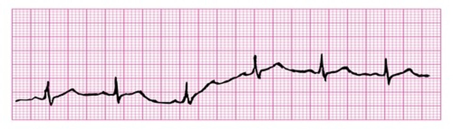
Wandering baseline
wavelike up-and-down movement throughout the tracing caused by improper electrode placement
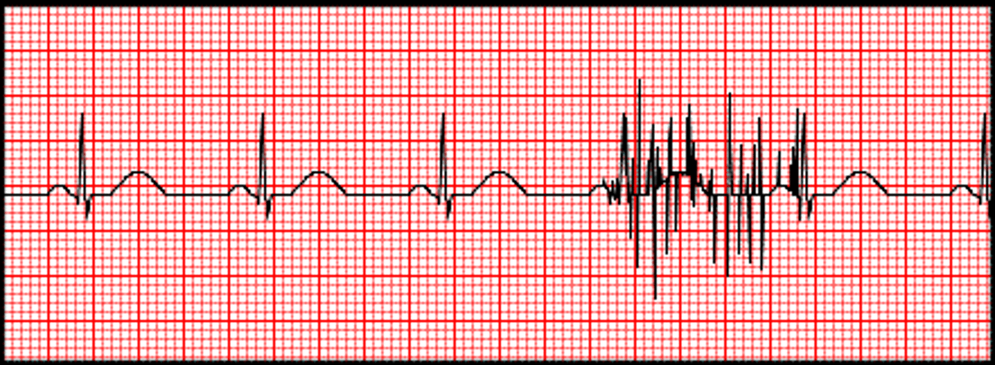
Somatic tremor
uneven spikes on EKG tracing caused by muscle movement
AC interference
electronic devices on or near a patient causing an electrical interference
Interrupted baseline
break in the baseline or a fully nonrecorded lead can be noted caused by broken wires or disconnected leads
When should an EKG tech monitor a patient heart rate until it reaches the target heart rate?
Stress testing
How to calculate target heart rate
(220 - age) * 0.85
During a stress test these symptoms are to be reported to the provider
dizziness, lightheadedness, nausea, severe SOB, tingling sensations, numbness, CP, extreme fatigue
Abnormal signs during stress testing are
abnormal vital signs, excessive tachycardia, hypotension, arrhythmias, PVCs, ventricular tachycardia, supraventricular tachycardia, heart blocks, T wave inversion, ST segment changes
Three domains of the NHA CET exam
Safety/Compliance & Coordinated Care, EKG Acquisition, EKG Analysis & Interpretation.
HIPAA regulation in healthcare
Protects patient privacy and medical records.
OSHA enforcement in healthcare settings
Workplace safety and infection control practices.
Scope of practice for EKG techs
The procedures and actions legally permitted for a technician to perform.
Universal precautions
Infection control methods treating all human blood and fluids as infectious.
Importance of communication in patient care
Ensures understanding, trust, and safety in care.
Factors affecting patient communication
Language, culture, religion, age, gender, and disability.
Normal vital signs indicators
Baseline for evaluating patient health and spotting abnormalities.
Patient preparation for stress testing
Wear comfortable clothing, avoid caffeine, follow fasting instructions.
Holter monitor usage
Continuous 24-48 hour monitoring of heart activity.