1.03 Parasternal Short Axis View (SAX)
0.0(0)
Card Sorting
1/11
There's no tags or description
Looks like no tags are added yet.
Last updated 9:07 PM on 5/14/25
Name | Mastery | Learn | Test | Matching | Spaced | Call with Kai |
|---|
No analytics yet
Send a link to your students to track their progress
12 Terms
1
New cards
Where is the echocardiographic window for getting a SAX?
In the parasternal space, LSB (left sternal border), and inbetween the 3rd and 5th intercostal space (same as LAX).
2
New cards
How do you get into a SAX view from a LAX view?
Rotate the transducer 90° clockwise.
3
New cards
The image pointer in SAX should be towards what side of the patient?
Their left.
4
New cards
You should adjust the transducer to ensure what?
The structures you want to look at are in the centre of the display.
5
New cards
What are the different levels that are investigated at the SAX view? (5 + one extra for learning purposes)
* Pulmonary artery bifurcation
* Short axis aortal/aortic valve
* SAX LVOT (this is the extra one)
* SAX Mitral valve
* SAX papillary muscles
* SAX apex
* Short axis aortal/aortic valve
* SAX LVOT (this is the extra one)
* SAX Mitral valve
* SAX papillary muscles
* SAX apex
6
New cards
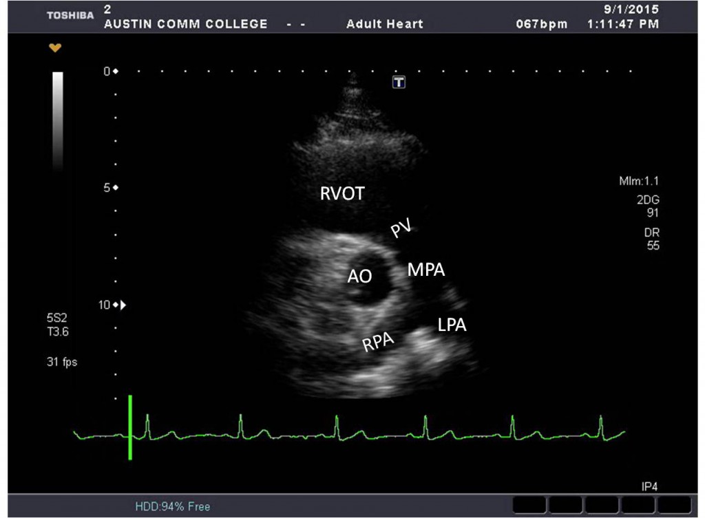
What structures are seen in the pulmonary bifurcation view? (transducer is positioned superiorly, anteriorly, and slightly left)
* RVOT
* MPA (RPA, LPA)
* PV
* Aorta (AO)
* Tricuspid valve (TV)
* Right atrium (RA)
* MPA (RPA, LPA)
* PV
* Aorta (AO)
* Tricuspid valve (TV)
* Right atrium (RA)
7
New cards
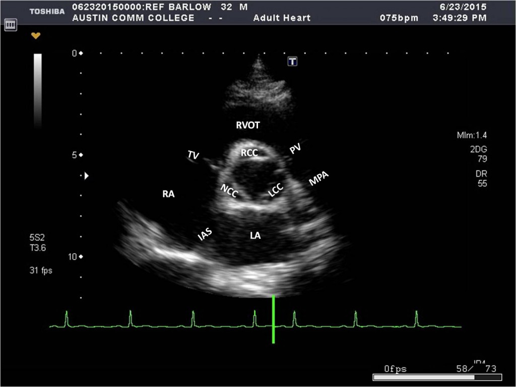
What structures are seen in the SAX level of the aorta/aortic valve? (transducer is angled inferiorly - can also slide inferiorly from the pulmonary bifurcation view)
* RVOT
* PV
* MPA
* AO and Aortic cusps (RCC, LCC, NCC)
* RA
* LA
* IAS (interatrial septum)
* TV (anterior and posterior leaflets)
* Occasionally the left main coronary artery (LMCA)
* PV
* MPA
* AO and Aortic cusps (RCC, LCC, NCC)
* RA
* LA
* IAS (interatrial septum)
* TV (anterior and posterior leaflets)
* Occasionally the left main coronary artery (LMCA)
8
New cards
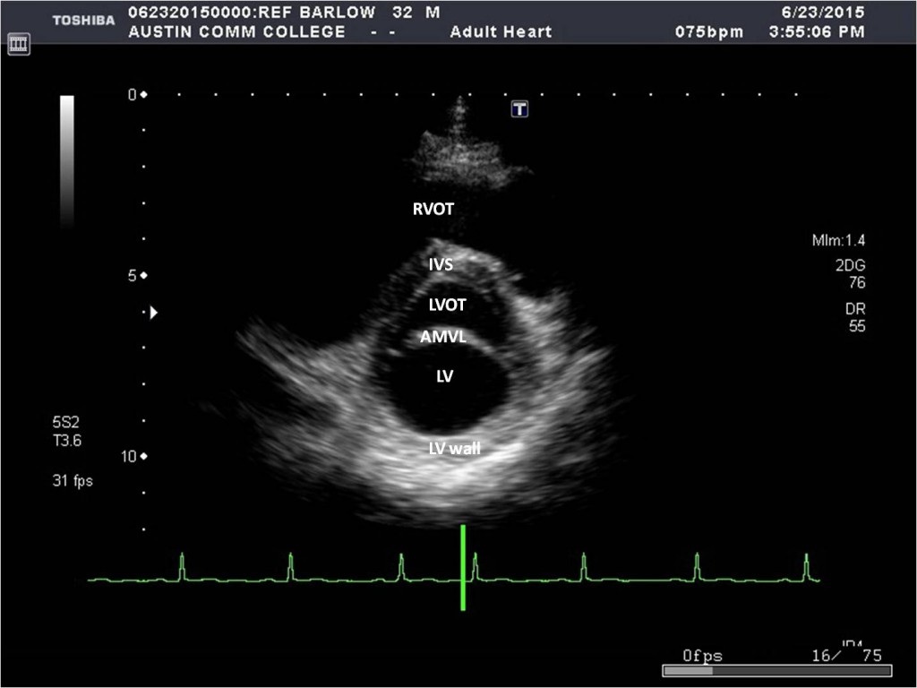
What structures are seen in the SAX level of the LVOT? (transducer is angled inferiorly or slides inferiorly from the aortic valve view)
* RV
* LVOT
* AMVL
* LA
* RA
* TV
* LVOT
* AMVL
* LA
* RA
* TV
9
New cards
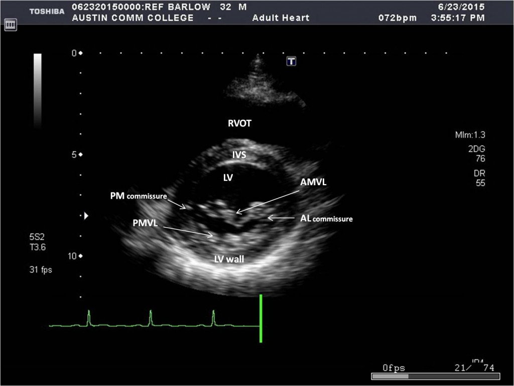
What structures are seen in the SAX level of the Mitral valve? (transducer is angled inferiorly or slides inferiorly from the LVOT view)
* LV
* RV
* Anterior mitral valve leaflet (AMVL)
* Posterior mitral valve leaflet (PMVL)
* IVS (interventricular spetum)
* Inferior LV wall
* RV
* Anterior mitral valve leaflet (AMVL)
* Posterior mitral valve leaflet (PMVL)
* IVS (interventricular spetum)
* Inferior LV wall
10
New cards
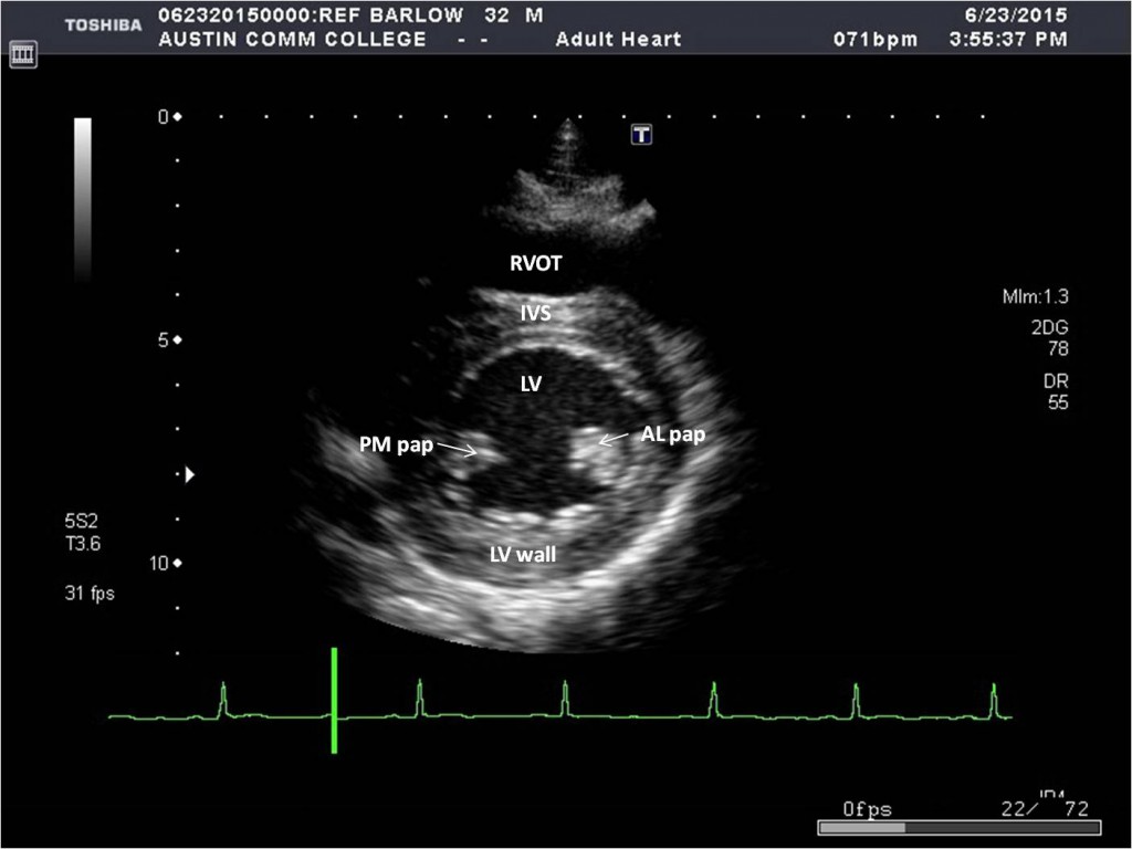
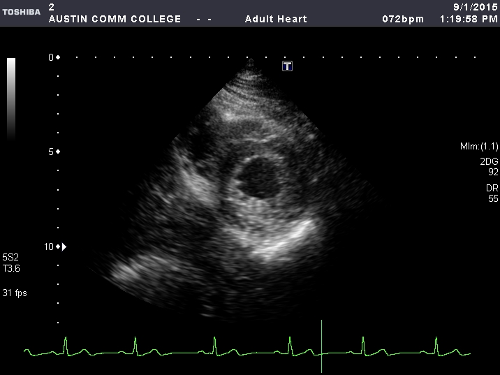
What structures are seen in the SAX level of the papillary muscles? (transducer is angled inferiorly or slides inferiorly from the MV view)
* LV
* RV
* IVS
* Anterolateral and posterolateral papillary muscles
* Inferior LV wall
* RV
* IVS
* Anterolateral and posterolateral papillary muscles
* Inferior LV wall
11
New cards
What structures are seen in the SAX level of the apex? (transducer is angled inferiorly or slides inferiorly from the papillary muscle view)
* LV apex
* RV apex
* IVS (apical segment)
* Anterior, lateral, and inferior LV walls
* RV apex
* IVS (apical segment)
* Anterior, lateral, and inferior LV walls
12
New cards
When doing an EKG, there are red, white, and black electrodes. Where are they placed?
Red - right arm
White - left chest (bottom left of tummy)
Black - left shoulder
White - left chest (bottom left of tummy)
Black - left shoulder