Methods of studying cells 3.2.1.3
 Call Kai
 Learn
Learn Practice Test
 Spaced Repetition
 Match
Match Flashcards
 Knowt Play
1/7

Earn XP
Description and Tags
Name | Mastery | Learn | Test | Matching | Spaced | Call with Kai | Chat |
|---|
No analytics yet
Send a link to your students to track their progress
8 Terms
Magnification definition
How many times larger an image is compared to the object.
Resolution definition
The ability to distinguish between two separate points.
Calculating magnification
Magnification = Image size / Actual size
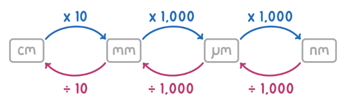
Unit conversions
Optical Microscopes
Light rays are shone through a thin, dead, stained specimen to magnify an image. Light rays have a relatively long wavelength so they have a low resolution. Low magnification. Cheap and can produce colourful images.
Transmission Electron Microscopes
Use electromagnets to transmit a beam of electrons through a specimen (denser parts absorb more electrons so appear darker). Produce high-resolution, high-magnification, 2D images that show internal organelles. Only thin, dead specimens can be observed (because of the vacuum).
Scanning Electron Microscopes
A beam of electrons is reflected off the surface of a dead specimen to produce an extremely high resolution, high magnification, 3D black and white image.
Cell fractionation
A method used to isolate components/organelles of a cell, so they can be studied.
Sample preparation - place sample in an ice-cold (slows enzymes), isotonic (prevents osmosis), buffered (prevent enzyme/protein denaturation) solution.
Homogenisation - physically breaking the plasma membrane (blender), allowing organelles to be released into the solution.
Filtration - passed through a gauze to remove large debris.
Ultracentrifugation - separates organelles based on their density by spinning the solution at various speeds. The pellet (bottom of tube) contains the heavier organelles and the supernatant (liquid above pellet) contains lighter organelles. The supernatant is then centrifuged again to produce the next dense organelle (nuclei, chloroplasts, mitochondria, lysosomes, endoplasmic reticulum, ribosomes).