🚦 Lecture 10: Signal Transduction + GPCR (G Protein-Coupled Receptors)
1/17
There's no tags or description
Looks like no tags are added yet.
Name | Mastery | Learn | Test | Matching | Spaced | Call with Kai |
|---|
No analytics yet
Send a link to your students to track their progress
18 Terms
Signal Transduction – How Cells Sense External Signals
Overview
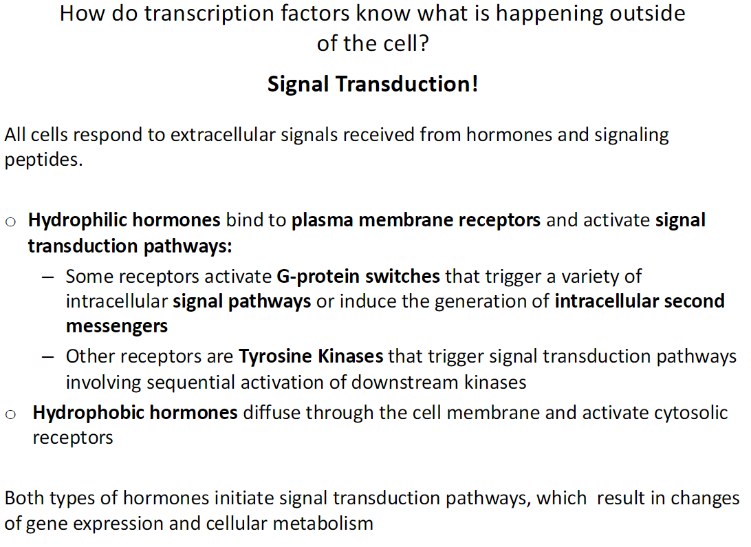
Transcription factors respond to extracellular signals from hormones and signaling peptides
Signal transduction connects external signals to changes in gene expression and metabolism
Hydrophilic Hormones
Cannot cross the cell membrane
Bind to plasma membrane receptors
Activate signal transduction pathways:
• Some receptors activate G-proteins → trigger various intracellular pathways or generate second messengers
• Tyrosine kinase receptors → sequential activation of downstream kinases
Hydrophobic Hormones
Diffuse through the cell membrane
Bind cytosolic receptors directly
Outcome
Both types of hormones initiate signal transduction
Lead to changes in gene expression and cellular metabolism
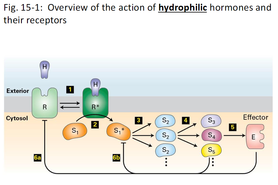
Signal Transduction Pathway – Hydrophilic Hormones *Don’t need to know
Step 1 – Ligand Binding
A receptor protein (R) on the plasma membrane binds a hydrophilic hormone (H)
Binding triggers a conformational change in the receptor
Step 2 – Receptor Activation
Activated receptor interacts with a signal transduction protein
Often a GTP-binding protein, kinase, or phosphatase
Step 3 – Signal Cascade
Signal transduction protein activates or inhibits other downstream signaling proteins
One protein can activate multiple downstream targets → signal amplification
Step 4 – Further Signaling
Activated proteins continue to transmit the signal through the pathway
Amplifies and diversifies the cellular response
Step 5 – Effector Activation
Some signaling proteins activate effector proteins (E)
Effectors can be enzymes, transcription factors, transport proteins, or ion channels
Effectors carry out the cellular response
Step 6 – Feedback Control
Proteins in the pathway or effectors can modify the receptor or early signaling proteins
Inhibits or blocks early steps in the pathway
Can trigger receptor degradation → reduces receptor numbers → lowers cell sensitivity to the ligand
Key Points
Signal amplification ensures a small number of ligands produces a large cellular response
Feedback mechanisms maintain control and prevent overstimulation
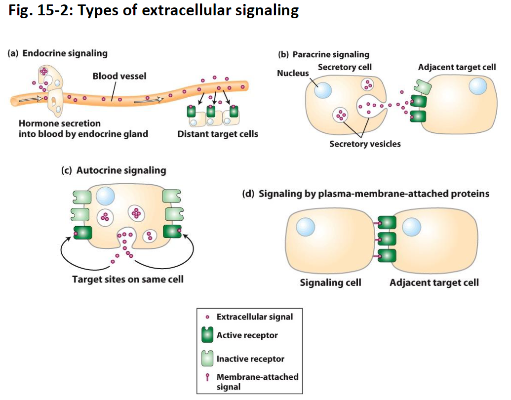
Cell-to-Cell Signaling – Types and Mechanisms
(a) Endocrine Signaling
Hormones are secreted by endocrine glands into the blood
Travel long distances to reach distant target cells
Etymology: endo- = inside, crine = secrete → “secrete inside” (into the blood)
(b) Paracrine Signaling
Signaling molecules are released by a cell to act on nearby adjacent cells
Short-distance signaling
Etymology: para- = beside/near → “secrete beside” (affects neighboring cells)
(c) Autocrine Signaling
A cell secretes signaling molecules that act on itself
Can regulate its own activity or gene expression
Etymology: auto- = self → “self-secretion” (acts on the same cell)
(d) Signaling by Plasma-Membrane-Attached Proteins
Proteins on the plasma membrane of one cell interact directly with receptors on an adjacent cell
Does not require secretion of molecules
Allows direct cell-to-cell communication
Key Points
Signaling distance varies: autocrine/paracrine = micrometers, endocrine = meters
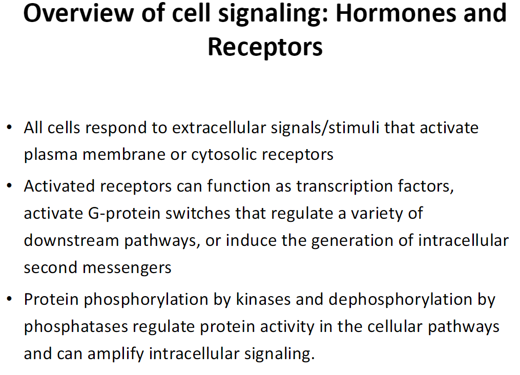
Overview of Cell Signaling – Hormones and Receptors
Signal Reception
All cells respond to extracellular signals or stimuli
Signals activate plasma membrane receptors or cytosolic receptors
Receptor Function
Activated receptors can:
• Act as transcription factors → change gene expression
• Activate G-protein switches → regulate multiple downstream pathways
• Generate intracellular second messengers → amplify and transmit the signal
Signal Regulation
Protein activity is controlled by:
• Phosphorylation by kinases
• Dephosphorylation by phosphatases
These modifications regulate signaling pathways and can amplify intracellular signals
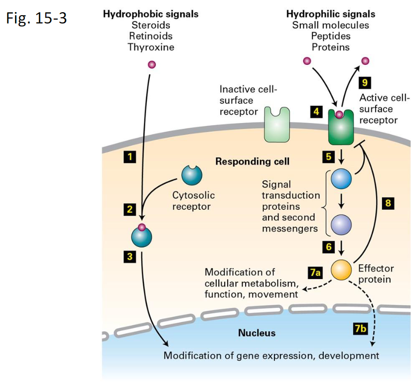
Overview of Cell Signaling – Hydrophobic vs Hydrophilic Signals
Hydrophobic Signals
Examples: steroids, retinoids, thyroxine
Steps:
Signal molecule diffuses through the plasma membrane
Binds to a cytosolic receptor → forms receptor-signal complex
Complex moves into the nucleus
Binds DNA transcription-control regions → activates or represses gene expression
Hydrophilic Signals
Examples: small molecules (adrenaline, acetylcholine), peptides (glucagon, yeast mating factors), proteins (insulin, growth hormone)
Steps:
Signal binds to an inactive cell-surface receptor (4)
Receptor undergoes conformational change → becomes active (4)
Activated receptor activates downstream signal transduction proteins or generates second messengers (5)
Signal transduction activates effector proteins (6)
a. Effector proteins stay in cytosol → modify enzymes → short-term changes in metabolism, movement, or function (7a)
b. Effector proteins enter the nucleus → long-term changes in gene expression (7b)
Termination / Down-Modulation:
6. Negative feedback from intracellular signalling molecules (8)
7. Removal of the extracellular signal (9)
Key Points:
Hydrophobic → cytosol receptor → nucleus → gene expression modification
Hydrophilic → membrane receptor → signal transduction → effector → cytosol or nucleus
Signal amplification and feedback control regulate strength and duration of response
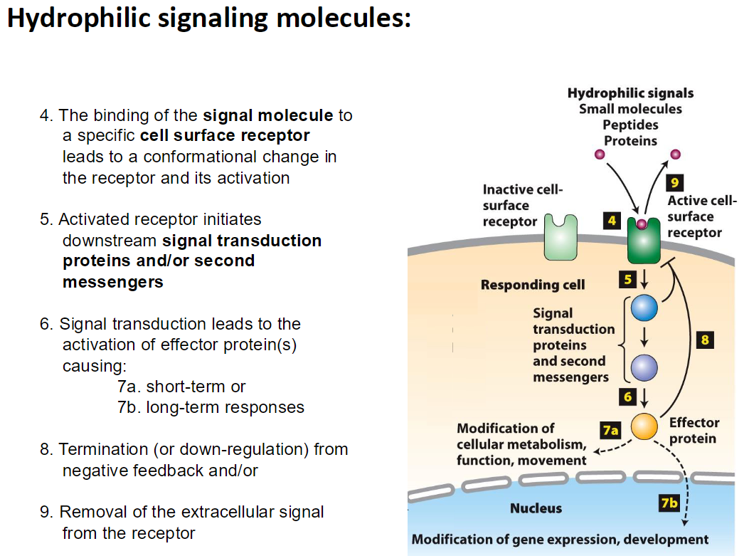
Hydrophilic Signaling – Stepwise Pathway
Steps:
4. Signal molecule binds to a specific cell-surface receptor → receptor undergoes conformational change → receptor becomes active
5. Activated receptor triggers downstream signal transduction proteins and/or generates second messengers
6. Signal transduction activates effector protein(s)
7a. Short-term response: cytosolic effectors modify enzymes → changes in metabolism, function, or movement
7b. Long-term response: effectors move into the nucleus → changes in gene expression
8. Termination / Down-regulation: negative feedback from intracellular signaling molecules inhibits early steps and/or:
9. Removal of the extracellular signal from the receptor ends the pathway
Key Points:
Hydrophilic signals cannot cross the membrane → require membrane receptors
Pathway can amplify the signal through multiple downstream proteins
Feedback ensures the response is controlled and reversible
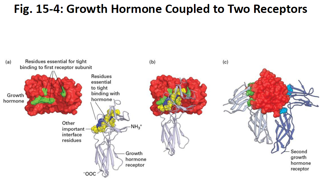
Growth Hormone Binding to Two Receptors (Steps from textbook, don’t need to know)
Overview
A single growth hormone (GH) protein binds simultaneously to two growth hormone receptors (GHRs)
Binding is mediated by multiple weak, noncovalent forces
Key Findings from Structural Studies
GH-Receptor Interface: 28 amino acids on GH interact with the first receptor
Critical Residues: Only 8 amino acids on GH contribute ~85% of the binding energy
• These 8 residues are distant in the primary sequence but adjacent in the folded 3D structureReceptor Contribution: Two tryptophan residues on the receptor provide most of the binding energy
• Other receptor amino acids at the interface also contribute
Binding Sequence
GH binds to the first receptor molecule (involving GH’s key residues and receptor interface residues)
A second receptor molecule binds on the opposite side of GH
• Uses the same critical amino acids on the receptor
• Interacts with different residues on GH compared to the first receptor
Key Points
- GH binding involves specific residues on both hormone and receptor
- Multiple weak interactions combine to create strong and specific binding
- Protein folding brings distant amino acids close together → essential for receptor interaction
Hormone Receptors – Activation of Effector Proteins
Overview
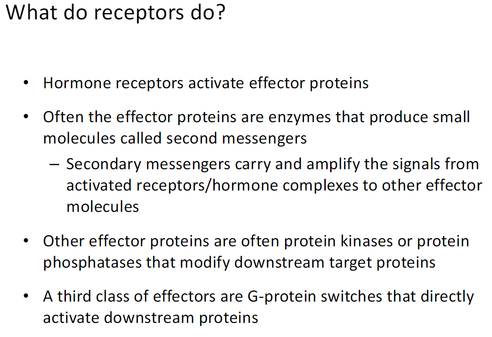
Receptors sense hormones and activate effector proteins inside the cell
Effector Proteins
Some are enzymes that make second messengers → carry and amplify the signal
Some are kinases or phosphatases → modify downstream proteins by adding or removing phosphate groups
Some are G-protein switches → directly activate downstream proteins
Outcome
Receptors transmit the hormone signal and control cellular responses like metabolism, function, or gene expression
Intracellular Second Messengers
Overview
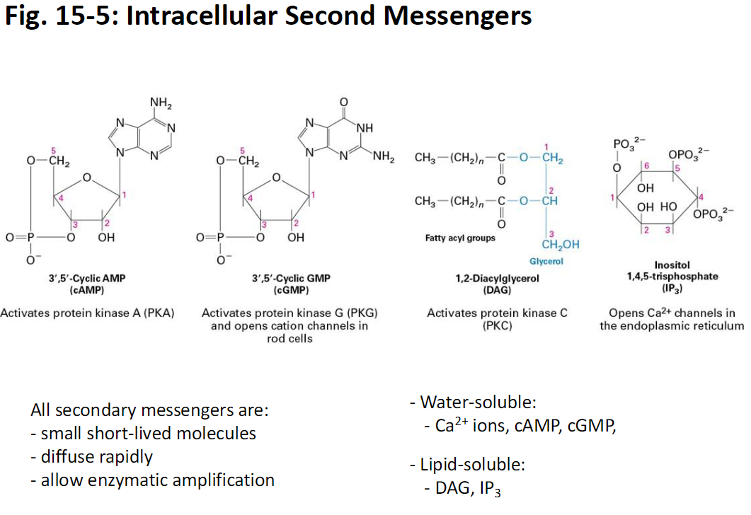
Second messengers are small molecules produced by effector proteins
They transmit and amplify signals from hormone-receptor complexes
Properties
Small and short-lived
Diffuse rapidly within the cell
Enable enzymatic amplification of the signal
Types
Water-soluble: Ca²⁺ ions, cAMP, cGMP → move freely in cytosol
Lipid-soluble: DAG, IP₃ → can interact with membrane-associated proteins
Outcome
Carry the signal from activated receptors to other proteins, amplifying the cellular response
Signal Transduction by Protein Phosphorylation
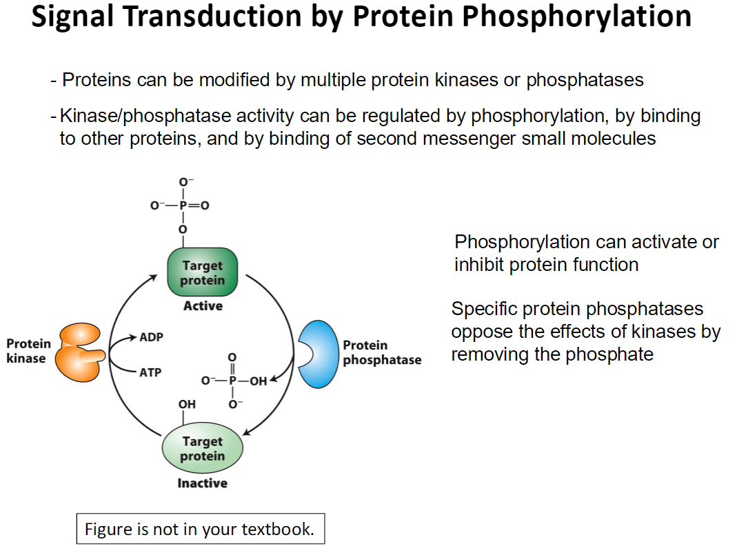
Overview
Proteins can be modified by multiple protein kinases or phosphatases
These modifications regulate protein activity and propagate signals in the cell
Regulation of Kinases and Phosphatases
Activity can be controlled by:
Phosphorylation of the kinase or phosphatase itself
Binding to other proteins
Binding of second messenger molecules
Effects of Phosphorylation
Phosphorylation can activate or inhibit a protein’s function
Specific protein phosphatases remove phosphate groups to reverse kinase effects
Outcome
Phosphorylation and dephosphorylation allow fine control of signaling pathways and amplify cellular responses
Protein Kinases and Phosphatases in Animal Cells
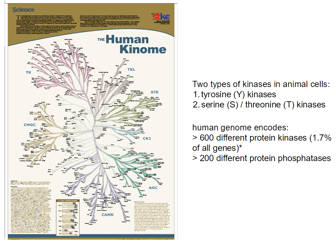
Kinase Types
Two main types of protein kinases in animal cells:
Tyrosine (Y) kinases
Serine (S) / Threonine (T) kinases
Human Genome
Encodes more than 600 different protein kinases (~1.7% of all genes)
Encodes over 200 different protein phosphatases
Overview
Kinases add phosphate groups to proteins, regulating their activity
Phosphatases remove phosphate groups, reversing kinase effects
Together, they control cellular signaling pathways precisely
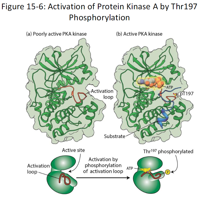
Activation of Protein Kinase A (PKA) by Phosphorylation *Don’t need to know
Overview
Protein kinase A (PKA) exists in an inactive, unphosphorylated form
Activation occurs through phosphorylation of a threonine residue (Thr197) in the activation loop
Mechanism
Phosphorylation at Thr197 causes a conformational change in the activation loop
This change allows PKA to bind ATP and substrate proteins, enabling its kinase activity
Phosphorylation also allows PKA to bind its inhibitory subunit R, which regulates activity via cAMP
Additional Notes
PKA can autophosphorylate Thr197
Other cellular kinases can also phosphorylate this residue
Similar phosphorylation-dependent activation occurs in many other protein kinases
Outcome
Phosphorylation of Thr197 switches PKA from inactive to active
Enables it to phosphorylate downstream target proteins and propagate cellular signals
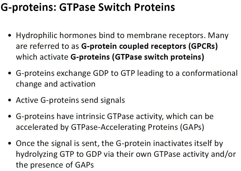
G-Proteins: GTPase Switch Proteins
Overview
Hydrophilic hormones bind to membrane receptors, often called G-protein coupled receptors (GPCRs)
These receptors activate G-proteins, which are GTPase switch proteins
Activation Mechanism
G-proteins exchange GDP for GTP, causing a conformational change and activation
Active G-proteins transmit signals to downstream effectors
Inactivation
G-proteins have intrinsic GTPase activity
GTPase-Accelerating Proteins (GAPs) can speed up GTP hydrolysis
Hydrolysis of GTP to GDP inactivates the G-protein and stops the signal
Outcome
G-proteins act as molecular switches, turning signaling on when bound to GTP and off when hydrolyzing it to GDP
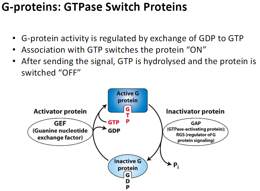
G-Proteins: GTPase Switch Proteins – Regulation
Overview
G-protein activity is controlled by the exchange of GDP for GTP
Activation
Binding of GTP switches the G-protein ON
Activator proteins called GEFs (Guanine nucleotide exchange factors) promote this GDP → GTP exchange
Active G-proteins transmit signals to downstream effectors
Inactivation
After signaling, GTP is hydrolyzed to GDP → switches the G-protein OFF
Inactivator proteins such as GAPs (GTPase-activating proteins) or RGS (regulators of G-protein signaling) accelerate this hydrolysis
Outcome
G-proteins act as molecular switches, cycling between active (GTP-bound) and inactive (GDP-bound) states to regulate signal transduction
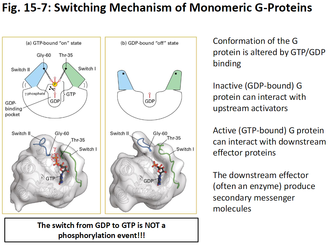
Switching Mechanism of Monomeric G-Proteins
Overview
G-protein conformation changes depending on whether GDP or GTP is bound
Inactive State
G-protein bound to GDP is inactive
Can interact with upstream activator proteins
Active State
G-protein bound to GTP is active
Can interact with downstream effector proteins, often enzymes
Effectors produce second messenger molecules to transmit and amplify the signal
Key Point
The switch from GDP to GTP is not a phosphorylation event
It is a conformational change caused by nucleotide exchange
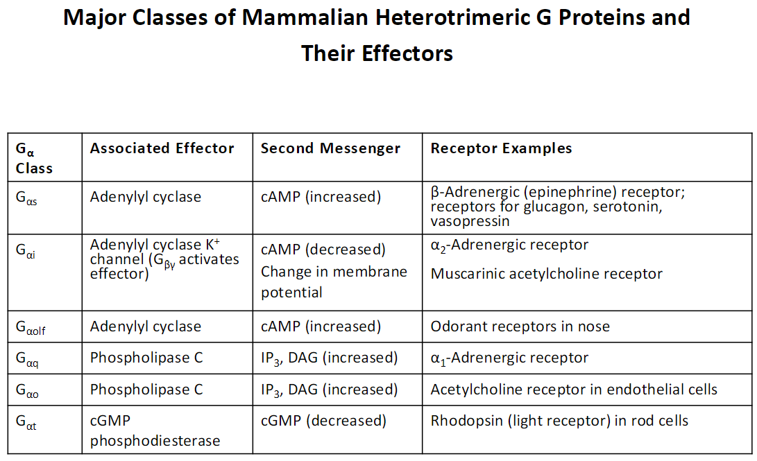
Major Classes of Mammalian Heterotrimeric G Proteins and Their Effectors *Just know if second messengers increase or decrease
Gαs
Effector: Adenylyl cyclase
Second Messenger: cAMP (increased)
Receptor Examples: β-Adrenergic (epinephrine) receptor, glucagon, serotonin, vasopressin receptors
Gαi
Effector: Adenylyl cyclase, K⁺ channels (Gβγ activates effector)
Second Messenger: cAMP (decreased), change in membrane potential
Receptor Examples: α2-Adrenergic receptor, muscarinic acetylcholine receptor
Gαolf
Effector: Adenylyl cyclase
Second Messenger: cAMP (increased)
Receptor Examples: Odorant receptors in the nose
Gαq
Effector: Phospholipase C
Second Messenger: IP₃, DAG (increased)
Receptor Examples: α1-Adrenergic receptor
Gαo
Effector: Phospholipase C
Second Messenger: IP₃, DAG (increased)
Receptor Examples: Acetylcholine receptor in endothelial cells
Gαt
Effector: cGMP phosphodiesterase
Second Messenger: cGMP (decreased)
Receptor Examples: Rhodopsin (light receptor) in rod cells
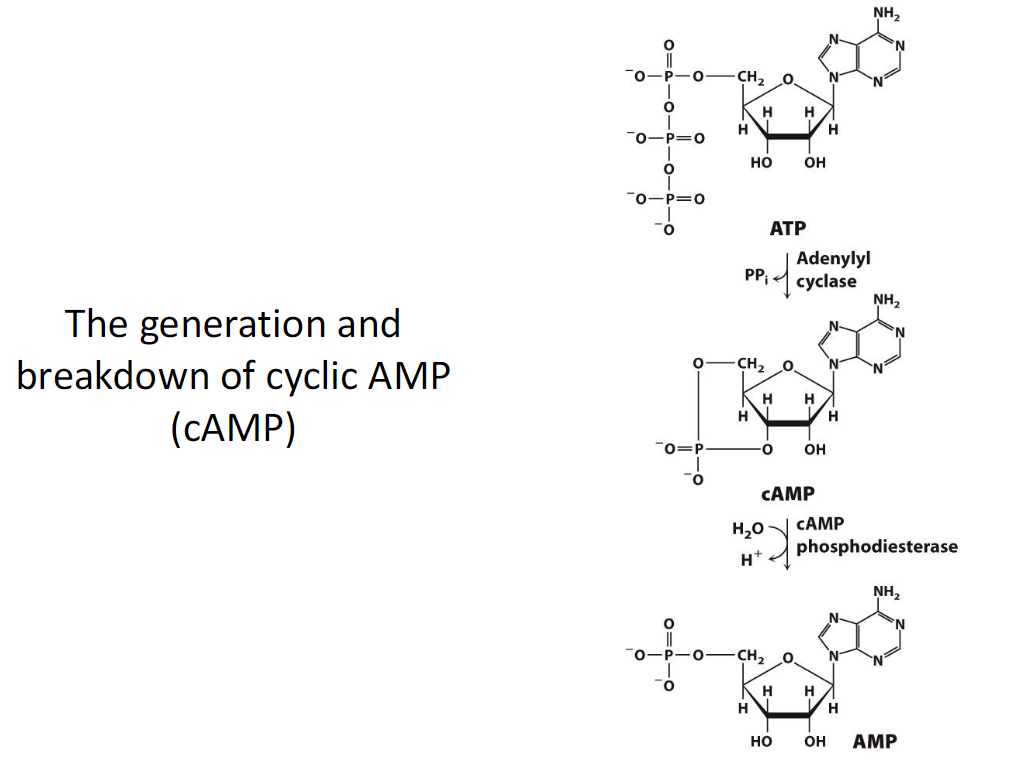
Generation and Breakdown of cAMP *Don’t need to know
Overview
cAMP (cyclic AMP) is a second messenger produced from ATP by the enzyme adenylyl cyclase
Generation
ATP is converted into cAMP by adenylyl cyclase when a G-protein (Gαs) activates it
cAMP carries and amplifies the signal to downstream effectors
Breakdown
cAMP phosphodiesterase converts cAMP into AMP, terminating the signal
This ensures that the signal is short-lived and tightly controlled
Outcome
cAMP levels regulate cellular responses by activating cAMP-dependent protein kinases (PKA) and other effectors
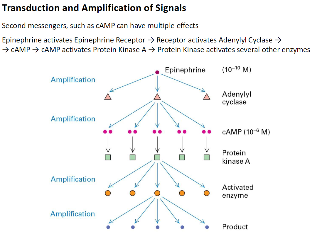
Transduction and Amplification of Signals
Overview
Second messengers, like cAMP, transmit and amplify signals from hormones
Example: Epinephrine Signaling
Epinephrine binds to its receptor → activates adenylyl cyclase → produces cAMP
cAMP activates Protein Kinase A (PKA) → PKA activates multiple downstream enzymes
Amplification
One epinephrine molecule (10⁻¹⁰ M) → many cAMP molecules (10⁻⁶ M)
Each PKA molecule can activate several enzymes → large-scale production of final products
Outcome
Signal amplification ensures that a small amount of hormone can trigger a strong cellular response