Chordates Lab 2
1/31
There's no tags or description
Looks like no tags are added yet.
Name | Mastery | Learn | Test | Matching | Spaced | Call with Kai |
|---|
No analytics yet
Send a link to your students to track their progress
32 Terms
What is cartilage
connective tissue formed from proteoglycan rich extracellular matrix
receives nutrients that diffuse from nearby vessels
not directly innervated
not vascularized
strong and durable, yet lightweight and flexible
Do hagfish and lampreys have bone?
they lack bone
vertebral column of lamprey is hardened by calcium deposits formed by
precipitation of calcium salts in the cartilage matrix.
How might the composition of the shark’s skeleton
affect their general movement pattern compared to a bony fish like the perch?
A shark’s skeleton, composed entirely of cartilage rather than bone, significantly enhances its mobility, speed, and agility compared to bony fish like the perch. The cartilaginous skeleton is approximately half as dense as bone, providing greater flexibility and allowing for more efficient, energy-saving movements.
Bone can form in two ways
in connective tissue (dermal)
or thru calcification of cartliage (endochondral):
ENDOCHONDRAL (replacement) BONE -
develops through the ossification of cartilage
precursors. This includes such things as the brain
case, axial and appendicular skeletons
Lamellar bone
mature bone, is stronger as its fibers are arranged in multiple specific layers
how is bone attatched to muscle? bone attacthed to other bone?
tendons and ligaments respectively
bone, tendon, and ligament are connective tissues.
Characteristics of bone, compare to cartilage
bone is vascularized and innervated, unlike cartilage, and responds to mechanical stress with increased depostition (creation of new bone), unlike cartilage which is flexible and flexes under pressure
more dense and less flexible than cartilage
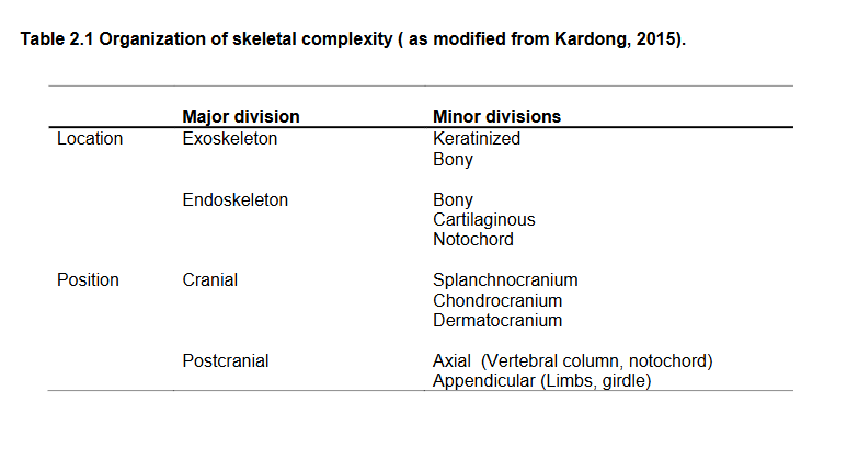
Organization of skeletal complexity graph
Braincase name
The braincase (the upper and back part of the skull, which forms a protective case around the brain) and sensory structures is referred to as the
Chondrocranium in cartilaginous fish
Neurocranium in bony fish
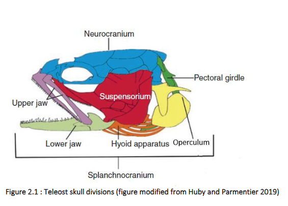
Teleost skull divisions graph
The neurocranium of bony fish includes dermal bone (formed on its own, didn’t replace cartilage). The splanchnocranium includes the framework for the jaws and gill arches and HYOID of gnathostomes which first arose to support the pharyngeal gill slits.
Mandible: lower jaw
Hyoid arch: the second of the gill arches of primitive jawless fish and supports the mouth of jawed fishes. The remainder of the primitive branchial (gill) arches have retained their original function.
Ceratobranchial: The gill arches of both cartilaginous and bony fish are composed of three elements. The longest element is called the CERATOBRANCHIAL which bears the gill rays which supports the soft tissue of the gills.
Other pieces in the head region of bony fish
Contd. from diagram previous.
Suspensorium: bones which attach the mandible to the skull, including the hyomandibular, pterygoids, and quadrate. The suspensorium articulates with the bones of the operculum.
Branchiostegal Rays: support the jaws of bony fish. Lie ventral to the operculum. They articulate with the bones of the hyoid arch/apparatus, which provides additional support to the jaw.
in tetrapods, the hyoid arch becomes the skeletal support for the muscles of the tongue.
Where do teeth form?
Teeth form within the epidermis and dermis and are located in the upper and lower jaws. The teeth secure the prey and create puncture wounds for entry of digestive juices in the digestive tract. Generally, fish do not masticate, or chew, their food.
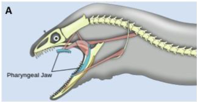
Pharyngeal teeth
additional teeth located on the first pharyngeal arch. Can be more conical and pointed, or more molar like.
What kind of teeth do fish and sharks have
Homodont teeth - no specialization, all the teeth resemble each other.
Sharks also have polyphyodont teeth that are replaced as they are lost
Purpose of vertebrae and how it develops
Vertebrae provide support for the body and protection for the spinal cord. They represent the fusion of elements which appeared separately during the evolutionary history of the vertebrates.
During embryogenesis of most verts the notochord becomes surrounded and constricted by blocks of bone or cartilage, the centra. Remenants of the notochord persist as intervertebral disks between successive vertebrae in bony fish. spinal chord is dorsal to notochord.
How is the spinal chord protected in vertebrates
protected within a canal in NEURAL ARCHES with dorsal NEURAL SPINS of the VERTEBRAE.
neural spines also provide areas of attachment for dorsal body musculature.
How are the artery and vein to the tail in fish protected
protected with a canal in the HAEMAL ARCHES and HAEMAL SPINES ventral to the centrum.
Noteable characters of the vertebrae of fish
fishes weight is supported by water, the vertebrae are discrete units.
Vertebrae of the trunk in sharks is composed of…
heamal plates rather than arches and spines
What connects to trunk vertebrae
On trunk vertebrae, there are dorsal and ventral RIBS which form in the myosepta that separate muscles.
What is the appendicular skeleton,
includes the limb girdles and limbs. in fish this includes the pelvic (posterior) and pectoral (anterior) fins.
The fins/limbs of all vertebrates articulate with the endochondral shoulder girdle.
the pectoral girdle of bony fish is connected to the skull via the POSTTEMPORAL bone.
the pelvic girdle of fish consists of a pair of endochondral bony plates embedded in the muscle.
The teleost caudal skeleton is derived almost entirely from skeletal elements ventral to the vertebral column
How are fins internally supported in chondrichthyans (not bony, cartilage fish) vs in bony fish?
Fins generally are supported internally by rays.
in condrichthyans such as the sharks and chimaeras, these rays are cartilaginous ceratotrichia.
in bony fishes, these rays are derived from embryological ACTINOTRICHIA and are branched at their tips.
Fin rays of both types are supported by PTERYGIOPHORES, often named for their position relative to the body (basal and radial). The base of each ray forms a ball and socket joint with the pterygiophores.
Can the spines and rays of fins move?
In the higher teleosts the rays of some fins have become fused and modified as SPINES which are unbranched. The spines and rays of the dorsal and anal fins and the pectoral rays each have individual sets of muscles which can erect, depress or incline single rays to either side
Characters of muscles
Muscles have one or more ORIGINS ( ends which remain stationary during contraction) and INSERT o n a b o n e t h a t w i l l b e m o v e d d u r i n g t h e c o n t r a c t i o n . Most muscles o r i g i n a t e a n d insert on bones, but some muscles originate or insert on FASCIA (connective tissue) or a seam where the left and the right halves of the muscle meet, the RAPHE.
Tendons are narrow bands of connective tissue that connect muscle to bone or another muscle. Some muscles have multiple points of insertion.
pg 8
Remember that muscle do their work when they contract. Joints tend to be moved via
antagonistic pairs of muscles. Some key muscle actions are depicted in Figure 2.5. Opposite actions
are performed by ANTAGONISTS- muscles whose actions counteract one another. Two very common
antagonistic actions are ABDUCTION- movement of a part away from a point of reference ( such as in
the elevation of the fish pectoral fun in the figure) and ADDUCTION –movement toward a point of
reference ( such as the depression of the fish fin in the figure. .
Visualizing the contraction of a muscle will help you to remember its action. Some muscles
are named for their action while others are named for their origins and insertions. Try to integrate these
factors in your exploration of muscle systems
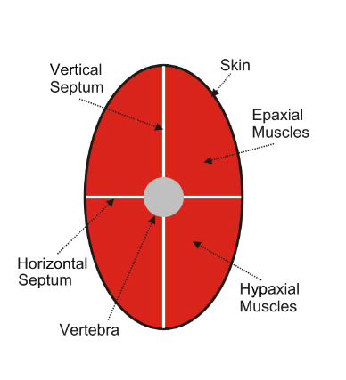
myomeres
The MYOMERES (with connective tissue separations between them called MYOSEPTA) associated with on the axial skeleton are the principal locomotory muscles of fish.
There are distinct dorsal (EPAXIAL) and ventral (HYPAXIAL) portions to each myomere,
separated along the body length by the HORIZONTAL SKELETOGENOUS SEPTUM.
The folds of themyomeres do not extend straight inwards but are angled so that internal parts of the myomeres extend more caudally or cranially than at the surface. This has the effect of cupping the myomeres inside one
another and if you cut a cross section through the tail, they appear to be a series of concentric rings or arcs.The dorsal end of each myomere attaches to successive vertebrae and the ventral ends insert on a band of connective tissue.
RED MUSCLE fibers, so named for the high concentration of myoglobin.
are used in normal active cruising.
How do fish swim via axial muscles
For many fish, and some amphibians and reptiles, swimming is via lateral undulation of the axial skeleton. Waves of contraction begin in the anterior trunk and moves toward the posterior, with left and right side waves alternating. As a series of myomeres contracts on one side of the body, the corresponding muscles on the opposite side relax, resulting in a bend in the fish’s body.
Sequential contraction and relaxation of pairs of myomeres results in the passage of a sine wave along the body. These waves push against the water resulting in forward motion. The caudal fin accelerates faster than the body and generates a great deal of force.
How do fast swimming fish have bodies that allow them to swim fast?
In fast swimmers, the body is rigid and the tail/caudal fin has a half moon shape. Tendons converge on the tails of these fish and the tail can oscillate laterally quickly to provide thrust.
What are the appendicular muscles and what do they do
The appendicular muscles of fish control the fins to provide lift, stability and maneuverability.
They generally do not provide propulsion (though there are some fish that use them for propulsion).
The muscles on the dorsal and ventral surfaces of the fins are antagonistic. The dorsal muscles elevate,while the ventral muscles depress or adduct, the fin.
Do fish use fins in propulsive swimming?
Some fish also use fins in propulsive swimming, which allows them to swim backward by reversing the direction of the waves in the fin. In general, this is not possible in sharks because the pectoral fins are rigid. Shark pectoral fins do provide lift.