Microbiology: Microscopy Techniques and Cell Structures Overview
1/282
There's no tags or description
Looks like no tags are added yet.
Name | Mastery | Learn | Test | Matching | Spaced | Call with Kai |
|---|
No analytics yet
Send a link to your students to track their progress
283 Terms
What are the units of measurement for microorganisms?
Microorganisms are measured in micrometers (µm) and nanometers (nm).
What is a simple microscope?
A simple microscope has only one lens and is similar to a magnifying glass.
What type of microscopy uses visible light to observe specimens?
Light microscopy.
Name one type of light microscopy.
Compound light microscopy.
What is total magnification in a compound microscope?
Total magnification = objective lens magnification × ocular lens magnification.
What is resolution in microscopy?
Resolution (resolving power) is the ability of lenses to distinguish two points or fine detail.
What is the limit of resolution for a compound light microscope?
The limit of resolution is 0.2 µm.
What is the maximum magnification of compound light microscopes?
Approximately 1500x.
What is the refractive index?
The refractive index is a measure of the light-bending ability of a medium.
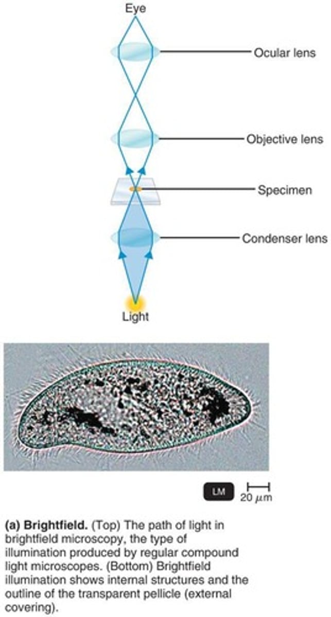
What is brightfield illumination?
Brightfield illumination shows dark objects against a bright background.
What is darkfield microscopy?
Darkfield microscopy allows light objects to be visible against a dark background.
What is the purpose of an opaque disk in darkfield microscopy?
The opaque disk eliminates all light in the center of the beam, allowing only light reflected off the specimen to enter the objective lens.
What is phase-contrast microscopy used for?
Phase-contrast microscopy allows detailed examination of living organisms and internal cell structures without fixation or staining.
How does differential interference contrast (DIC) microscopy enhance images?
DIC microscopy uses two light beams and prisms to split light beams, giving more contrast and color to the specimen.
What is fluorescence microscopy?
Fluorescence microscopy uses UV light to excite fluorescent substances that emit visible light.
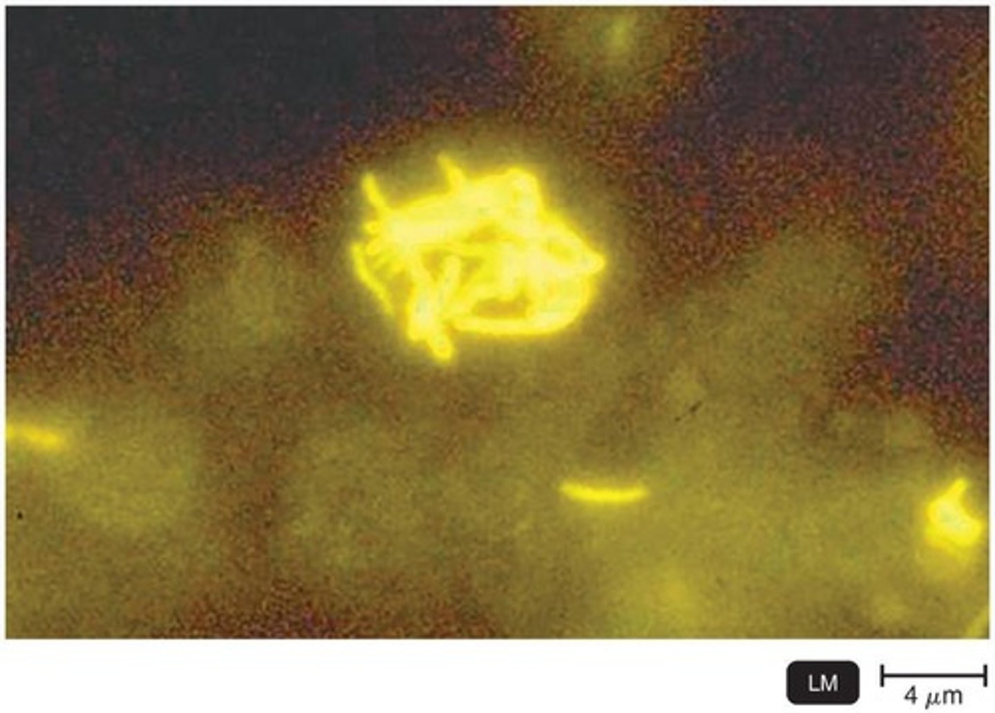
What is the fluorescent-antibody (FA) technique?
The FA technique uses antibodies tagged with fluorochromes to detect specific microbial pathogens.
What is the role of fluorochromes in fluorescence microscopy?
Fluorochromes stain cells, allowing them to fluoresce against a dark background.
What is the significance of using immersion oil in microscopy?
Immersion oil is used to prevent light from refracting, improving resolution.
What type of microscopy is useful for viewing live unstained microorganisms?
Darkfield microscopy.
What does phase-contrast microscopy combine to form an image?
It combines direct rays and diffracted rays.
What is the appearance of images in DIC microscopy?
Images may be brightly colored and appear three-dimensional.
What is the main advantage of phase-contrast microscopy?
It allows examination of living cells without fixation or staining.
What is the purpose of staining cells with fluorescent dyes?
To enable visualization of cells that do not naturally fluoresce.
What is the role of antibodies in the fluorescent-antibody technique?
Antibodies specifically bind to microbial pathogens and fluoresce under UV light.
What type of light does fluorescence microscopy use?
UV (short wavelength) light.
What is the benefit of using shorter wavelengths of light in microscopy?
Shorter wavelengths provide greater resolution.
What is the primary limitation of brightfield microscopy?
It may be difficult to view unstained cells due to a lack of contrast.
What is the main feature of darkfield microscopy?
It allows visualization of light objects against a dark background.
What is confocal microscopy?
A technique that uses short-wavelength light to excite a single plane of a specimen, producing exceptionally clear two-dimensional images.
How does two-photon microscopy work?
It uses two photons of long-wavelength light to excite fluorochrome dyes, allowing for the study of living cells up to 1 millimeter deep.
What is the purpose of super-resolution light microscopy?
It uses two laser beams to produce images with higher resolution by scanning the specimen nm by nm.
What does scanning acoustic microscopy measure?
It measures sound waves reflected from a specimen, used to study cells attached to surfaces like cancer cells and bacterial biofilms.
What is the advantage of electron microscopy over light microscopy?
Electron microscopy uses electrons instead of light, providing greater resolution for images too small to be seen with light microscopes.
What is transmission electron microscopy?
A method where a beam of electrons passes through ultrathin sections of a specimen, producing high-magnification images.
What is the magnification range of transmission electron microscopy?
10,000 to 10,000,000x.
What is scanning electron microscopy?
A technique where an electron beam scans the surface of a specimen, providing a three-dimensional view.
What is the resolution limit of scanning electron microscopy?
0.5 nm.
What does scanned probe microscopy examine?
It uses probes to examine the surface of specimens with electric current without modifying the specimen.
What is scanning tunneling microscopy?
A technique that uses a tungsten probe to scan a specimen, revealing surface details down to the atomic level.
What is atomic force microscopy?
A method that uses a metal-and-diamond probe to produce three-dimensional images at near atomic detail.
What is the purpose of preparing smears for staining?
To color microorganisms with a dye that emphasizes certain structures.
What is the role of fixing a smear?
It attaches microorganisms to the slide, kills them, and preserves parts of microbes with minimal distortion.
What are the two methods for fixing a smear?
Heat fixation (passing through a flame) and chemical fixation (using methanol).
What is a simple stain?
The use of a single basic dye to highlight the entire microorganism for visualization.
What are differential stains used for?
To distinguish between different types of bacteria.
What does the Gram stain classify?
Bacteria into gram-positive (purple) and gram-negative (pink/red) based on cell wall structure.

What is the primary stain used in the Gram stain procedure?
Crystal Violet.
What is the function of the mordant in the Gram stain?
Gram's iodine is used to fix the primary stain to the bacterial cell wall.
What is the decolorizing agent in the Gram stain?
Alcohol or acetone-alcohol.
What is the counterstain used in the Gram stain?
Safranin, which stains gram-negative bacteria pink/red.
What happens to gram-positive bacteria during the Gram stain process?
They retain the purple color due to their thick peptidoglycan cell walls.
What happens to gram-negative bacteria during the Gram stain process?
They lose the purple color and take on the pink/red color from the counterstain.
What is the Gram stain used for?
It is one of the most important staining techniques in medical microbiology, used to detect bacteria in clinical specimens and often the first step in identifying an unknown bacterium.
When are Gram stains most reliable?
Gram stains are most consistent when used on young, actively growing bacteria.
What does the Acid-Fast stain bind to?
It binds only to bacteria that have a waxy material in their cell walls, which is not decolorized by acid-alcohol.
Which bacteria are identified using the Acid-Fast stain?
Mycobacterium and Nocardia.
What is the primary stain used in Acid-Fast staining?
Carbolfuchsin.
What color do Acid-Fast bacteria appear after staining?
Red.
What is the purpose of a capsule stain?
To distinguish parts of microorganisms, specifically to visualize capsules that do not accept most dyes.
How is a capsule visualized in a negative stain?
A suspension of India ink or nigrosin contrasts the background with the capsule, which appears as a halo around the stained bacterial cell.
What are endospores?
Resistant, dormant structures inside some cells that cannot be stained by ordinary methods.
What is the primary stain used in the Schaeffer-Fulton endospore stain?
Malachite green, usually applied with heat to help dye penetrate the endospore.
What color do endospores appear after Schaeffer-Fulton staining?
Green within red or pink cells.
What is the purpose of flagella staining?
To visualize flagella, which are structures of locomotion that are too slender to be viewed with a light microscope unless stained.
What is used in flagella staining to thicken the appearance of flagella?
A mordant and carbolfuchsin.
What does the term 'brightfield' refer to in microscopy?
A method where light moves upward through the condenser lens in two V-shaped beams, crossing through the specimen and refracting through the objective lens.
What is the role of the diaphragm in a microscope?
It controls the amount of light entering the condenser.
What is the function of the ocular lens in a microscope?
It remagnifies the image formed by the objective lens.
What is the purpose of the condenser in a microscope?
To focus light through the specimen.
What is the range of organisms typically studied in microbiology?
From 10 micrometers to 10 nanometers.
What is the typical size range of a tick?
1 millimeter to 10 millimeters.
What does a scale bar in microscopy indicate?
It provides a reference for the actual size of the specimen being viewed.
What is the typical magnification range of a light microscope?
200 nanometers to 10 millimeters.
What is the function of the coarse focusing knob on a microscope?
To make large adjustments to the focus of the microscope.
What does the fine focusing knob do?
It allows for smaller, more precise adjustments to the focus.
What is the role of the arm in a microscope?
It provides a handle-like structure for carrying or moving the microscope.
What is the purpose of the body tube in a microscope?
It transmits the image from the objective lens to the ocular lens.
What is the function of the stage in a microscope?
To hold the microscope slide in position.
What is the purpose of the illuminator in a microscope?
It serves as the light source for illuminating the specimen.
What type of microscopy allows only light reflected by the specimen to reach the objective lens?
Darkfield microscopy
In darkfield microscopy, what is the appearance of the edges of the cell?
Bright against a black background
What is the scale used in the micrograph of Paramecium in darkfield microscopy?
20 micrometers
What differentiates direct light rays from reflected or diffracted light rays in microscopy?
Direct rays are unaltered by the specimen, while reflected rays are altered.
What microscopy technique shows greater differentiation of internal structures?
Phase-contrast microscopy
What is the scale used in the micrograph of Paramecium in phase-contrast microscopy?
20 micrometers
What is visible in the transmission electron micrograph (TEM) of Paramecium?
Internal structures of the specimen
What is the scale used in the colorized transmission electron micrograph of Paramecium?
30 micrometers
How does a scanning electron microscope (SEM) capture images?
By knocking electrons from the surface of the specimen and collecting secondary electrons.
What is the scale used in the colorized scanning electron micrograph of Paramecium?
30 micrometers
What does a scanning tunneling microscopy (STM) image of DNA show?
DNA as a long filamentous coiled structure.
What is the scale bar measurement in the STM image of DNA?
8 nanometers
What are the steps in the Gram staining process?
1. Apply crystal violet, 2. Apply iodine, 3. Alcohol wash, 4. Apply safranin.
What color indicates gram-positive bacteria after Gram staining?
Purple
What color indicates gram-negative bacteria after Gram staining?
Pink
What is the scale used in the micrograph showing clusters of cocci and rods?
5 micrometers
What is the appearance of Bacillus pumilus in the stained LM micrograph?
Numerous rod-shaped bacterial cells with flagellum projections.
What is the scale bar measurement in the micrograph of Bacillus pumilus?
7.5 micrometers
What is the significance of the opaque disk in the light path of microscopy?
It blocks direct light, allowing only reflected light to be captured.
What type of lens focuses the image in a transmission electron microscope?
Magnetic lenses
What is the role of the annular diaphragm in microscopy?
It helps in controlling the light path and enhancing contrast.