ORAL PATHOLOGY LAB FINAL
1/151
Earn XP
Description and Tags
Name | Mastery | Learn | Test | Matching | Spaced |
|---|
No study sessions yet.
152 Terms
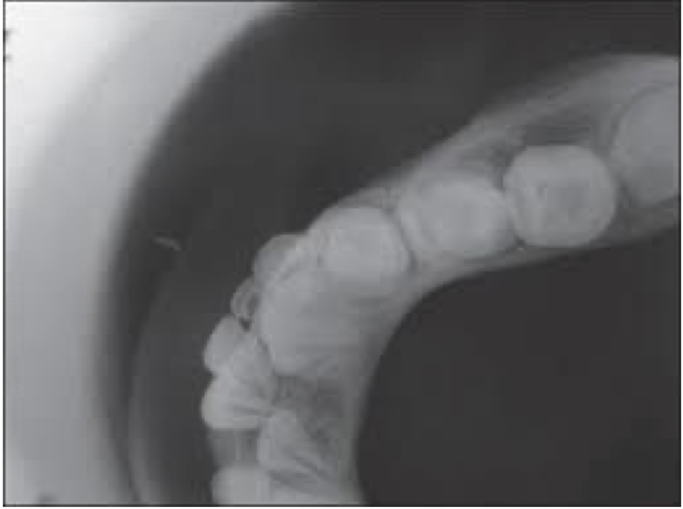
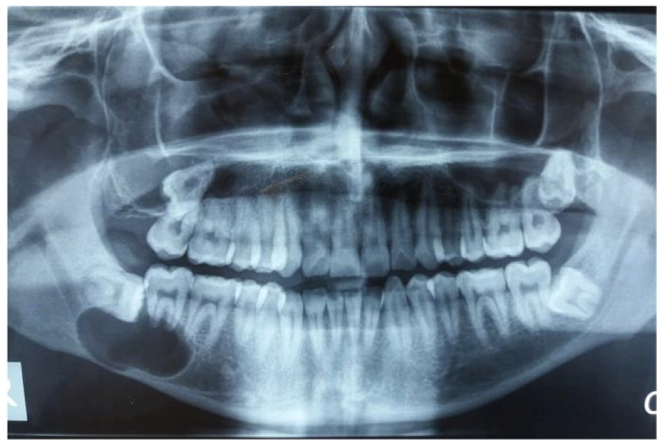
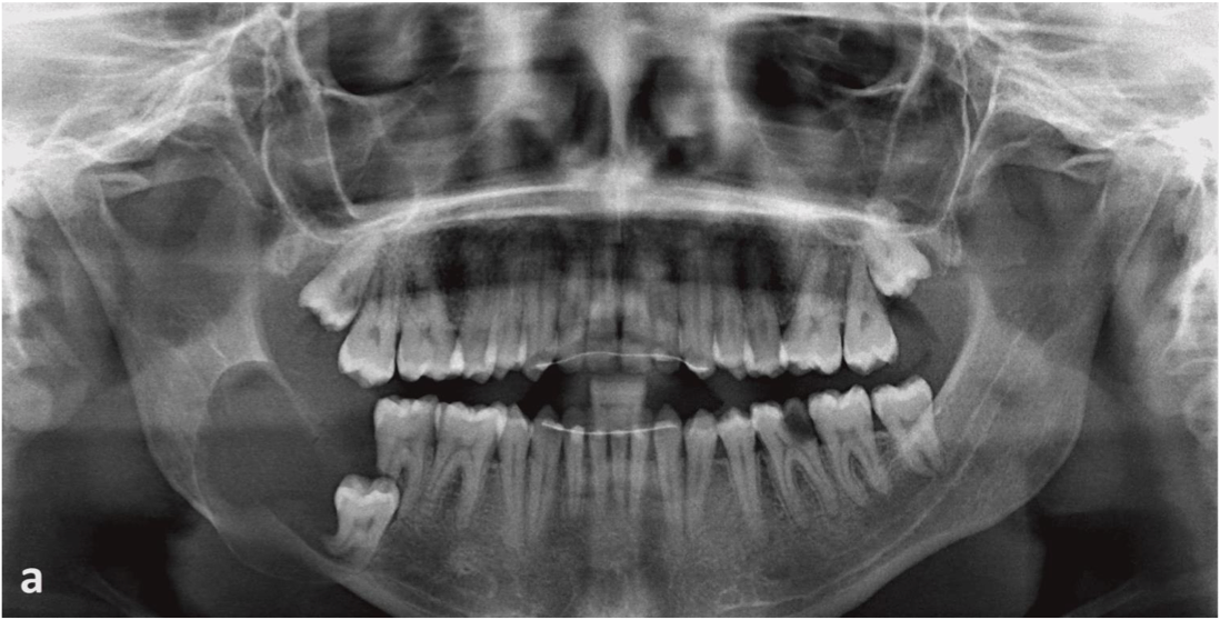
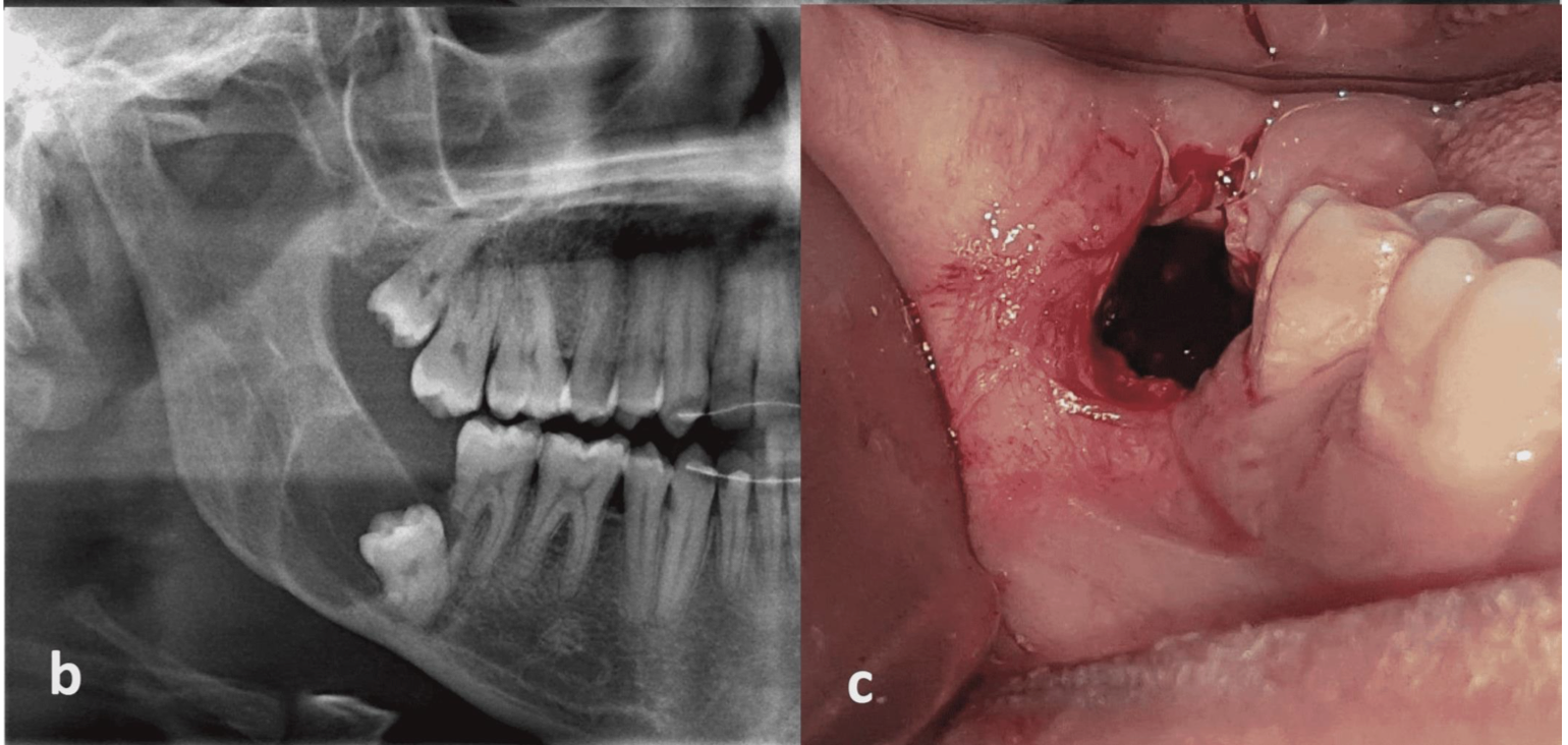
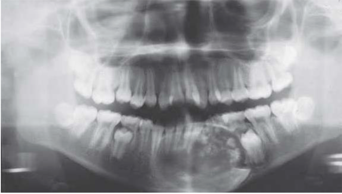
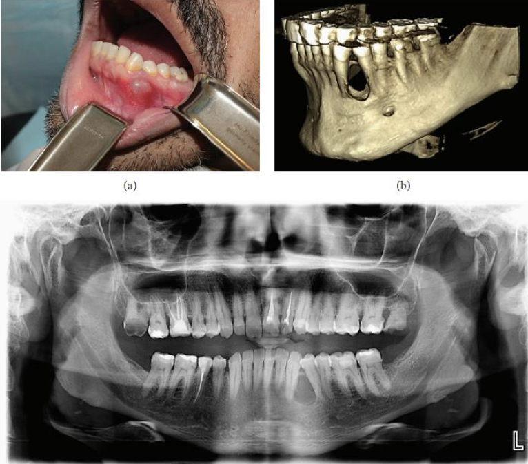
A 9 year old with deep caries on 46 with mild pain. An OPG was taken and provided below
Garres Osteomyleitis
Occlusal Radiograph
Garres Osteomyelitis
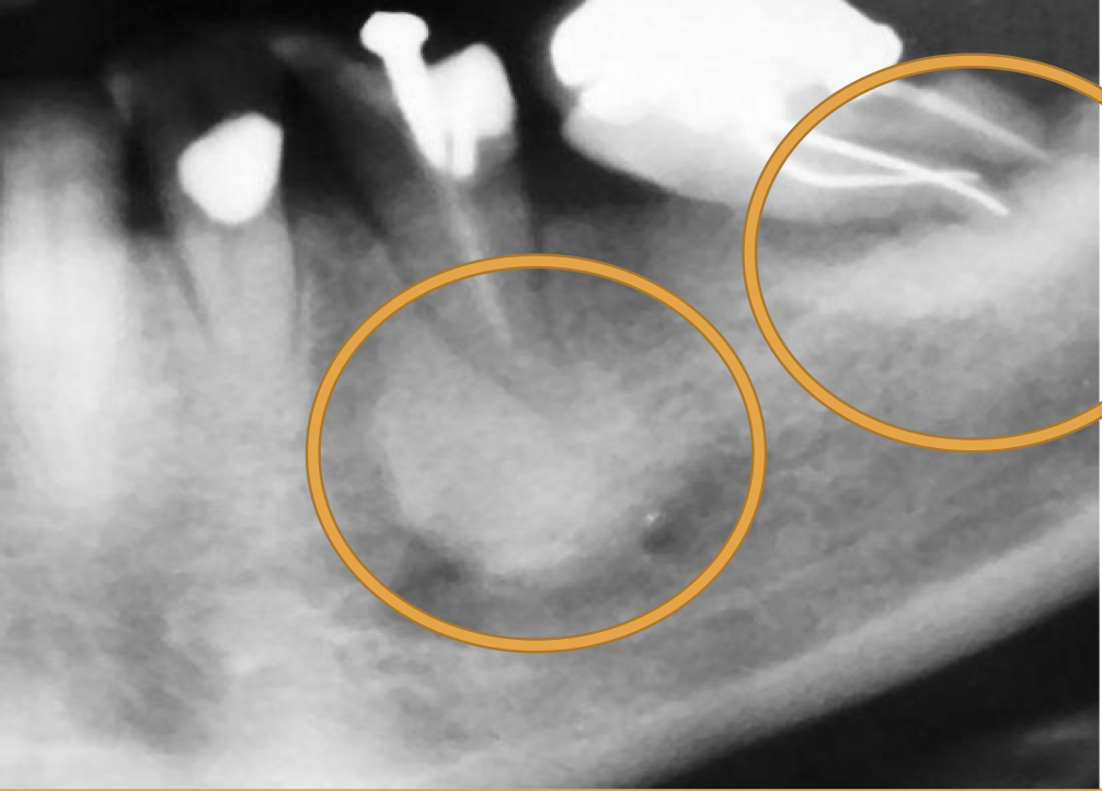
A 45 year old with severe pain and swelling in 35 region of 2 weeks duration. The patient also presents with fever chills, and lymph node enlargement on same side. Intraorally accumulation of pus along with fetid odor could be noted. Provide differential diagnoses
Acute Suppurative Osteomyelitis
Periapical Abscess
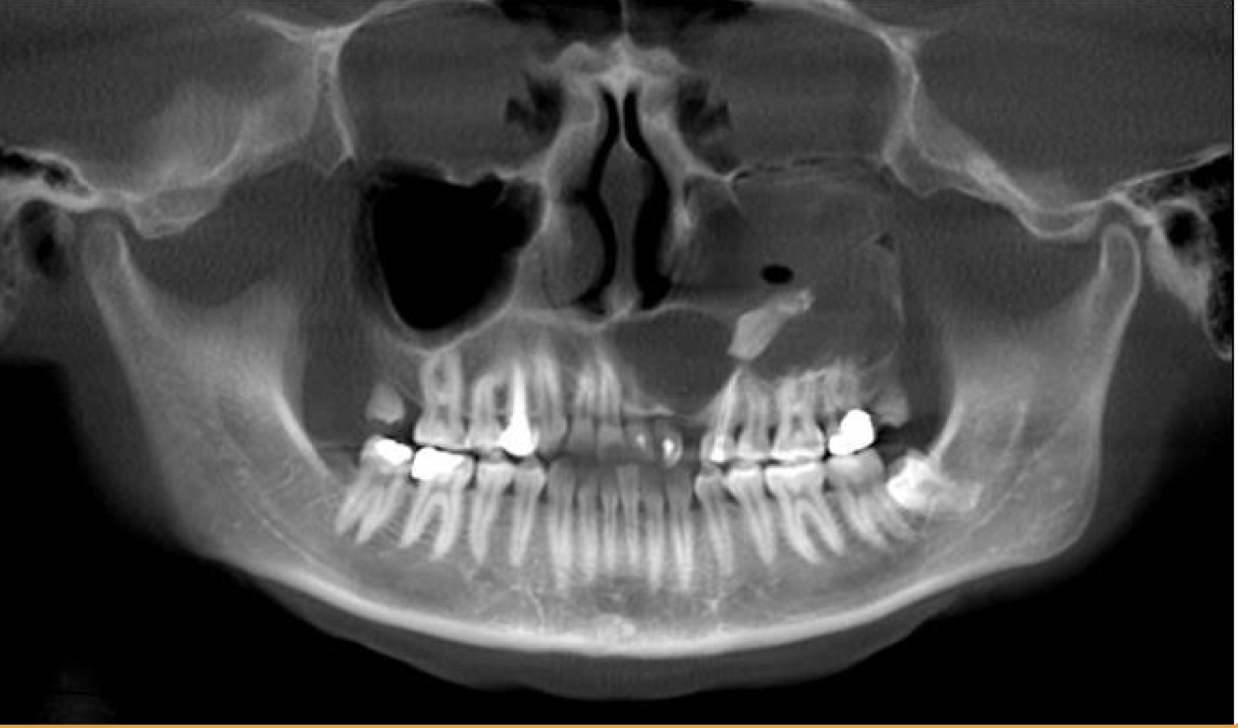
An OPG was taken and provided below. Describe the name of appearance and provide diagnosis
Acute Osteomyelitis (moth eaten appearance)
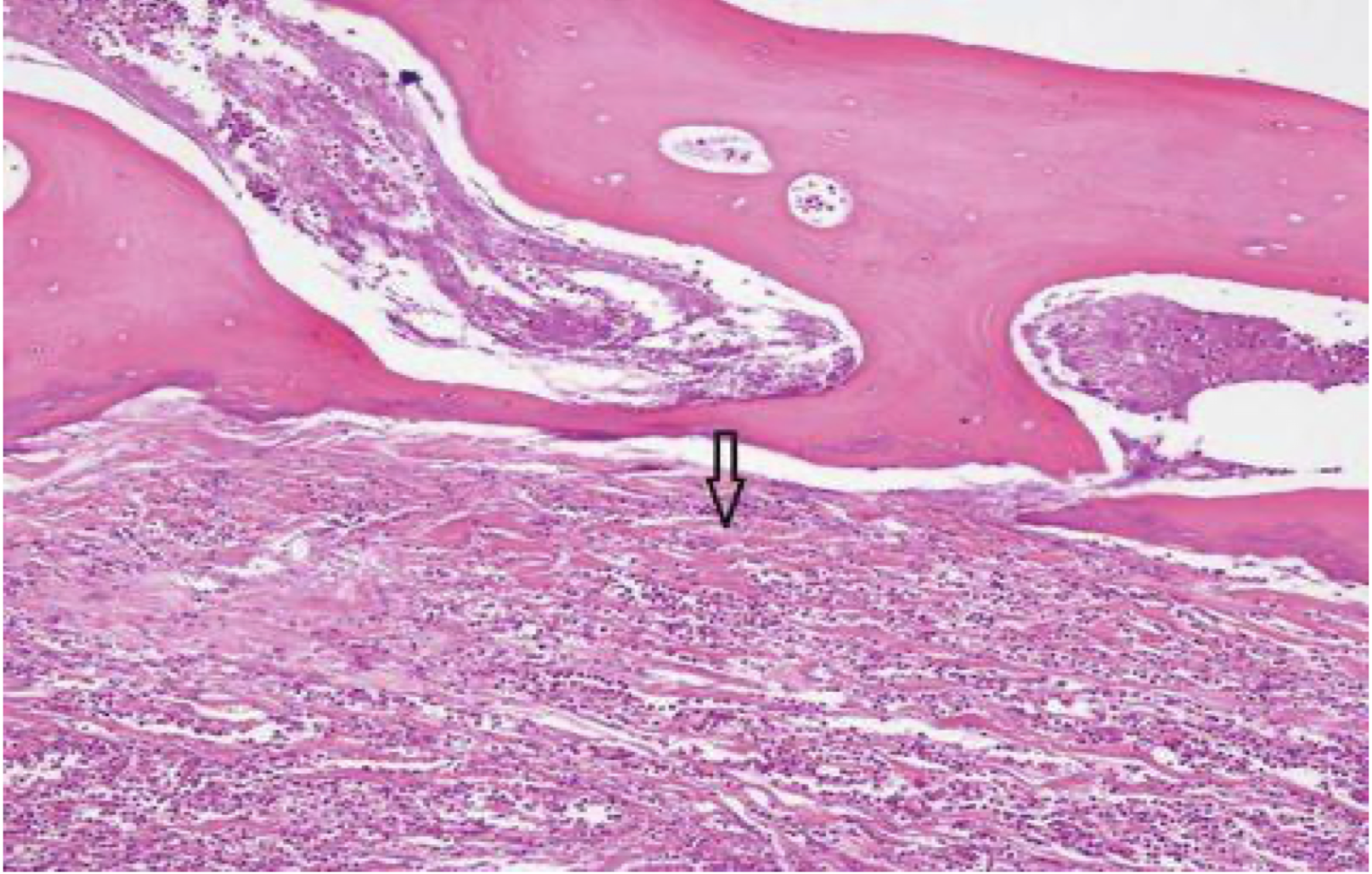
Osteomyelitis
Bone + CT
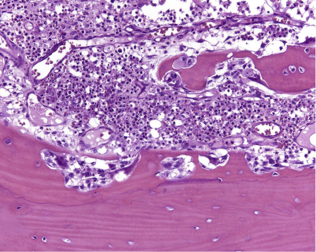
Acute Osteomyelitis
Chronic Osteomyelitis
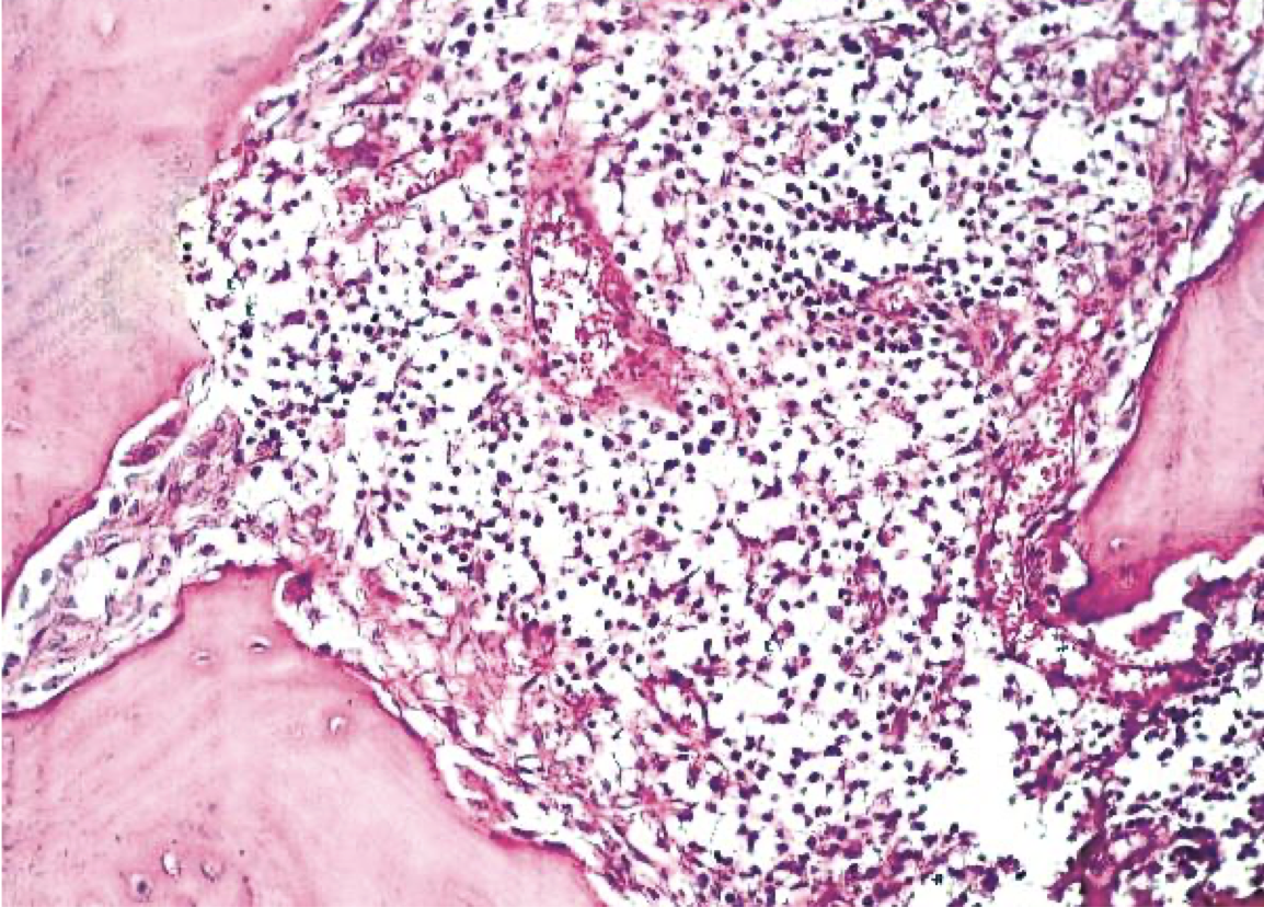
Condensing Osteitis
Condensing Osteitis
Condensing Osteitis
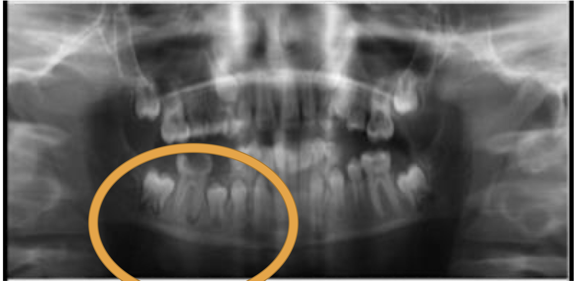
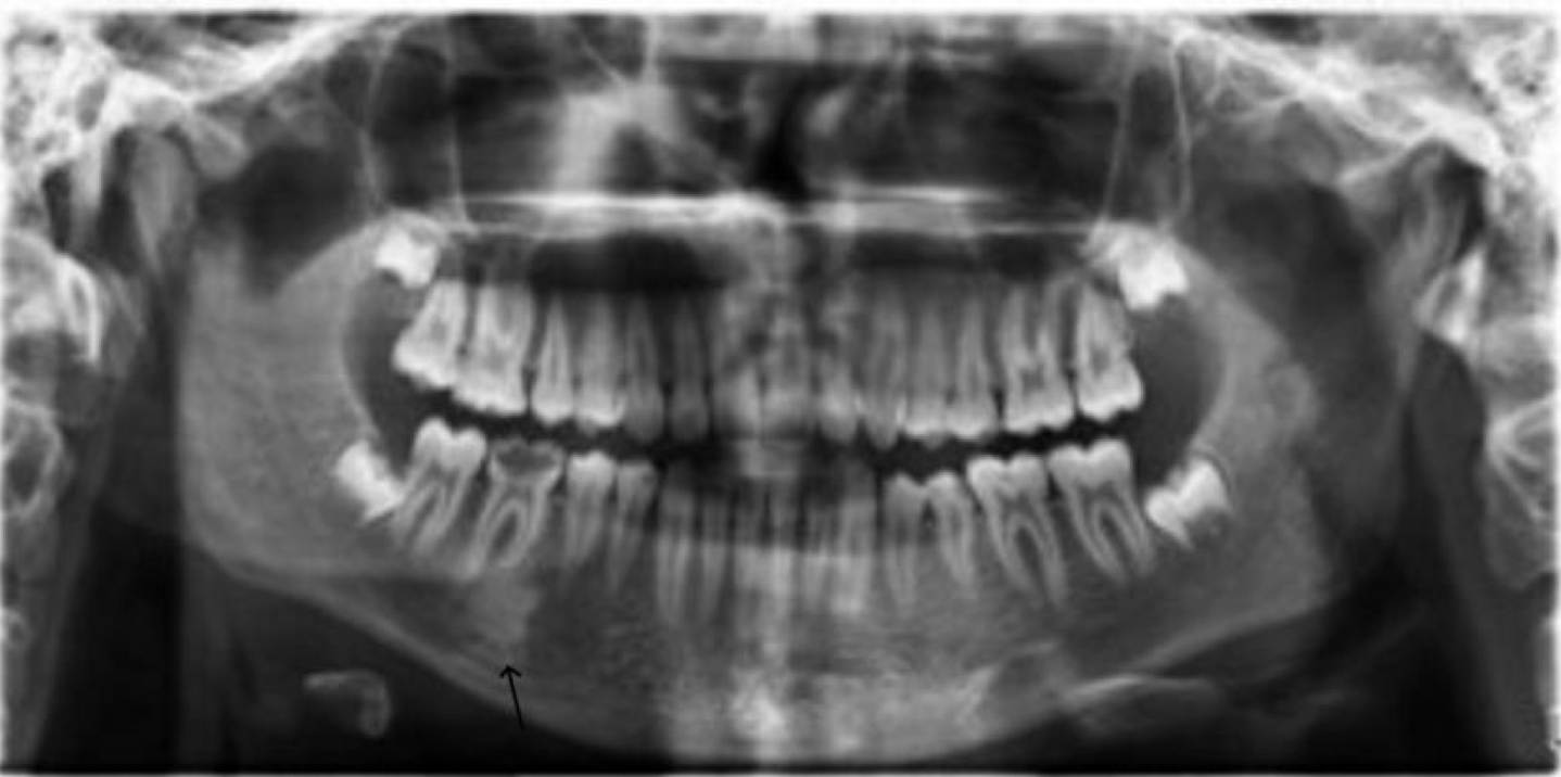
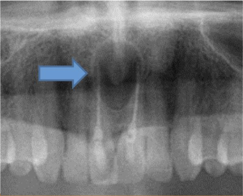
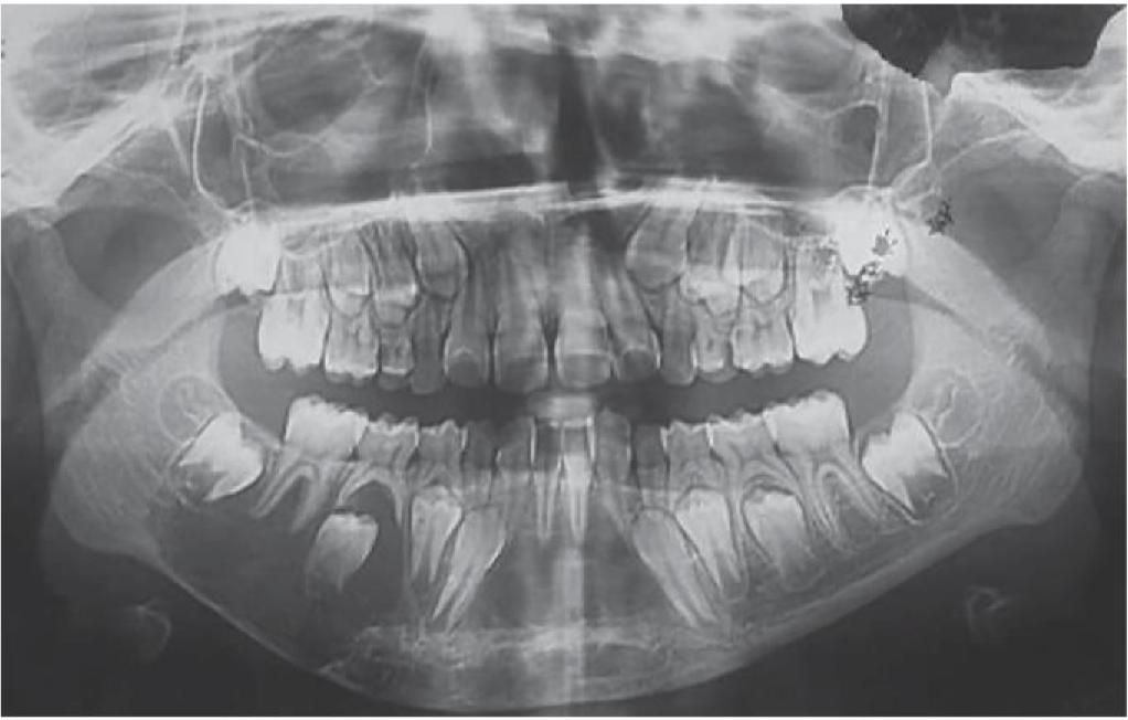
A 19 year old patient present with mild swelling of lower jaw. Intra orally a small swelling was visible over left third molar region. A radiograph taken. Provide Differential diagnosis
OKC
Dentigerous Cyst
OKC / dentigerous cyst
A 35 year old patient present with mild swelling of lower jaw. Intra orally a small swelling was visible over left third molar region.
OKC
OKC
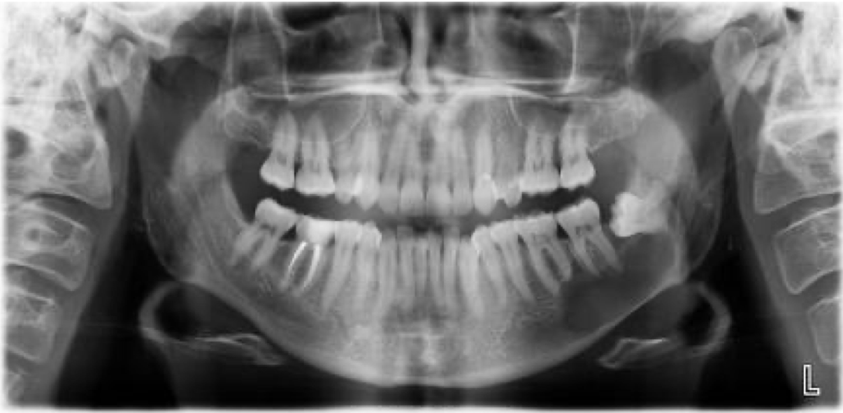
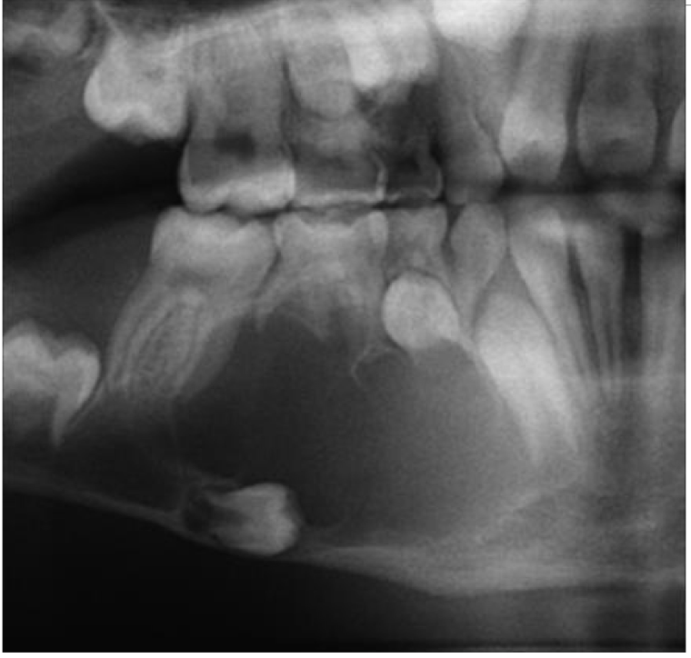
A 25 year old patient present with mild swelling of lower jaw. Intra orally a small swelling was visible over right third molar region. A radiograph taken. Provide Differential diagnosis and justify
OKC
Ameloblastoma
Multilocular Radiolucency
OKC
A 25 year old patient present with mild swelling of lower jaw. Intra orally a small swelling was visible over right molar region. A radiograph taken. Provide diagnosis and justify
Orthokeratinized Odontogenic Cyst
diagnose and justify
Orthokeratinized Odontogenic Cyst
Because of the keratin layer
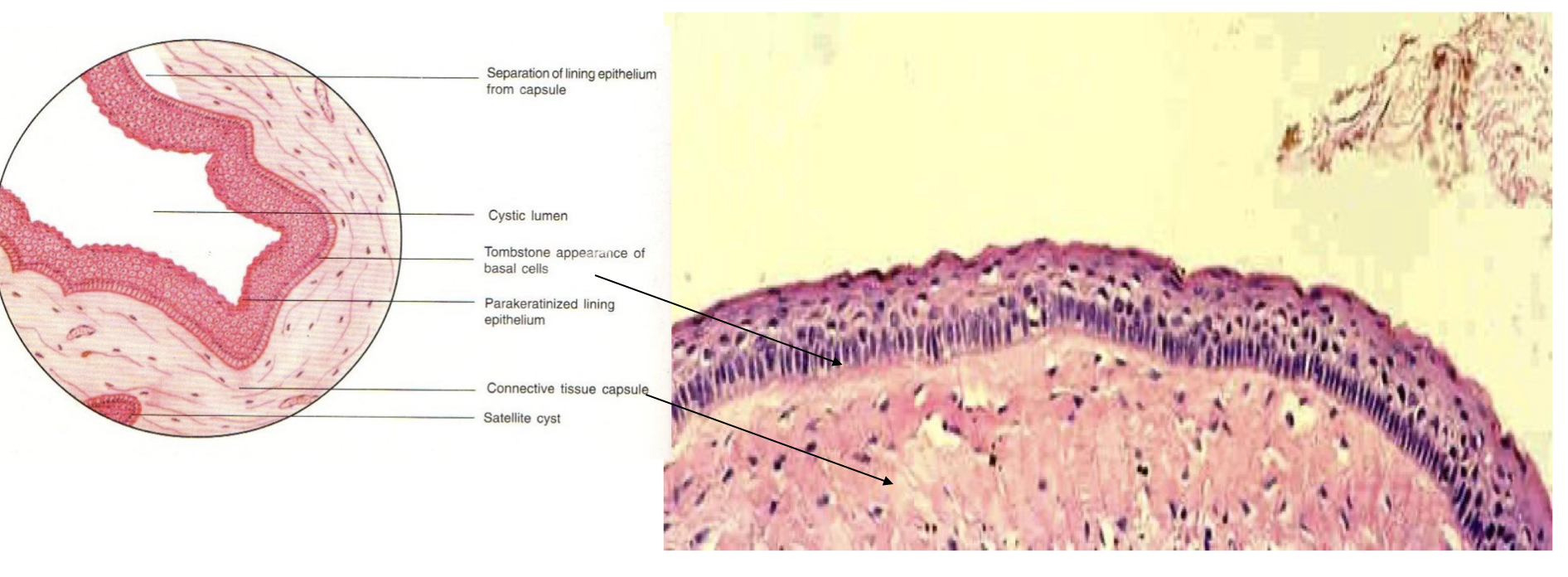
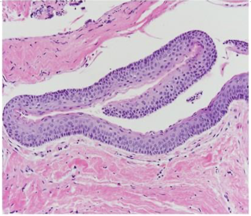
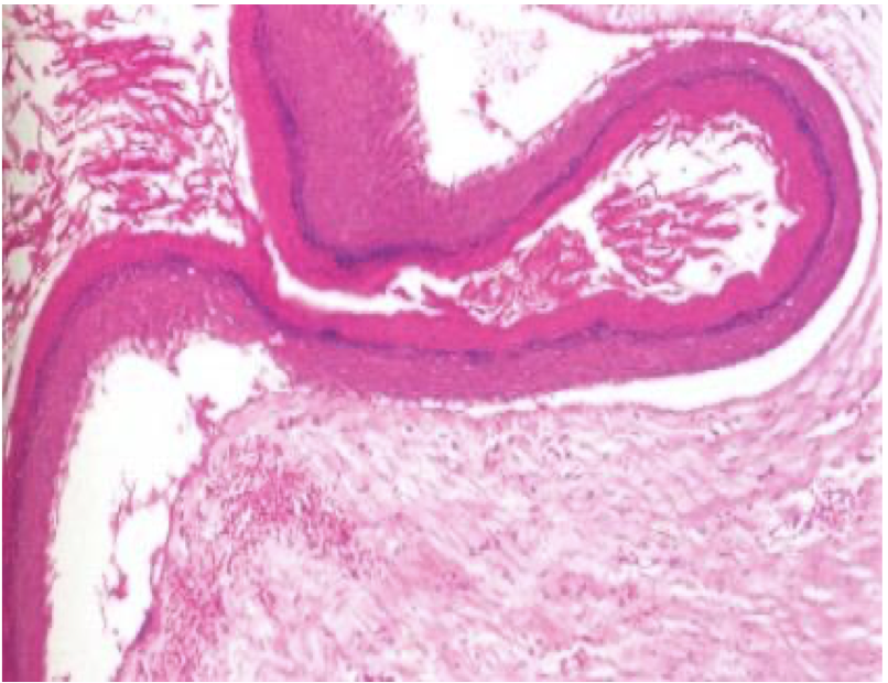
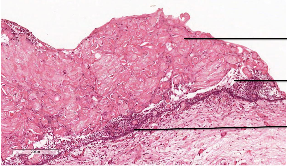
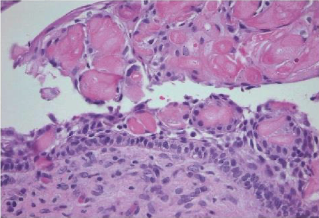
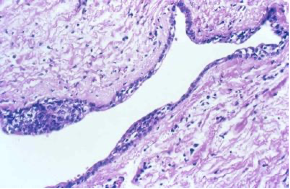
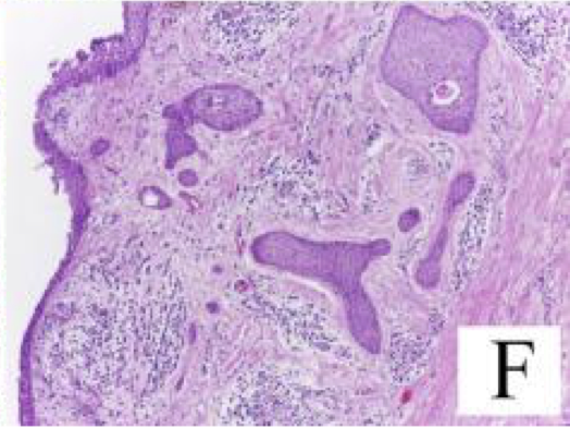
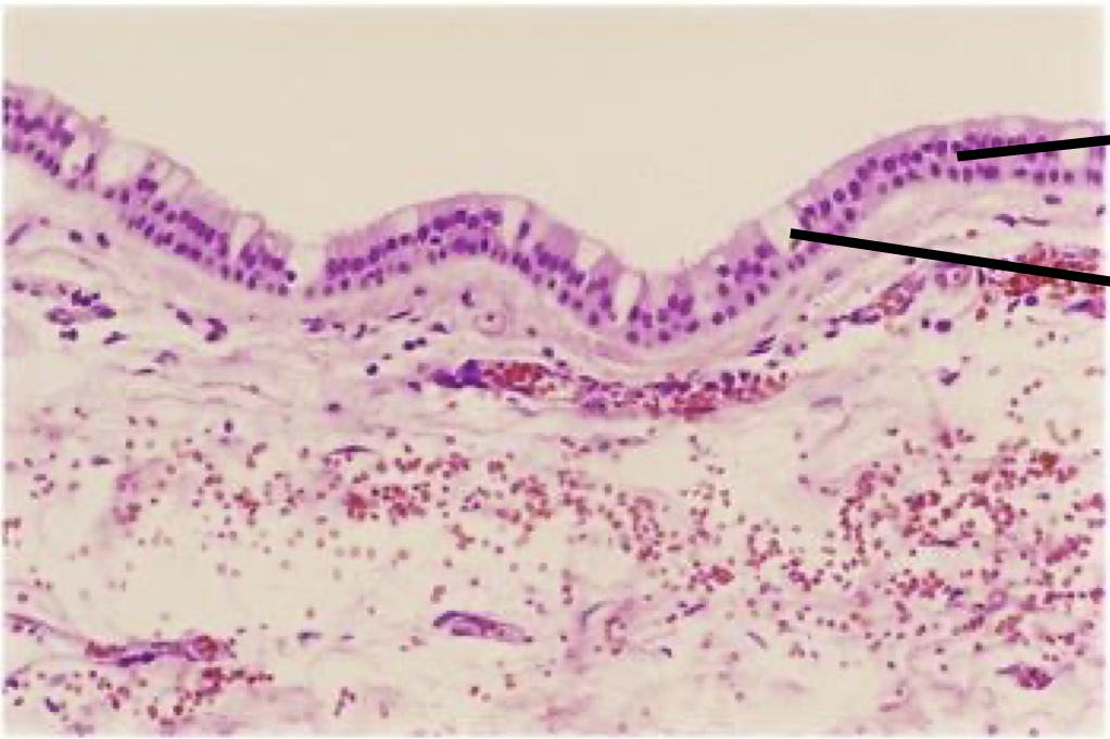
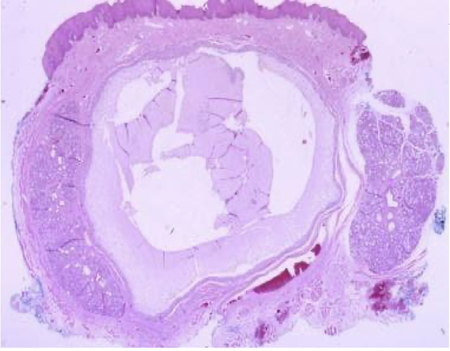
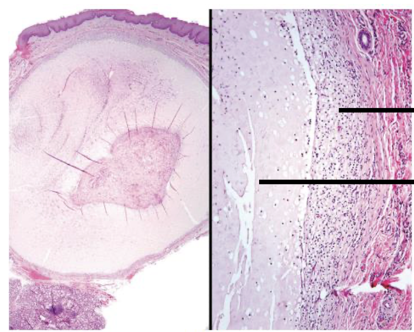
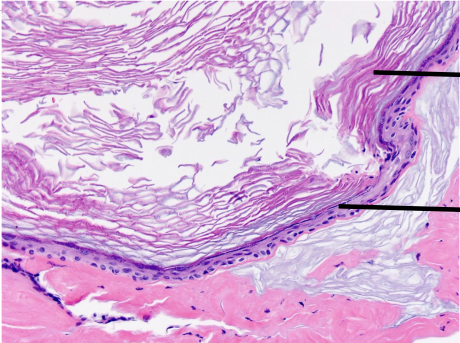
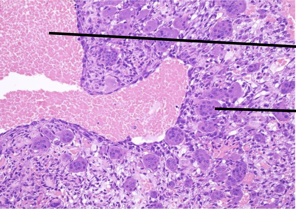
A 55 year old patient present with mild swelling of upper jaw. A biopsy was taken and the image is provided below.
OKC (inflamed towards one side)
Lining Epithelium - OKC
Basal Cell Budding - OKC
Daughter Cyst - OKC
Odontogenic Epithelial Rests - OKC
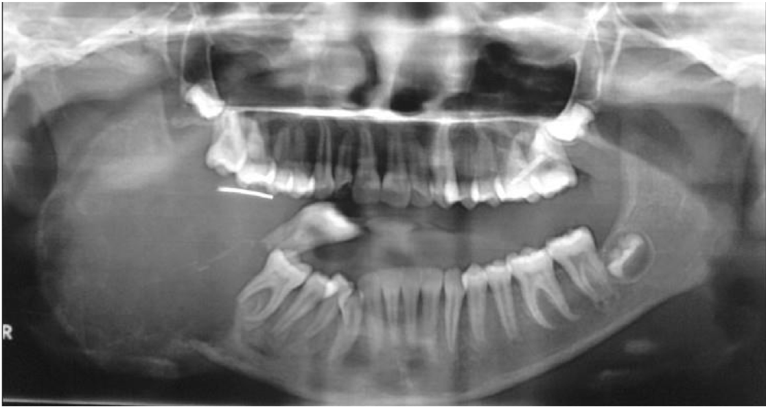
A 25 year old patient present with mild swelling of lower jaw. Intra orally a small swelling was visible over right third molar region. A radiograph taken. Provide Differential diagnosis
Dentigerous Cyst
OKC
Epithelial Lining - Dentigerous Cyst
Cystic Lining
Marsipulization/shrunken cyst
A 25 year old patient present with mild swelling of front jaw. Intra orally a small swelling was visible left vestibular region molar region and 23 was missing. A radiograph taken. Provide Differential diagnosis
Dentigerous cyst
Adenomatoid
odontogenic tumor
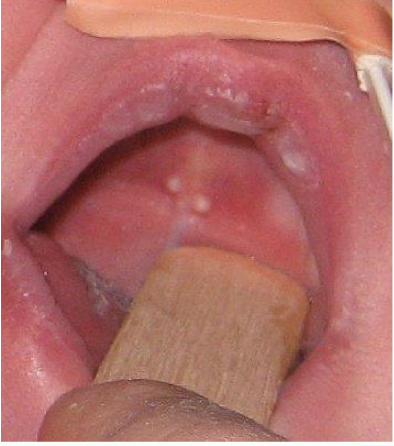
The mother of a 2 month old baby has a concern of white nodules in the mouth of her kid. The clinical image is shown below. There is no pain or any other symptoms. Provide diagnosis
Epstein Pearls
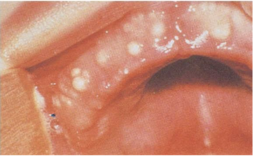
The mother of a 1 month old baby has a concern of white nodules in the mouth of her kid. The clinical image is shown below. There is no pain or any other symptoms. Provide diagnosis
Bohn Nodules
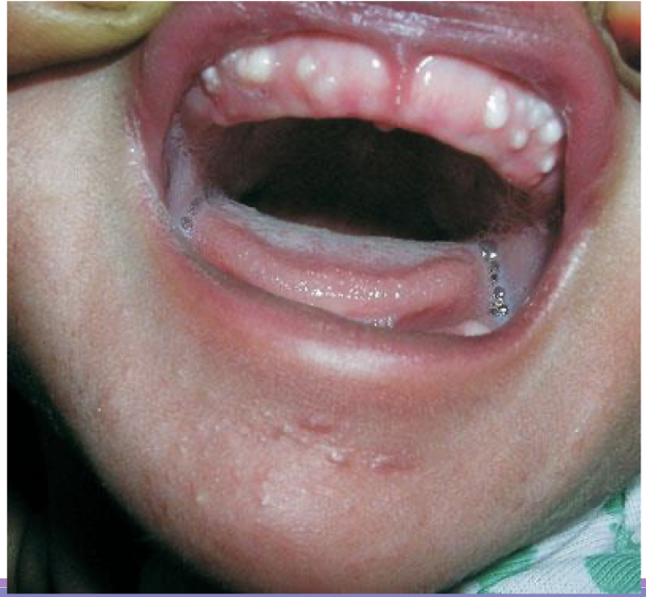
A full term newborn boy, weighing 3 kg, born out of an uncomplicated pregnancy, was brought to us for evaluation of a few small, white and round bumps on the gingival surface. Examination of the oral cavity showed multiple, firm, pearly-white papules measuring 2 to 4 mm in diameter, grouped over the vestibular aspect of the alveolar ridge of the maxillary arch
Bohn Nodules
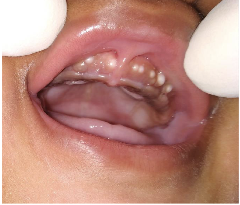
The mother of a 1 month old baby has a concern of white nodules in the mouth of her kid. The clinical image is shown below. There is no pain or any other symptoms. Provide diagnosis and management
Bohn Nodule
No Treatment
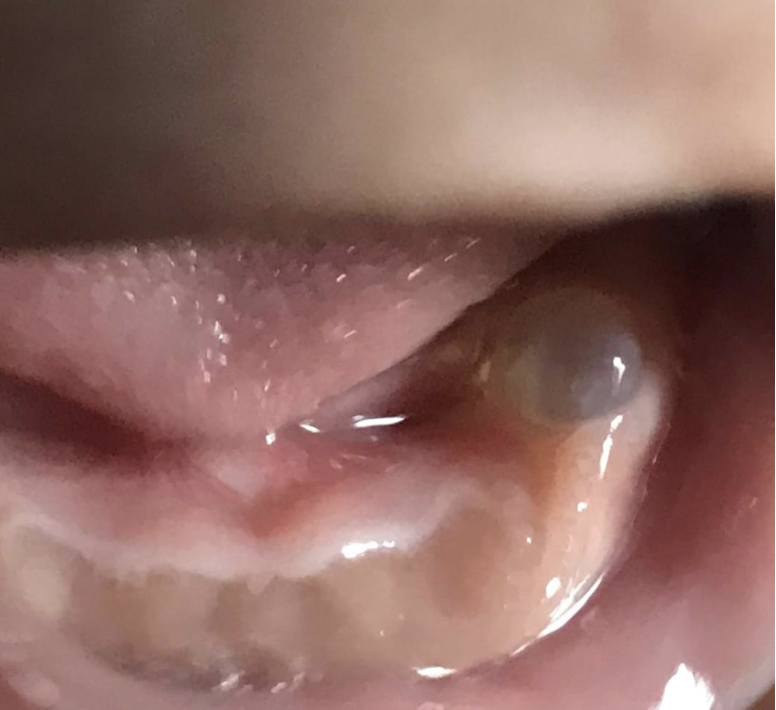
The mother of a 1 month old baby has a concern of a bubble in the mouth of her kid. The clinical image is shown below. There is no pain or any other symptoms. Provide diagnosis and synonym
Gingival cyst of newborn/infant
Dental lamina cyst of infants
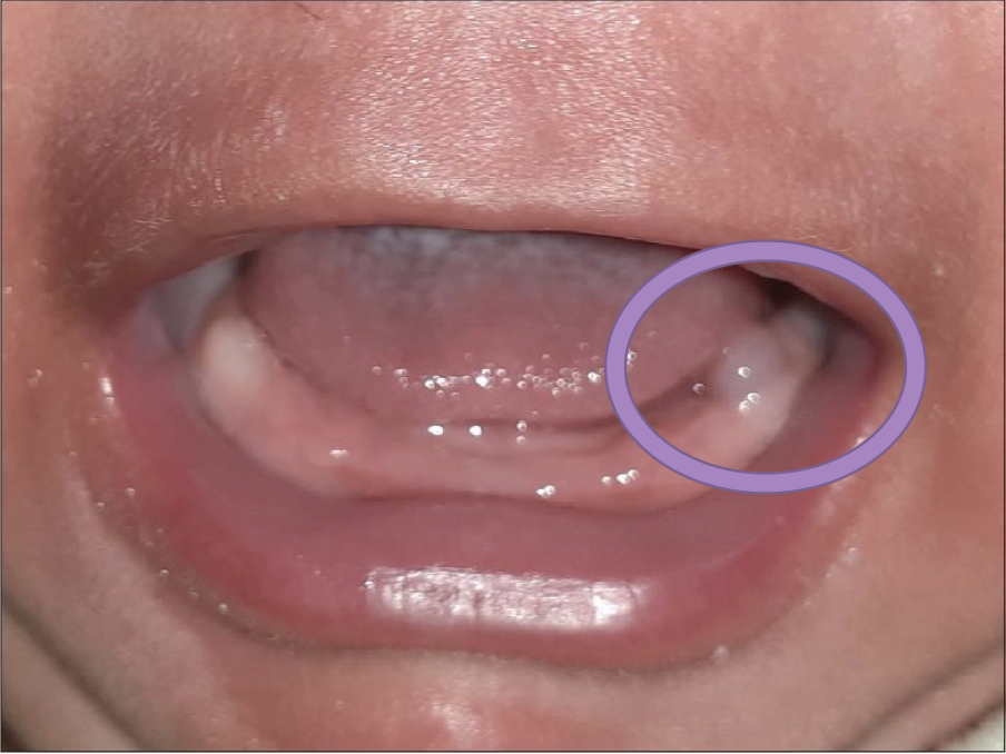
Spot the cyst and provide diagnosis
Gingival cyst of newborn/infant
Dental lamina cyst of infants
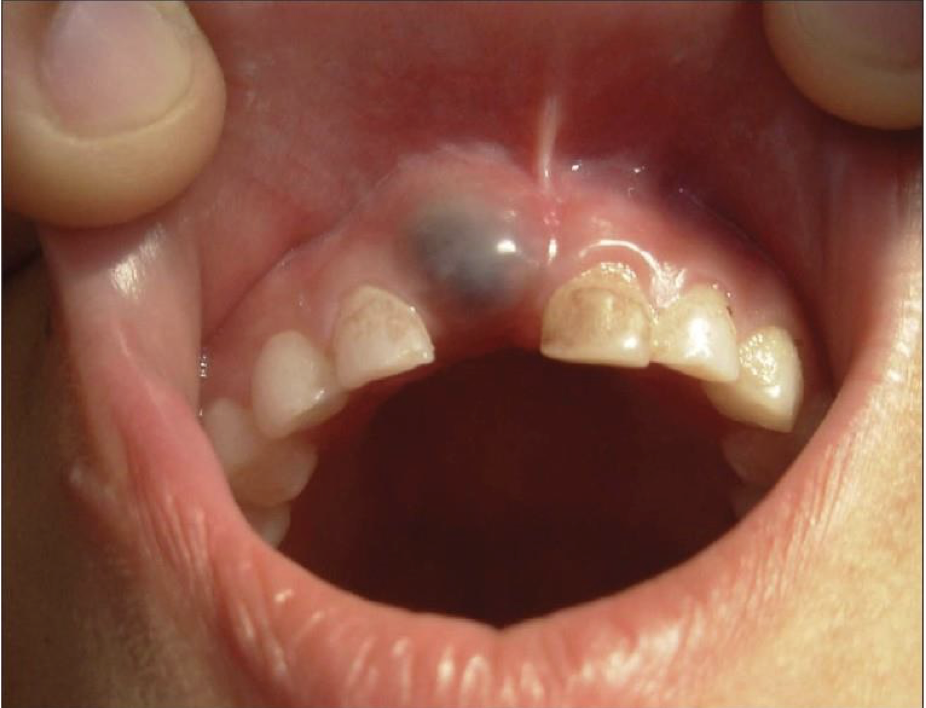
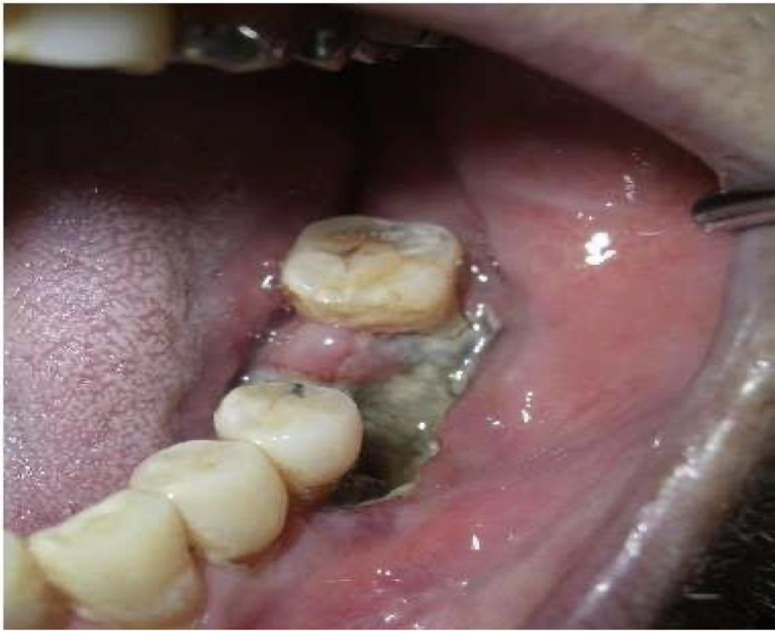
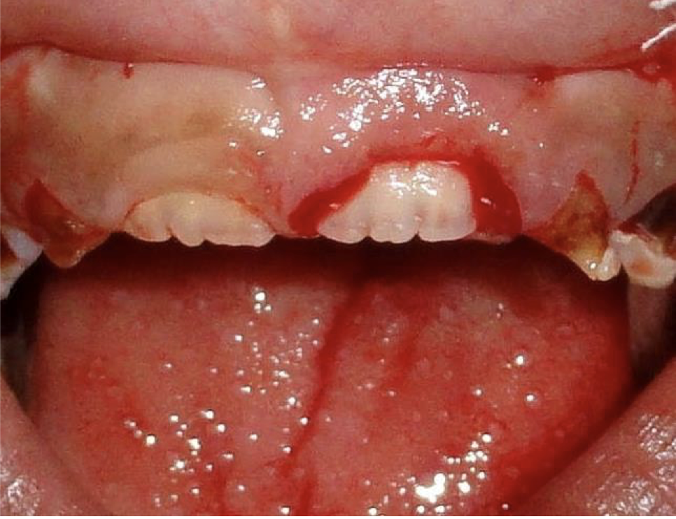
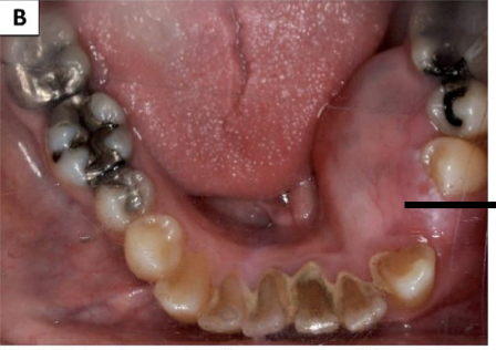
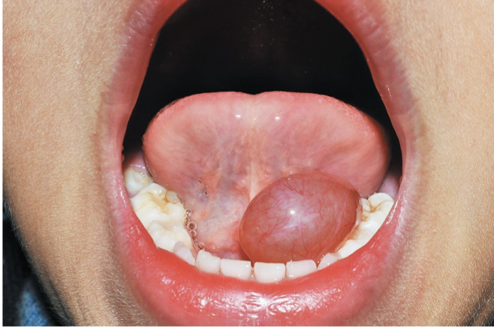
12-year old male patient, which presented as a swelling involving the gingiva in the region of 11 of 5 year duration
Eruption Cyst
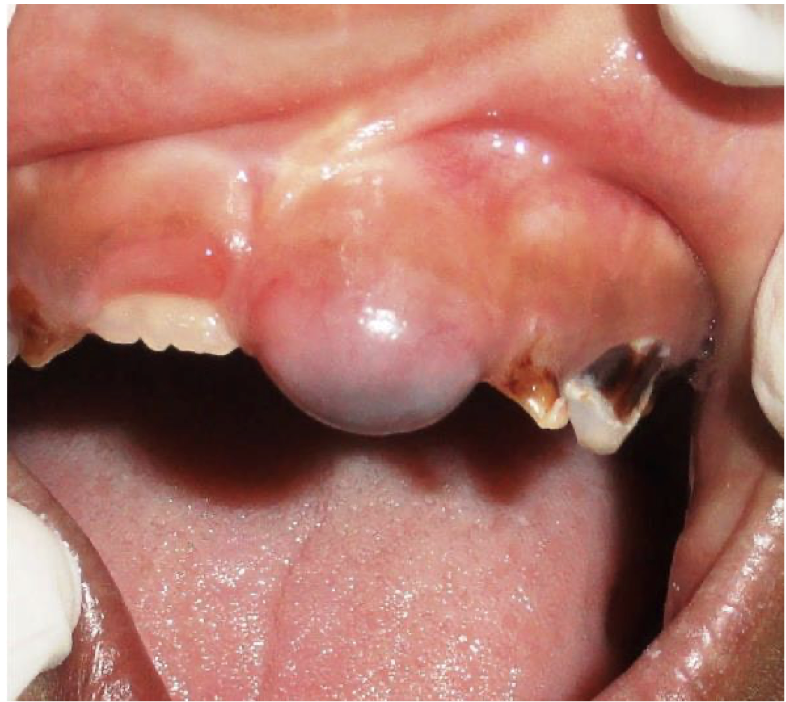
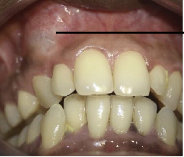
7-year old male patient, which presented as a swelling involving the gingiva overlying the crown of 21
Eruption Cyst
Surgical Incision of Eruption Cyst
A 30 year old female presented with complaint of swelling in the lower jaw. Intraorally a swelling was not over the left vestibule spanning over 43 to 35. Provide differential diagnosis.
Calcifying odontogenic cyst
Calcifying epithelial odontogenic tumor
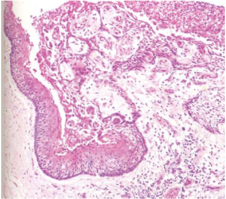
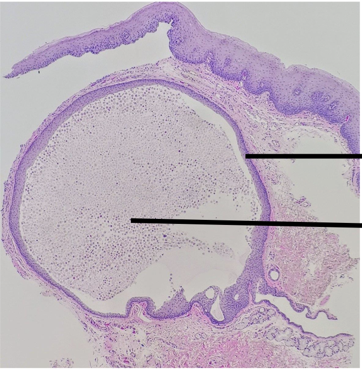
Label top to bottom and name the disease
Ghost Cells
Odontogenic epithelial lining with stellate reticulum like cells
Ameloblasts like basal cells
Calcifying Odontogenic Cyst
Calcifying Odontogenic Cyst
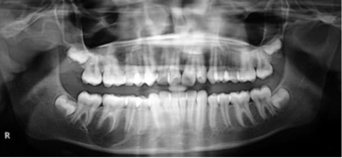
A 17-year-old patient referred by his orthodontist following the fortuitous discovery of a mixed radiolucent/radiopaque image in the right jaw. Provide DD
Calcifying odontogenic cyst
Calcifying epithelial odontogenic tumor
Calcifying odontogenic cyst
Ghost Cells
Calcifying Odontogenic Cyst
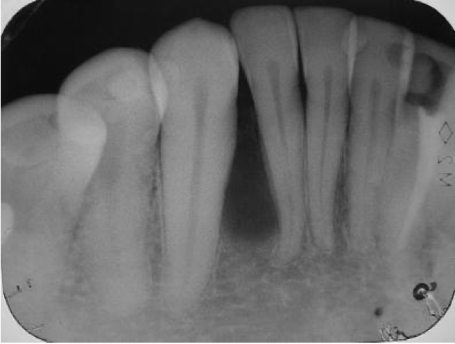
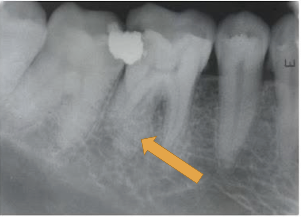
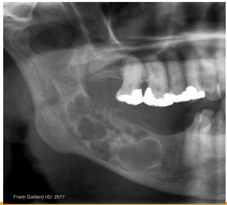
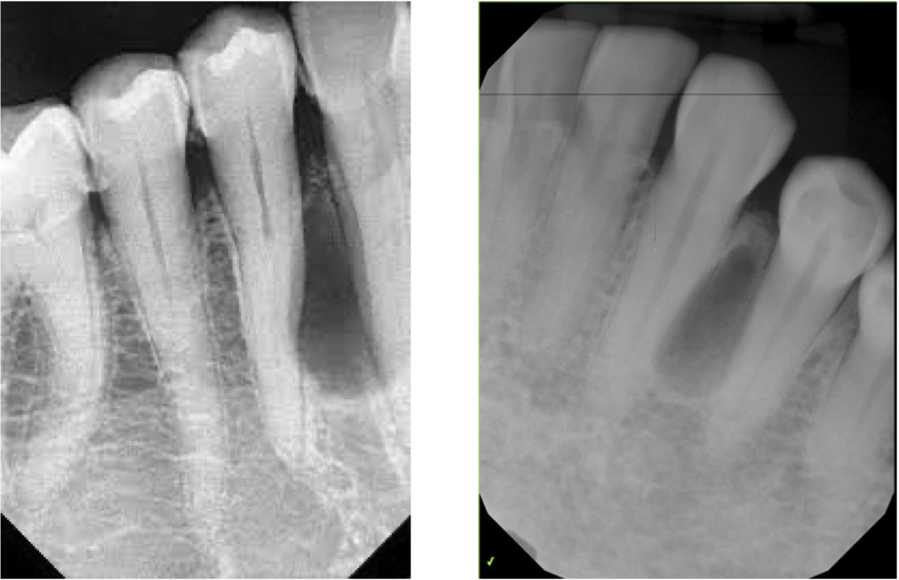
A 51-year-old male patient presented with a well-delimited, radiolucent, mandibular lesion, located between the roots of the right lower lateral incisor and canine and evidenced during routine radiographic examination. Provide 2 differential diagnosis
Lateral Periodontic Cyst
OKC
1-2 layer reduced enamel epithelium
Lateral Periodontal Cyst
A 43-year-old male patient with a complaint of painless swelling in the left mandibular premolar region. Although the history revealed a presence of the swelling for more than one year without any associated symptoms, the patient preferred to obtain clinical consultation as he was concerned about the swelling. The patient had no previous history of dental treatment except for periodic oral prophylaxis. Provide two differential diagnosis
Lateral periodontal cyst
OKC
Provide 2 DD
Lateral periodontal cyst
OKC
Lateral Periodontal Cyst
Botroid odontogenic cyst
Botroid odontogenic cyst
Botroid odontogenic cyst
Botroid odontogenic cyst
Botroid odontogenic cyst
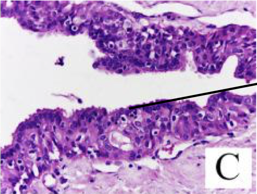
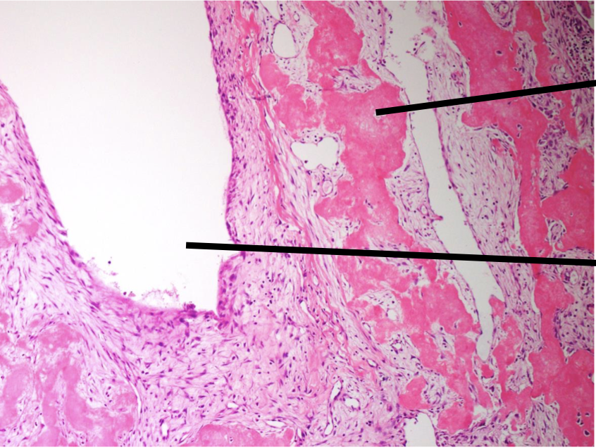
A 35-year-old woman presented with the chief complain of asymptomatic swelling in the lower front region of jaw since 4 months. On clinical examination, hard swelling was present on body of mandible in relation to 33 to 43, obliterating the buccal vestibule and measuring about 1.5 cm in diameter. On palpation, the lesion was nontender, with no discharge, and the overlying mucosa was normal. Provide DD
OKC
Glandular odontogenic cyst
Odontogenic tumor
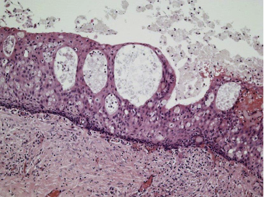
Glandular odontogenic cyst
Mucous cell/goblet cell
Glandular odontogenic cyst
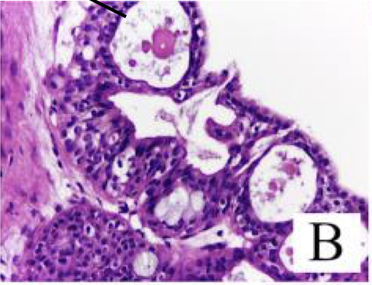
Microcyst
Glandular odontogenic cyst
Cilia
Glandular Odontogenic Cyst
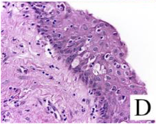
Glandular Odontogenic Cyst
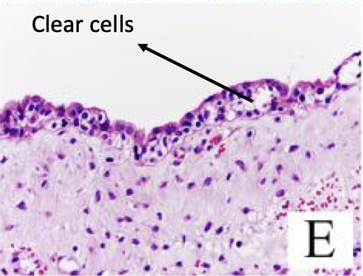
Clear Cells
Glandular Odontogenic Cyst
Glandular Odontogenic Cyst
Characteristic of cysts and tumors of jaw
Buccal Expansion
Characteristic of cysts and tumors of jaw
Lingual Expansion
Buccal Expansion
Palatal Expansion
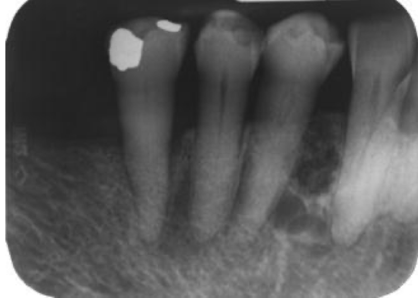
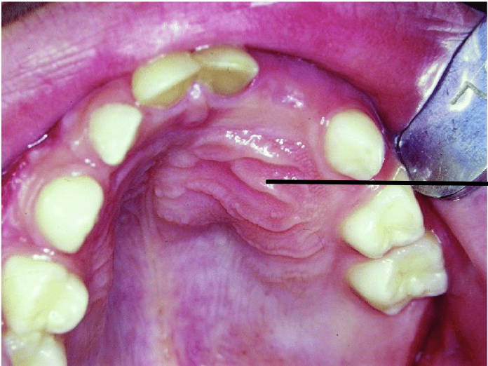
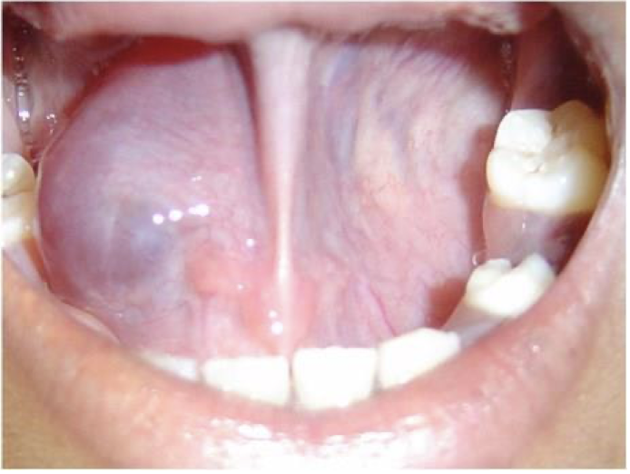
32 year old male patient with complaint of swelling in midline of palate of 3 months duration. There is no pain or any other symptoms. Provide differential diagnosis and justify
Periapical Cyst
Incisve Canal Cyst
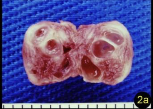
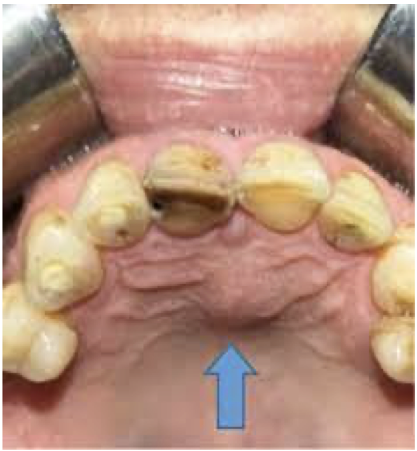
62 year old male patient with complaint of swelling in midline of palate. Provide differential diagnosis and justify
Incisive Canal Cyst
OKC
Pear shaped and in between central incisors
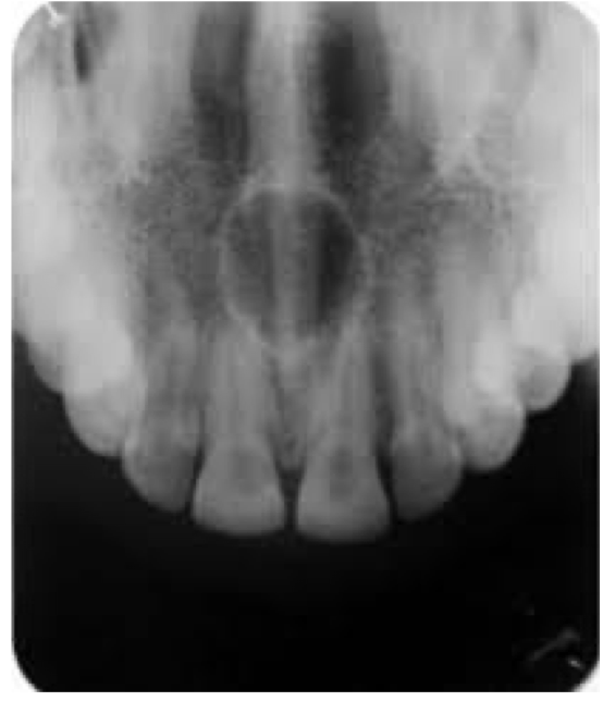
A 23 year old presenting with a cystic swelling in midline in the anterior region of palate. Provide differential diagnosis
Incisive Canal Cyst
OKC
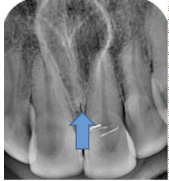
A 52 year old presenting with a cystic swelling in midline in the anterior region of palate. Provide differential diagnosis
Periapical cyst
Incisive canal cyst
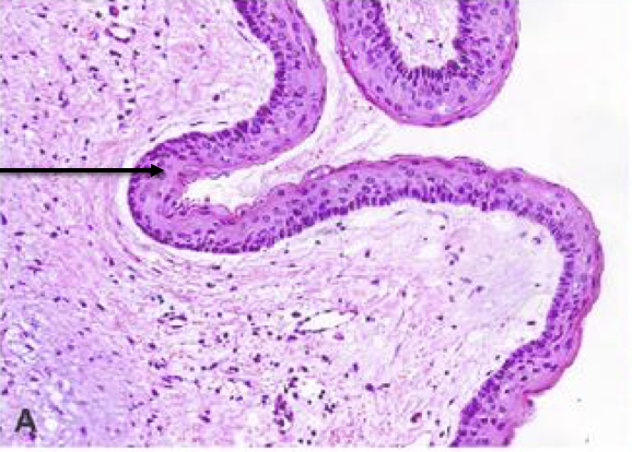
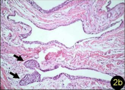
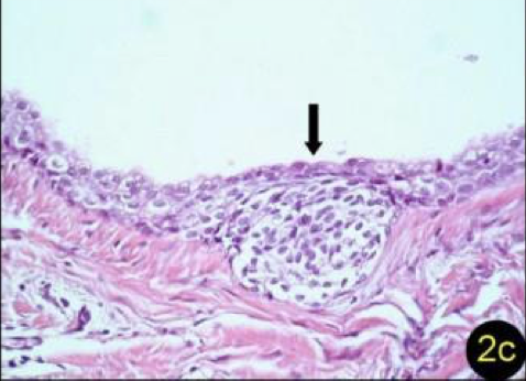
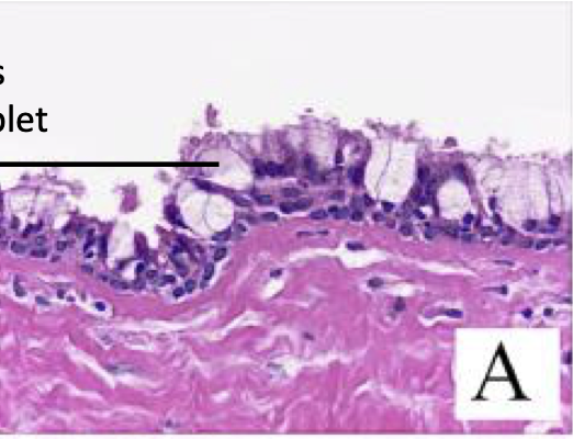
Label top to bottom and diagnose
Pseudostratified ciliated columnar epithelium
goblet cells/mucous cells
Incisive Canal Cyst
A 31 year old presenting with a cystic swelling in lateral aspect between lateral incisors and canine. Provide differential diagnosis
globulo- maxillary cyst?
OKC
Lateral periodontal cyst
Provide DD
globulo- maxillary cyst?
OKC
Lateral periodontal cyst
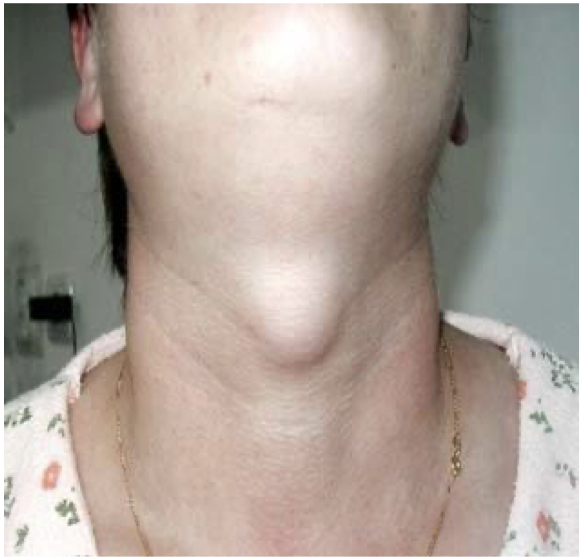
A 45 year old presented with a cystic swelling in midline of the neck. Provide DD
Thyroglossal duct cyst
Dermoid/epidermoid cyst
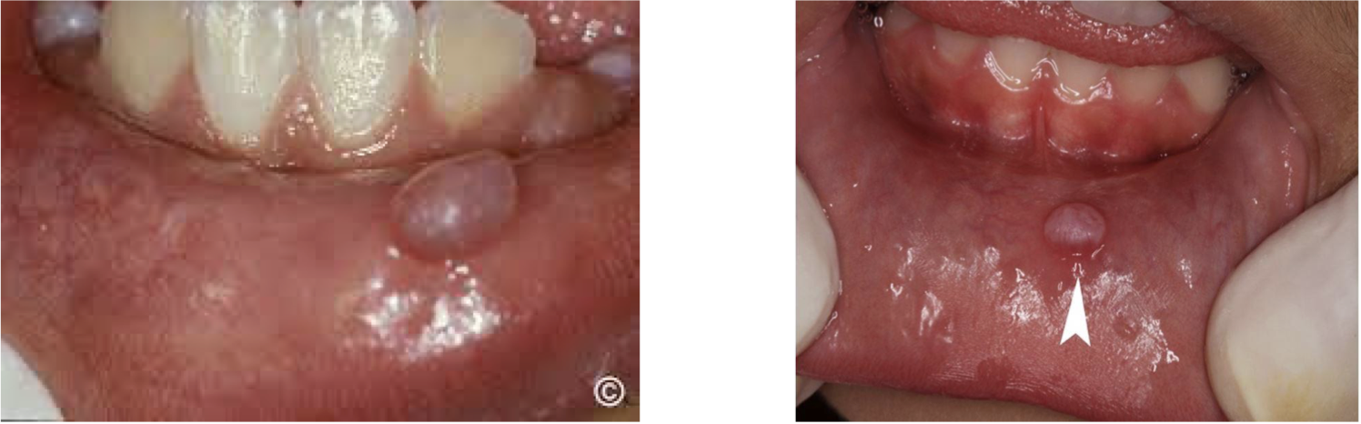
Diagnose
Mucocele
Mucocele - Extravasation type
Mucocele - Extravasation Type
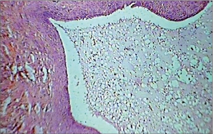
Mucocele - Retention Type
Label top to bottom and diagnose
Lining Epithelium
Mucin
Mucocele - Retention Type
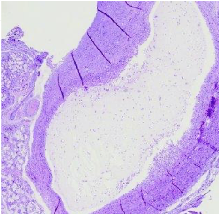
Label top to bottom and diagnose
Mucinophages
Mucin
Mucocele - Extravasation Type
Ranula
Ranula
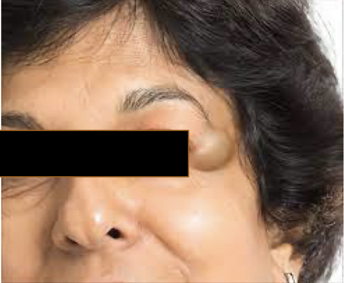
A 35 year presented with cystic swelling on lateral part of forehead of 2 cm diameter. Provide DD
Dermoid Cyst
Epidermoid Cyst
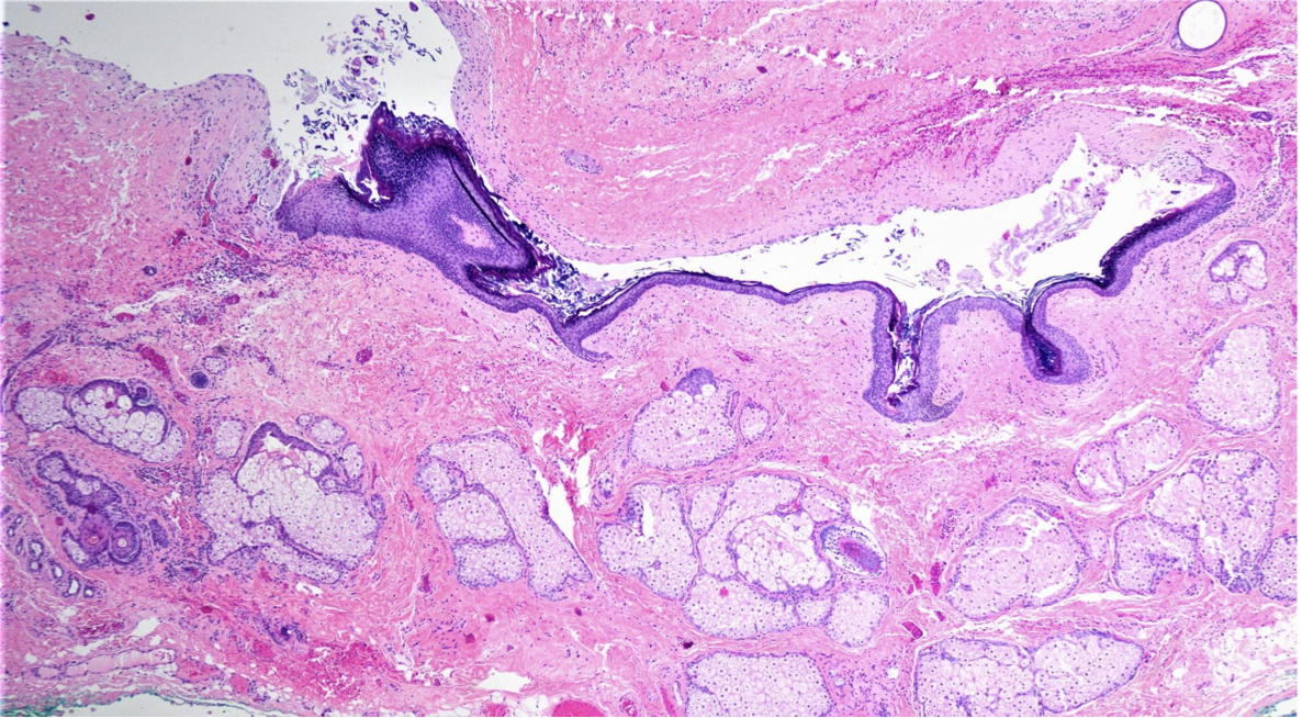
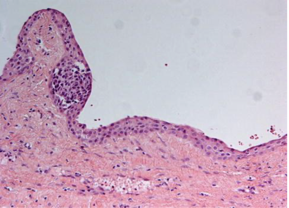
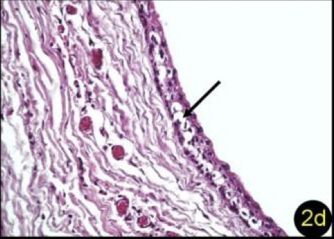
A 35 year presented with cystic swelling on lateral part of forehead of 2 cm diameter. The microscopic image is shown below.
Dermoid Cyst
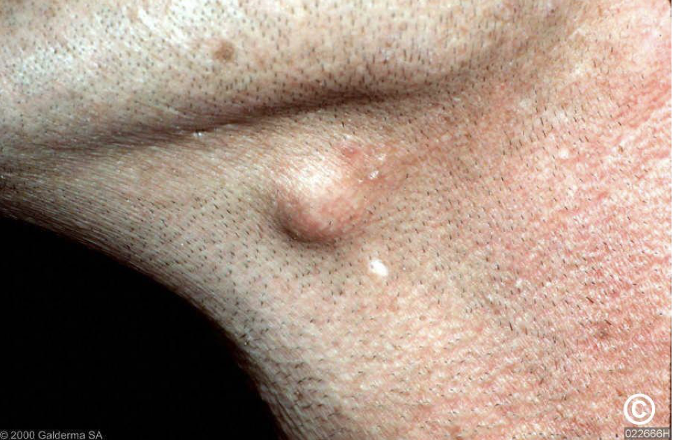
A 35 year presented with cystic swelling on left side of the neck of 2 cm diameter. Provide DD
Dermoid Cyst
Epidermoid Cyst
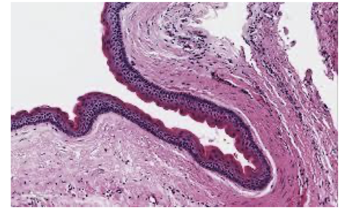
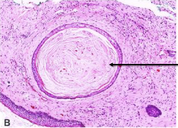
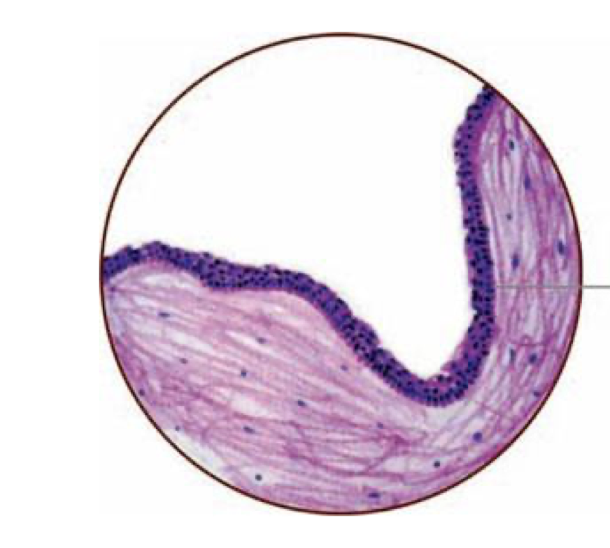
Lable top to bottom and diagnose
Keratin
Orthokeratinised stratified squamous epithelium
Epidermoid Cyst
OKC
Odontogenic Tumor
Lable top to bottom and diagnose
Bone
Cavity without lining epithelium
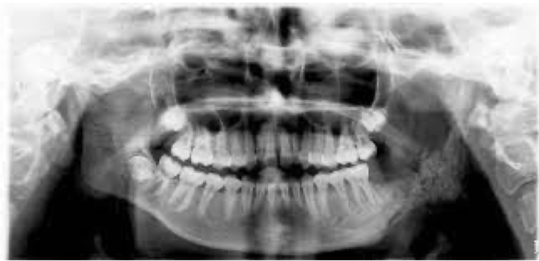
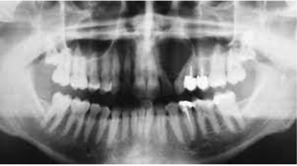
A 20 year old boy presented with swelling of the lower face.
Provide DD
Anyeursmal Bone Cyst
Ameloblastoma
A 9 year old boy presented with swelling of the lower face. Provide DD
Aneurysmal Bone Cyst
OKC
Ameloblastoma
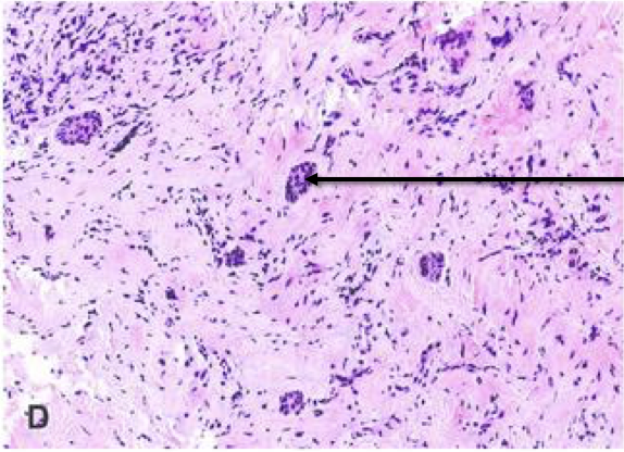
Label from top to bottom and diagnose
Blood filled sinuses/spaces
Giant Cells
Anyeursmal Bone Cyst
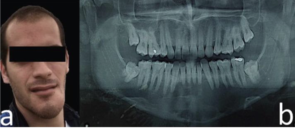
A 18 year old boy presented with cystic swelling of mandible. Provide DD and justify
ABC
Ameloblastoma
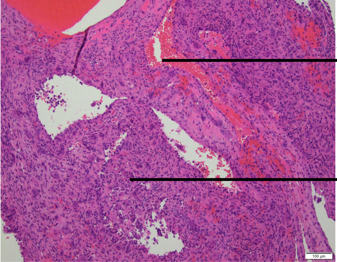
Label from top to bottom and diagnose
Blood filled sinuses/spaces
Giant Cells
Anyeursmal Bone Cyst
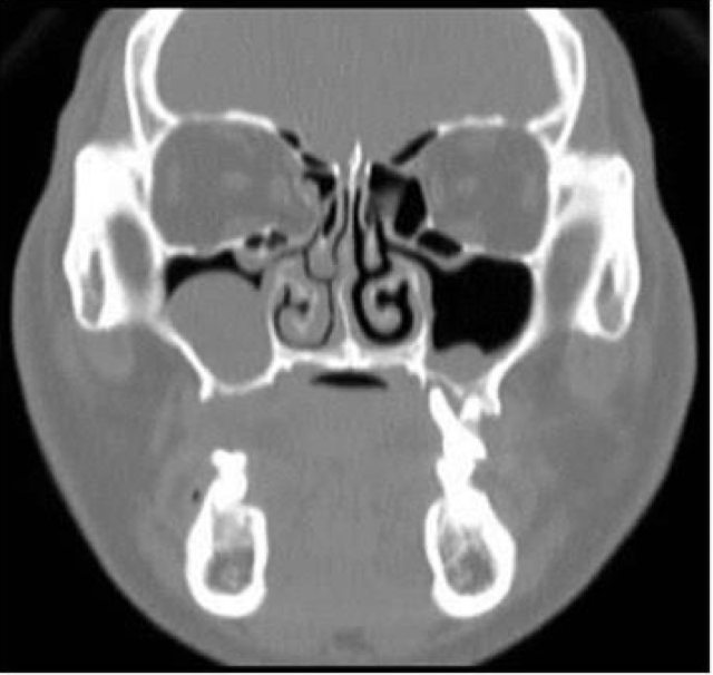
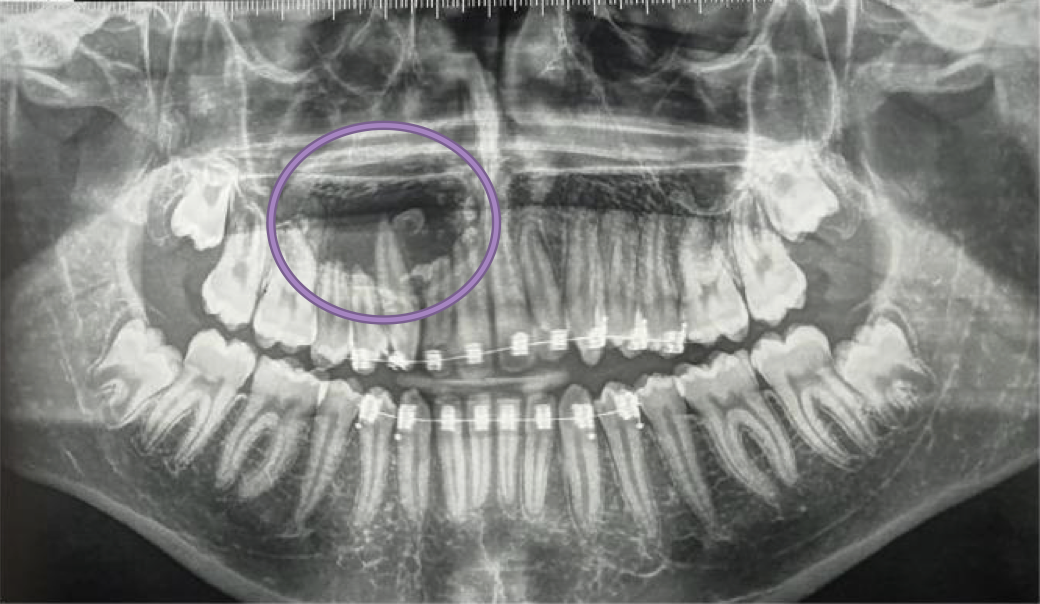
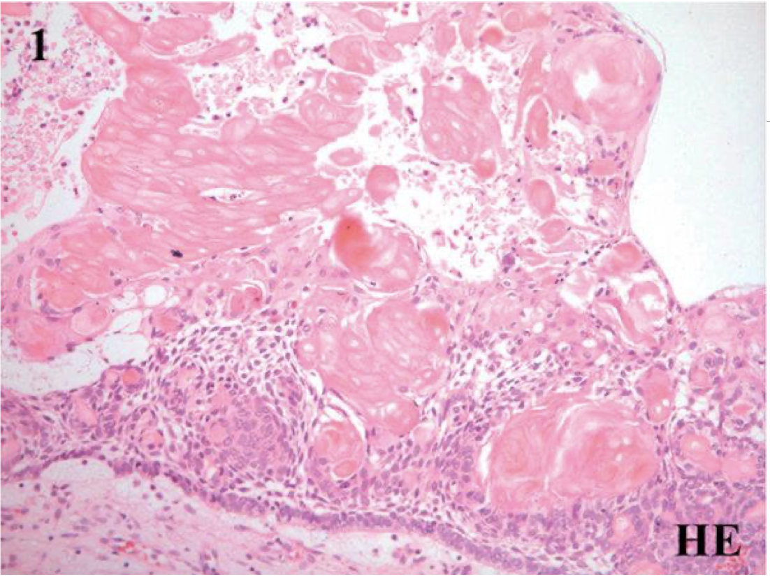
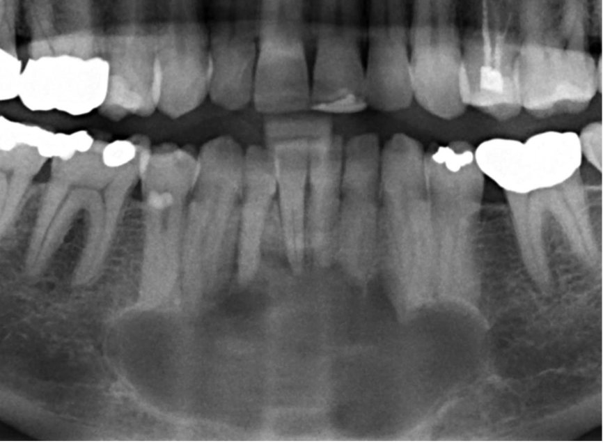
An 18-year-old woman presented with unilateral rhinorrhea for past 2 months. She had a history of road accident 12 months months ago and trauma to the maxillary bone and was surgically fixed. Provide DD
Mucosal cyst
Surgical cyst of maxillary antrum
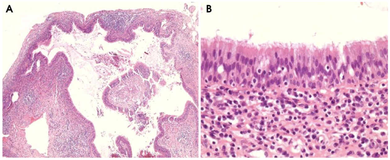
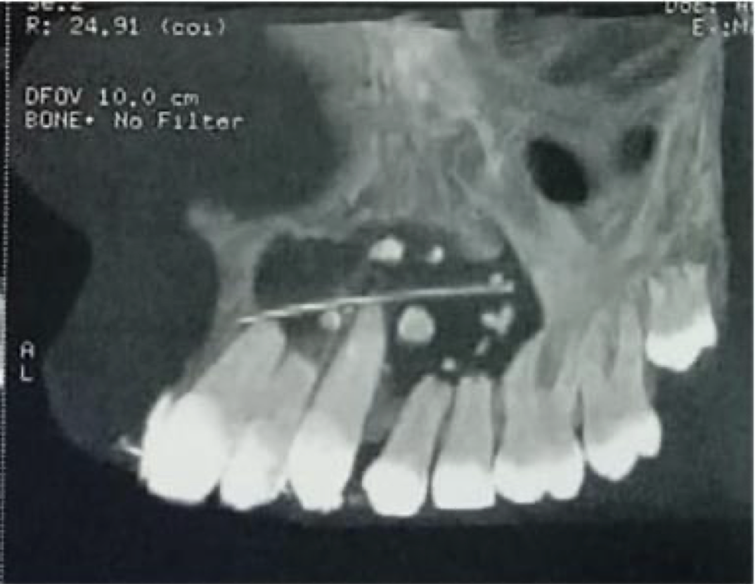
A 42-year-old woman came complaining of intermittent stinging pain in her right cheek. There were no clinically abnormal findings in physical and oral examinations. The patient only had a history of orthognathic surgery on both the maxilla and the mandible performed about 21 years prior to this visit
Surgical cyst of maxillary antrum
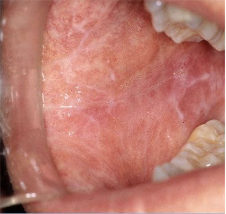
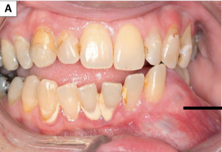
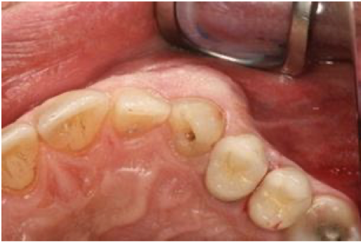
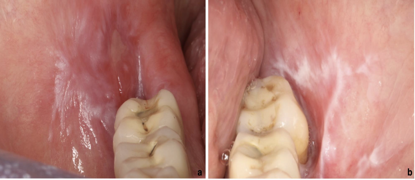
A 45 year old patient presented with bilateral white lace like patches throughout buccal mucosa. There is mild burning sensation on eating. What is your diagnosis? Provide one differential diagnosis
Lichen planus (reticular)
DD: Lichenoid reaction
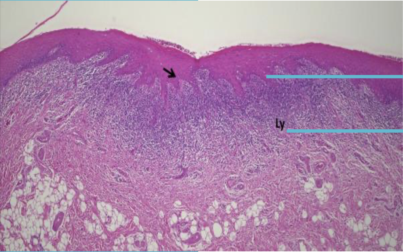
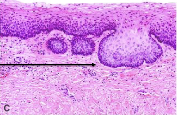
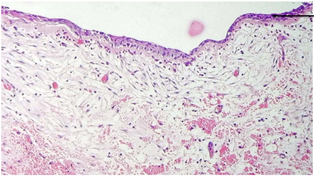
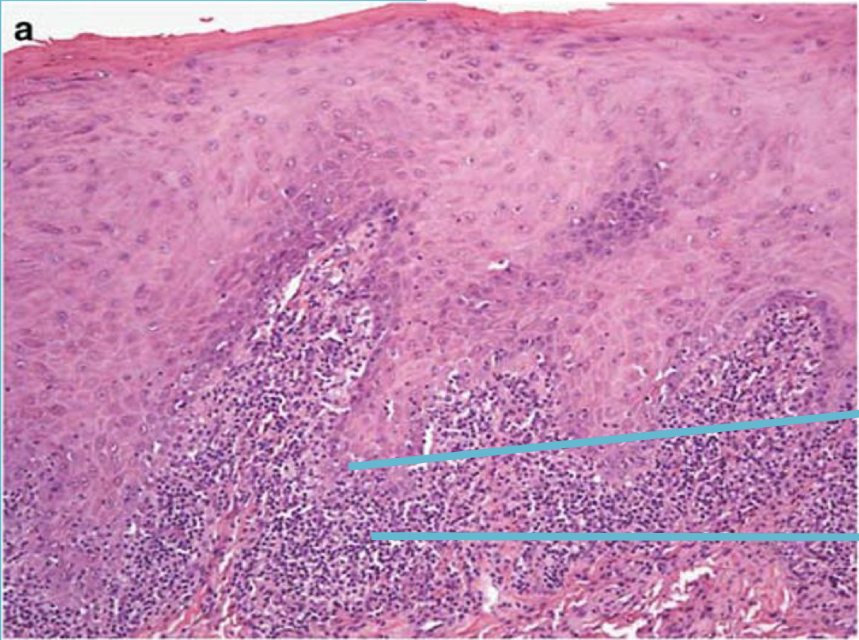
Label from top to bottom and diagnose
Saw tooth rete pegs
juxta-epithelial band of inflammatory cells
Lichen Planus
Label from top to bottom and diagnose
Liquefactive degeneration of basal cells
Juxta epithelial band of inflammatory cells
Lichen planus
A 45 year old patient presented with bilateral white lace like patches thoroughout buccal mucosa. There is severe burning sensation. What is your diagnosis?
Lichen planus (Erosive)
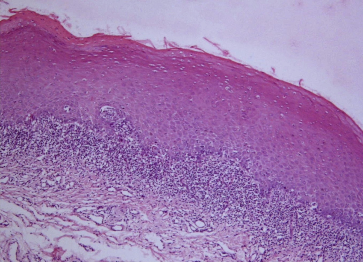
A 52year old patient presented with white lesions on tongue on both sides. There is severe burning sensation. A biopsy was taken and the histopathological image is shown here. What is your diagnosis?
Lichen Planus
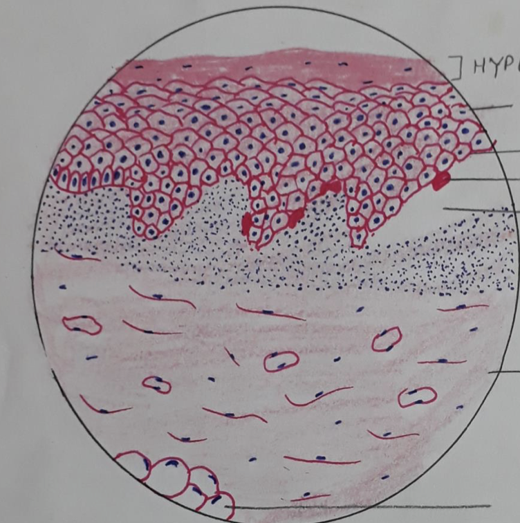
Label from Top to Bottom and Diagnose
Hyperparakeratinosis
Acanthosis
Basal Cell Degeneration
Colloid Bodies
Max-Joseph Space
Juxtaepithelial dense band of lymphocytes
Connective tissues
Deeper Adipocytes
Lichen Planus - Features