AP Psychology- Biological Bases of Behavior
1/138
Earn XP
Description and Tags
unit 2
Name | Mastery | Learn | Test | Matching | Spaced | Call with Kai |
|---|
No analytics yet
Send a link to your students to track their progress
139 Terms
Franz Gall
In 1800, Franz Gall suggested that bumps of the skull represented mental abilities; his theory, though incorrect, nevertheless proposed that different mental abilities were modular
Biological psychology
Branch of psychology concerned with the links between biology and behvaior; Some biological psychologists call themselves behavioral neuroscientists, neuropsychologists, behavior geneticists, physiological psychologists, or biopsychologists
Neural communication
Neurobiologists and other investigators understand that humans and animals operate similarly when processing information
Similarities in the brain regions are all engaged in information processing
Neurons
the messengers;About 100 billion neurons (nerve cells) in the human brain;
Neurons have many of the same features as other cells: Nucleus, Cytoplasm,
Cell membrane;
What makes neurons unique is their shape and function
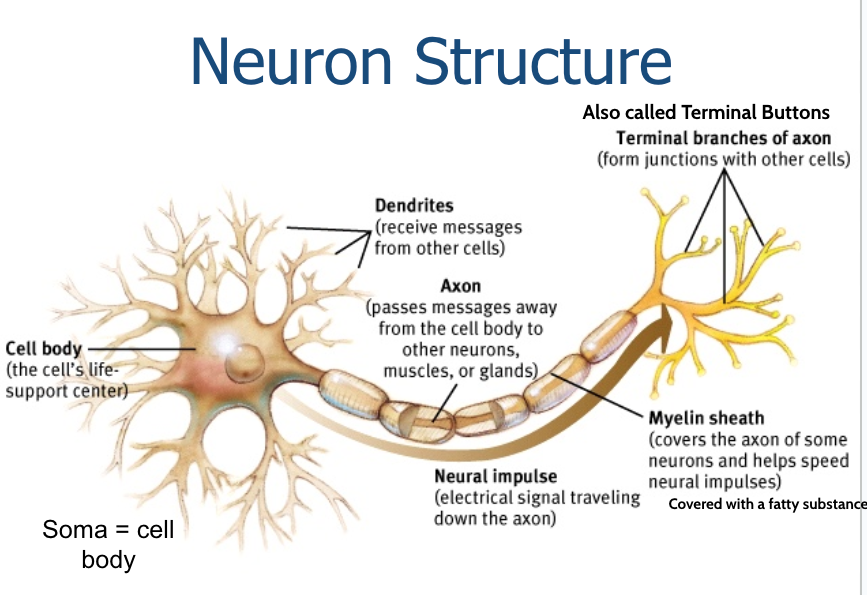
Neuron Structure
cell body (the cell's life support center); dendrites (receive messages from other cells); axon (passes messages away from the cell body to other neurons, muscles, or glands); myelin sheath (covers the axon of some neurons and helps speed neural impulses, covered w/ a fatty substance); terminal branches of axon/ terminal buttons (form junctions with other cells); neural impulse (electrical signal traveling down the axon)
Glial Cells
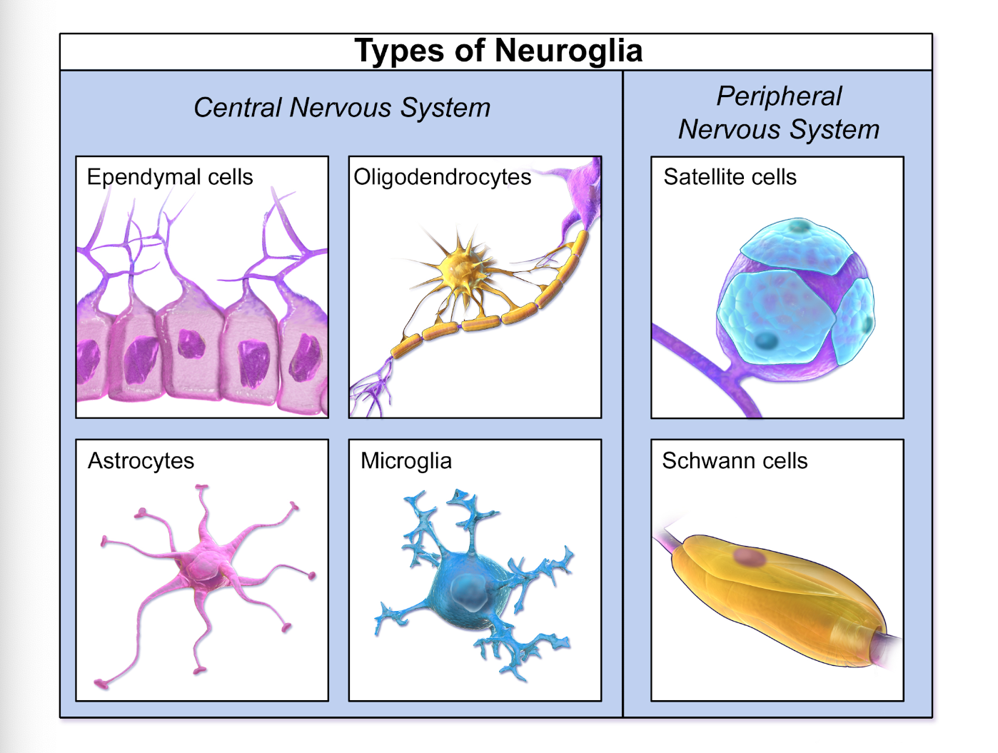
cells that insulate and support neurons
create the myelin sheath
remove waste products
provide nourishment
prevent harmful substances from entering the brain
types: astrocytes provide nutrition to neurons
Oligodendrocytes and Schwann cells insulate neurons as myelin
Schwann cells
A Schwann cell is a type of glial cell found in the peripheral nervous system
responsible for producing the myelin sheath
Schwann cells also play a role in nerve regeneration and support the overall health and function of neurons
Types of Neuroglia
polarized membrane
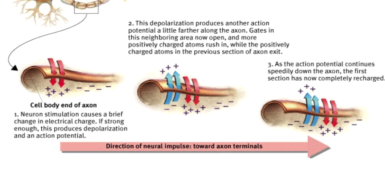
a lipid membrane that has a positive electrical charge on one side and a negative charge on another side, which produces the resting potential in living cells
Depolarization
the process that carries the neural impulse through the axon, action potential is what must happen for the process to occur
Sodium Potassium pump
pumps Na+ ions out from the inside of the neuron, making them ready for another action potential; K+ ions remain inside the axon
resting potential/resting state
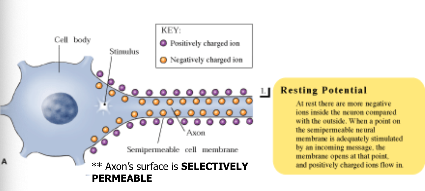
Neuron is not transmitting information- it is resting
Membrane Potential over time
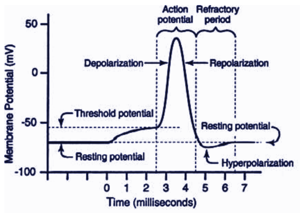
Resting potential/state
The resting potential of a neuron is the value its membrane potential keeps as long as it is not receiving stimulation or undergoing an action potential
action potential
An action potential occurs when a neuron transmits an electrical charge down its axon, which terminates in the release of chemical signals in the form of neurotransmitters; a brief electrical charge that travels down an axon, caused by the movement of positively charged ions in and out of the membrane
synapse
a junction between the axon tip of the sending neuron and the dendrite or cell body of the receiving neuron; this tiny gap is called the synaptic gap or cleft
neurotransmitters
small chemicals released from the sending neuron travel across the synapse and bind to receptor sites on the receiving neuron, thereby influencing it to generate an action potential, this is how neurons communicate
reuptake
neurotransmitters in the synapse are reabsorbed into the sending neurons through the process of re-uptake; this process applies the breaks on neurotransmitter response
all-or-none response
when the depolarizing current exceeds the firing threshold/absolute threshold, a neuron will fire; if the depolarizing current fails to exceed the threshold, a neuron will not fire
intensity of an action potential remains the same throughout the length of the axon
firing threshold/absolute threshold
the level of depolarization at which a neuron fires
refractory period
after a neuron fires an action potential, it pauses for a short period to recharge itself to fire again
neurotransmitter release
action potential causes vesicle to open
neurotransmitter released into the synapse
locks onto the receptor molecule in postsynaptic membrane
neurotransmitter reuptake in vesicles
Lock and Key Machanism
neurotransmitters bind to the receptors of the receiving neuron in a key-lock mechanism; neurotransmitters fit like chemical keys in chemical lock
excitatory neurotransmitters
-the key fits and ‘opens’ the receiving neuron
-activation of the receptor causes depolarization of the membrane and promotes an action potential in the receiving neuron
inhibitory neurotransmitters
-the key fits in but only stops and other keys
-activation of the receptor causes hyperpolarization and depresses the generation of action potential
acetylcholine
-excitatory
-function: motor movement (causes muscles to contract) and memory
-lack of acetylcholine: Alzheimer’s, paralysis
dopamine
-both excitatory and inhibitory
-function: motor movement, alertness, & pleasure
-lack of dopamine: Parkinson’s disease
-too much dopamine: schizophrenia
GABA
-the most common inhibitory neurotransmitters in the brain
-functions: controls a wide variety of functions
-lack of GABA: Huntington’s disease, anxiety, epilepsy, insomnia
Glutamate
-the most common excitatory neurotransmitter in the brain
-function: memory
-too much glutamate: ALS (Lou Gehrig’s Disease), migraines, seizures
endorphins
-inhibitory
-function: alleviating pain
-structurally very similar to opioids (opium, morphine, heroine, etc.)
-we’ve become addicted to endorphin-caused feelings
-lack of endorphins: chronic pain disorders
Serotonin
-inhibitory in pain pathways
-functions: sleep, mood, appetite, and sensory perception (“mood and food”)
-lack of serotonin: depression
-too much: anxiety, limits dreaming
substance p
-excitatory
-P stands for pain
-responsible for the sending of pain messages throughout our body
Norepinephrine (AKA Noradrenaline)
-excitatory
-functions: controls alertness and arousal, also functions as a hormone
-lack of Norepinephrine: can lead to depressed moods
neurotransmissions and drugs
-Drugs can affect synapses at a variety of sites and in a variety of ways
-drugs mimic NT
-increasing the number of synapses/firing
-release of NT from neurons with or without synapses/firing
-produce more/less NT than what is normal
-prevent vesicles from releasing NT
-block reuptake of NT into sending neuron
agonists
-agonists excite & mimic neurotransmitters
-structurally similar to neurotransmitters and mimics its effects on the receiving neuron
-ex) Morphine mimics the action of endorphins by stimulating receptors in brain areas involved in mood and pain sensations
antagonists
-antagonists inhibit & bloc neurotransmitters
-structure similar enough to neurotransmitter and occupies its receptor site and blocks it’s action, but not similar enough to stimulate the receptor
-curare poisoning paralyzes its victims by blocking ACh receptors involved in muscle movement
tolerance
builds with repeated use, requires increasing amount
physical dependence
intense cravings and physiological need
withdraw
painful side effects from stopping after long term usage
depressants
-alchohol (stimulates GABA)
-Barbiturates (tranquilizers)
-Benzodiazepines (anti-anxiety)
-opiates
opiates
-opium, morphine, oxycodone, heroine
-suppress pain, induce sleep, induce state of euphoria
-antagonist for endorphins; highly addictive
-leads brain to stop producing own endorphins
Hallucinogens
-LSD, mushrooms, peyote
-alter perception and create dramatic hallucinations
-disrupts transmissions of serotonin
-THC is also a hallucinogen (in marijuana)
Stimulants
-nicotine, caffeine
-MDMA- affects serotonin
-Cocaine and Amphetamines; increase dopamine and norepinephrine activities, elevated mood and energy; can lead to long-lasting damage to dopamine neurons
Reuptake Inhibitors
SSRIs: selective serotonin reuptake inhibitor
can be done with other neurotransmitters as well
Mirror Neurons
neurons that fire when you do an action, or you watch someone do an action; neural basis of ‘observational learning’ & empathy
nerves
-neural cables containing many axons
-part of the peripheral nervous system
-connect the central nervous system with muscles, glands, and sense organs
sensory (afferent neurons)
INPUT from sensory organs to the brain and spinal cord
sensory vs motor neurons mnemonic- SAME
Motor (efferent) neurons
OUTPUT from the brain and spinal cord, to the muscles and glands
sensory vs motor neurons mnemonic- SAME
effector cells
respond to stimulus at the terminal end of neuron
interneurons
carry information between other neurons only found in the brain and spinal cord
autonomic nervous system
-”involuntary”
-regulates the function of our internal organs, such as the heart, stomach, lungs, intestines
-part of the peripheral nervous system and it also controls some of the muscles within the body
-regulates involuntarily responses
ex.) we do not notice when blood vessels change size or when our heart beats faster
somatic nervous system
-”voluntary”
-regulates voluntary movements
-part of the peripheral nervous system, connects the brain to the motor neurons such as those found in the skeletal muscles
-we are in control of this system (voluntary) and we use it when we want to make our muscles move
autonomic nervous system
sympathetic vs parasympathetic
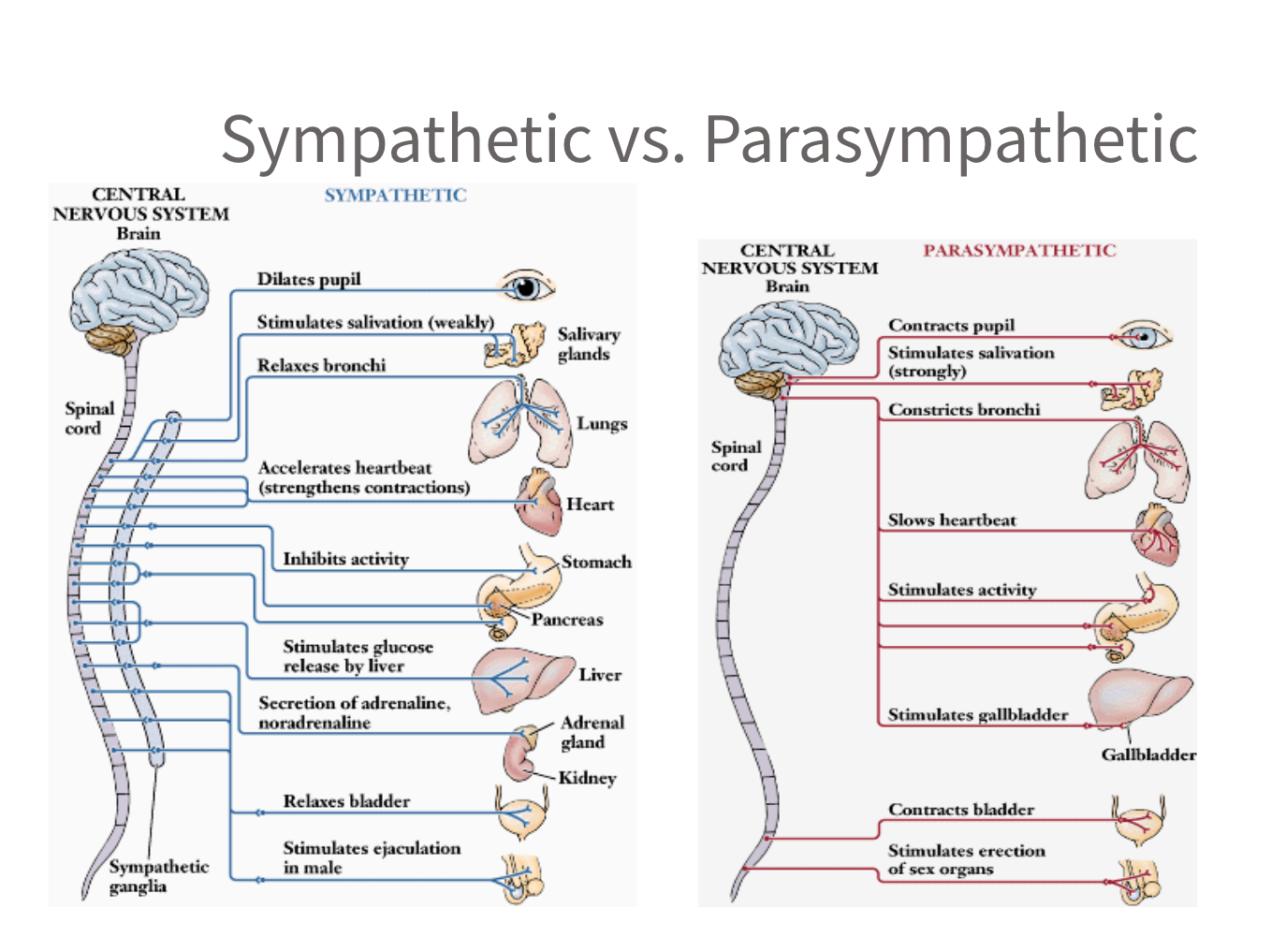
sympathetic nervous system
-”arouses”
-prepares the body for "fight or flight" responses
-It activates during times of stress or danger, increasing heart rate, dilating pupils, and releasing adrenaline
-It also inhibits non-essential functions like digestion
parasympathetic nervous system
-”calms”
-helps regulate the body's rest and digest
-responsible for conserving energy, promoting digestion, and promoting relaxation
-works in opposition to the sympathetic nervous system
central nervous sytem
the brian and spinal cord
brain
part of the CNS that plays important roles in sensation, movement, and information processing
spinal cord & spinal cord reflexes
-plays a role in body reflexes and in communication between the brain and the peripheral nervous system
-simple reflexes are controlled by interneurons in your spinal cord
endocrine system
-body’s “slow” chemical communication system
-communication is carried out by your hormones in your blood that is synthesized by a set of glands
hormones affect on behvaior
-growth of bodily structures such as muscles and bones, differences between males and females
-metabolism
-energy levels
-preparing the body for stressful situations
-mood
pituitary gland- “master gland”
-secretes many different hormones, some of which affect other glands
-controlled by the hypothalamus
-secretes hormones which control the output of hormones by other endocrine glands
-monitors hormone levels to prevent imbalances
-Human growth hormone (HGH): growth
-oxytocin: boding (released frequently by touch)
-cortisol: stress hormone
-norepinephrine is also a hormone released by adrenal glands
thyroid gland
affects metabolism, among other things
imbalance in thyroid can affect mood, weight
parathyroids
help regulate the level of calcium in the blood
Adrenal glands
-inner part, called the medulla, helps trigger the “fight or flight” response
-releases adrenaline
pancreas
regulates the level of sugar in the blood
ovary
secretes female sex hormones
testis
secretes male sex hormones
Brain lesion
Experimentally destroys brain tissue to study animal behaviors after such destruction
Autopsy
post mortem study of a brain to compare brain changes with behavior
Clinical observation
-shed light on a number of brain disorders
-alterations in brain morphology due to neurological and psychiatric diseases are now being catalogued
Electroencephalogram (EEG)
An amplified recording of the electrical waves sweeping across the brain’s surface, measured by electrodes placed on the scalp
CT (computed tomography scan)
A series of X-ray photographs taken from different angles and combined by computer into a composite representation of a slice through the body; also called CAT scan
PET (positron emission tomography) scan
A visual display of brain activity that detects where a radioactive form of glucose goes while the brain performs a given task
MRI (magnetic resonance imaging)
Uses magnetic fields and radio waves to produce computer-generated images that distinguish among different types of brain tissue; top images show ventricular enlargement in a schizophrenic patient; bottom image shows brain regions when a participant lies
fMRI (functional magnetic resonance imaging)
Can reveal brain functioning and structure by examining changes in blood flow based on different activities
brainstem
-Oldest part of the brain
-beginning where the spinal cord swells and enters the skull
-responsible for automatic survival functions
-medulla: basic life functioning and reflexes
-pons: sleep/wake cycle
Hindbrain
-brain stem
-reticular formation
-cerebellum
-thalamus
Reticular formation
Alertness/attention to incoming stimuli
cerebellum
Balance, voluntary movement, procedural learning
Thalamus
Sensory relay station except smell
The limbic system
-a doughnut-shaped system of neural structures at the border of the brainstem and cerebrum, associated with emotions such as fear, aggression and drives for food and sex
-includes hippocampus, amygdala, and hypothalamus
Amygdala
Consists of two almond-shaped neural clusters linked to the emotion of fear and agression
Hypothalamus
-lies below (hypo) the thalamus
-controls 4 f’s (fight, flight, feeding, and fornicating)
-body temperature
-circadian rhythms
-helps govern the endocrine system via the pituitary gland (brain region controlling the pituitary gland)
-hypothalamic centers are ventromedial and lateral
Ventromedial hypothalamus (VMH)
suppresses hunger (stimulation)
Lateral hypothalamus
increases hunger
hypothalamus & hormones
orexin increase- hypothalamus-increases hunger
ghrelin increase- stomach- increases hunger
insulin increase-pancreas-increases hunger
leptin increase-fat cells-decreases hunger
PPY increase-digestive tract-decreases hunger
Hippocampus
-structure that contributes to the formation of memories
-damage to the hippocampus has been implicated in the memory loss associated with Alzheimer’s
-case studies
frontal lobes
decision making, planning, movement
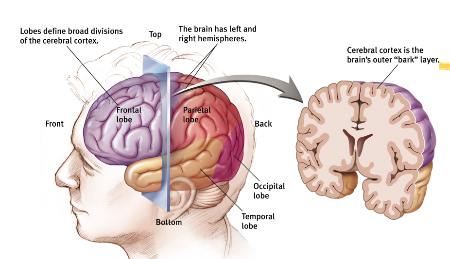
parietal lobes
include the somatosensory cortex (touch); spatial processing (location)
occipital lobes
include the visual areas which receive visual information from the opposite visual field
temporal lobes
auditory processing, facial and object recognition, olfactory cortex
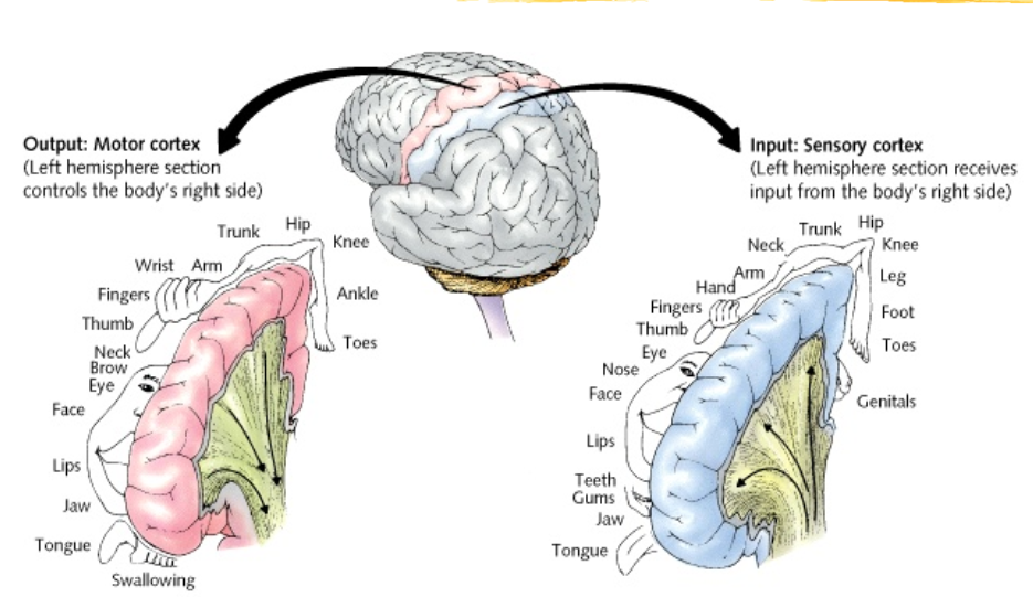
motor cortex
area at the rear of the frontal lobes that controls voluntary movements
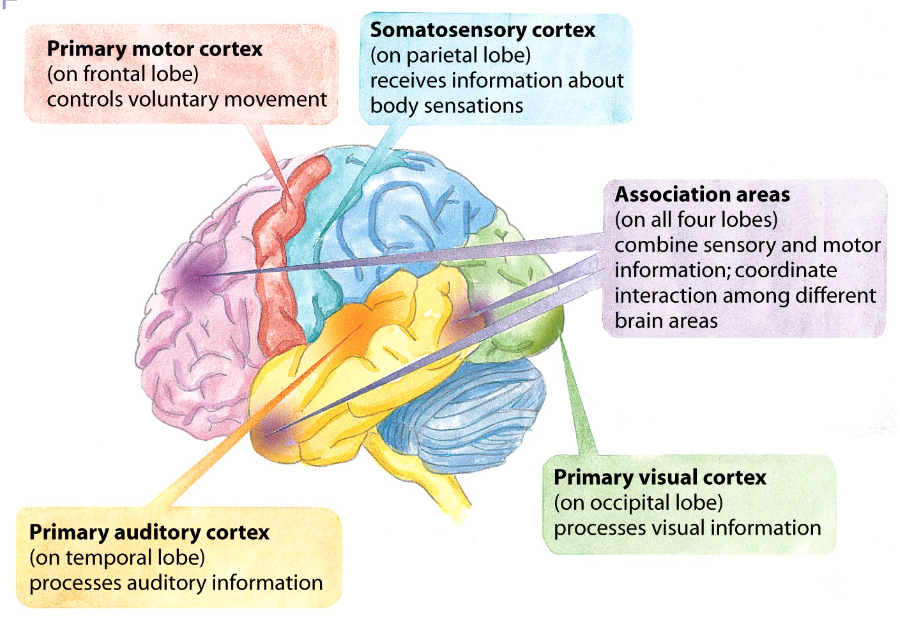
somatosensory cortex
area at the front of the parietal lobes that registers and processes body sensations, sense of touch, temperature, and pain
motor vs sensory cortex brain controls
association areas
-areas of the cerebral cortex that are not involved in primary motor or sensory functions; involved in higher mental functions such as learning, remembering, thinking and speaking
-Phineas Gage
-more intelligent animals have increased “uncommitted” or association areas of the cortex
language
-aphasia: an impairment of language, usually caused by left hemisphere damage to Broca’s area (impaired speaking) or to Wernicke’s area (impaired understanding)
broca’s area
controls speech muscles via the motor cortex
wernicke’s area
interprets auditory code
the homunculus
representation of how large body parts would be if they were to scale of how much the brain controls
plasticity
the brain’s ability to modify itself after some type of injury or illness