Eye Anatomy
0.0(0)
Studied by 9 peopleCard Sorting
1/12
Earn XP
Description and Tags
Last updated 5:26 PM on 11/17/22
Name | Mastery | Learn | Test | Matching | Spaced | Call with Kai | Chat |
|---|
No analytics yet
Send a link to your students to track their progress
13 Terms
1
New cards
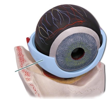
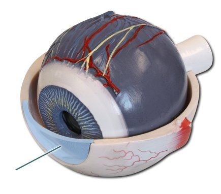
sclera
Makes up most of the fibrous layer; white
2
New cards
cornea
Clear anterior portion of fibrous layer
3
New cards
ciliary muscle
A circular muscle that relaxes or tightens to change the shape of the lens.
4
New cards
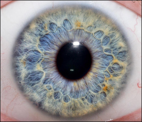
Iris
Circular and radial muscles that adjust the size of the pupil
5
New cards
Rods
photoreceptive cells that are adjusted for dim light; perceive gray tones
6
New cards
Cones
photoreceptive cells that are adjusted for bright light; perceive colors
7
New cards
Aqueous humor
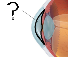
Between cornea and lens; supplies nutrients and maintains pressure
8
New cards
vitreous body
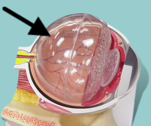
Gel-like, between lens and posterior; maintains eye shape
9
New cards
Eyelashes
Located on the margin of the eyelids. Prevent foreign substances from entering the eye.
10
New cards
Myopia
Difficulty focusing distant objects (nearsightedness)
11
New cards
Hyperopia
Difficulty focusing nearby objects (farsightedness)
12
New cards
tear duct
Small holes in the inside corners of the top and bottom eyelids. The small holes bring tears to keep the cornea moist and clear and constantly flow, washing away things like dust. The tear then carry away any accessible liquid in your eye.
13
New cards
Retina
Light sensitive layer of the eye; contains rods and cones