DEN: Ch 7&8 Cells & Basic Tissues
1/143
There's no tags or description
Looks like no tags are added yet.
Name | Mastery | Learn | Test | Matching | Spaced | Call with Kai |
|---|
No analytics yet
Send a link to your students to track their progress
144 Terms
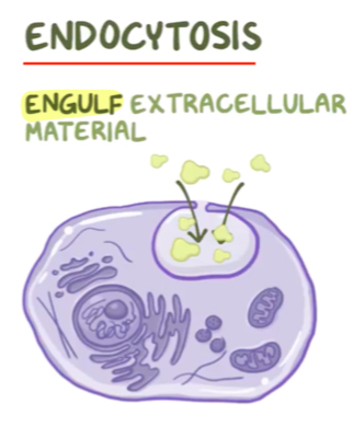
Endocytosis
Engulf extracellular material
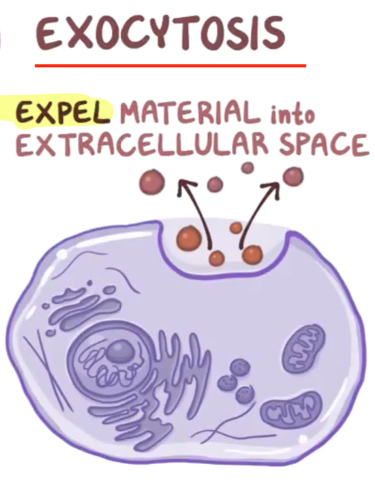
Exocytosis
Expel material into extracellular space
Macrophages perform __________
Phagocytosis
Phagocytosis
Eating bacteria, germs, virus
Pseudopods
The “hands” of macrophages that “hug” bacteria
What does a lysosome contain
Digestive enzymes
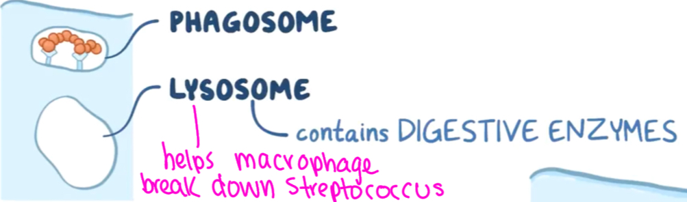
Phagosome
Area of macrophage where bacteria is encapsulated

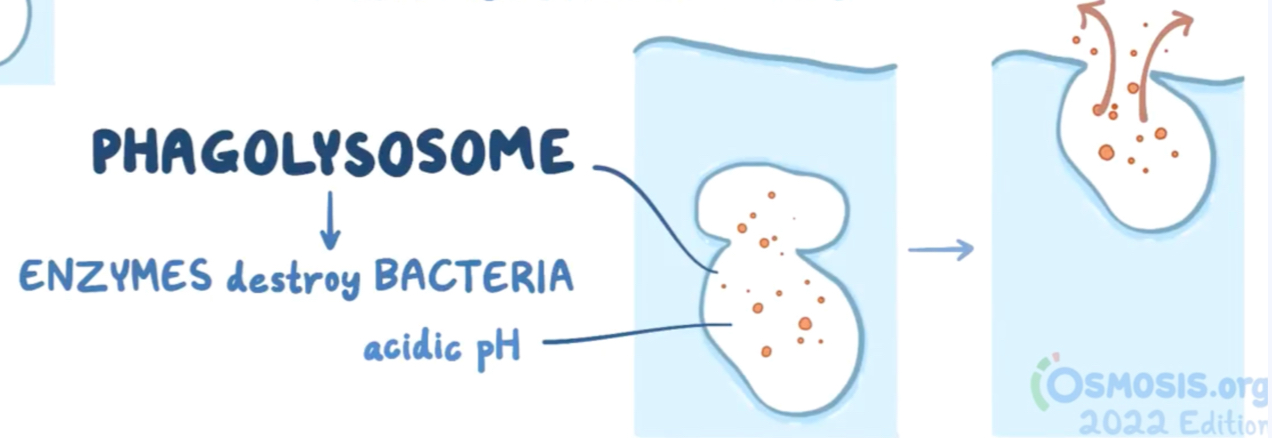
Phagolysosome
Enzymes destroy bacteria
Cell membrane function
Outer cell layer
Controls what goes in and out
Phospholipid bilayer
Cytoskeleton
Framework of cell
Gives cell its shape
Structural stability
Golgi complex
Packages proteins, fats, hormones
“Post office”
Rough endoplasmic reticulum
Aids in making proteins, lipids, steroids
Nucleus
Control center holding DNA
Mitochondria
POWERHOUSE
Makes energy
Aerobic respiration
Smooth endoplasmic reticulum
Aids in making fats removes harmful substances
Cell division for tissue cells
Reproduce themselves and replaces the dead tissue cells
End result of cellular division
Two identical daughter cells
Phases of cell division
Interphase
Prophase
Prometaphase
Metaphase
Anaphase
Telophase
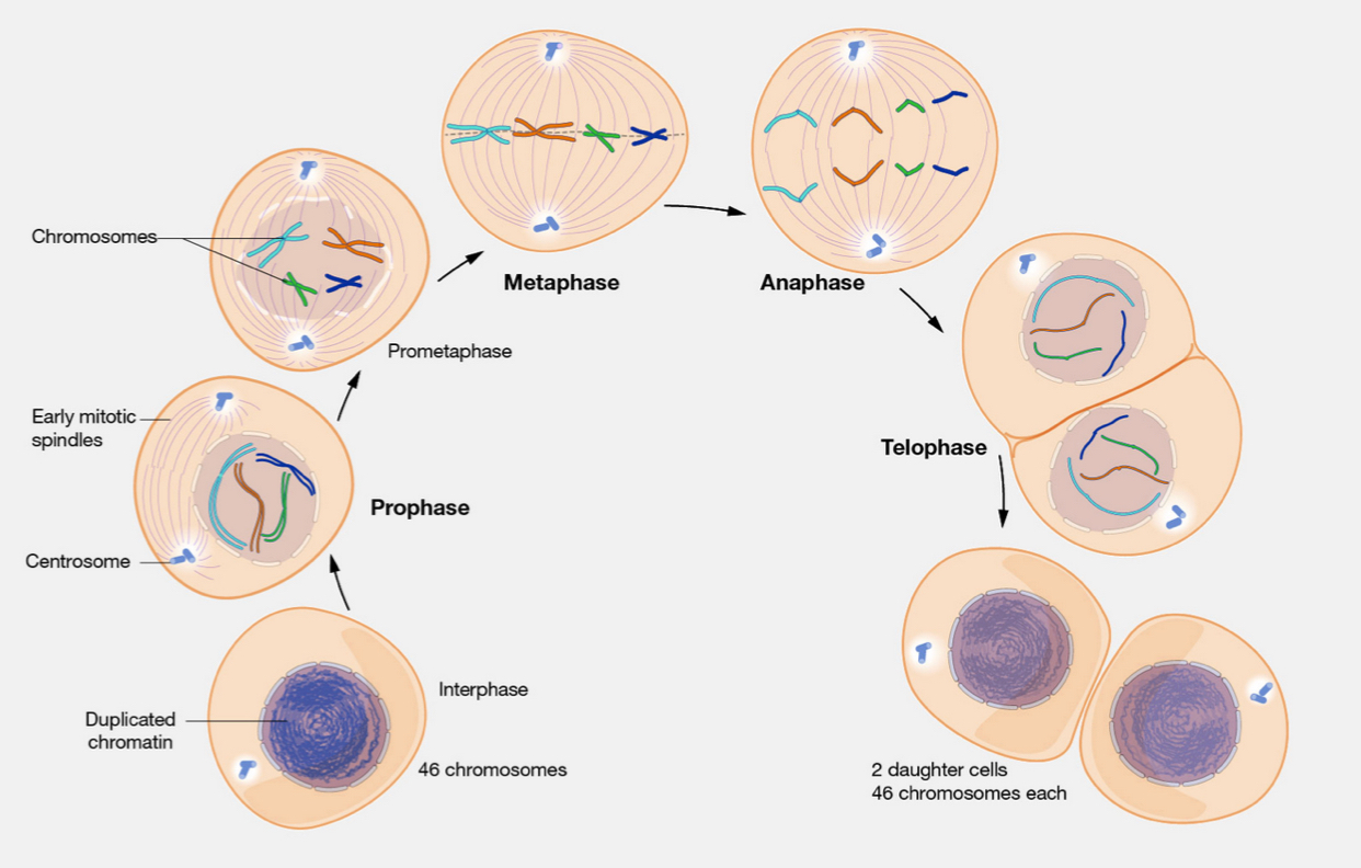
First phase of cell division
Interphase
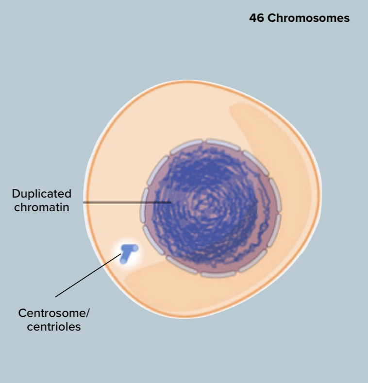
Interphase
Chromatin is duplicating
Cell is growing

Second phase of cell division
Prophase
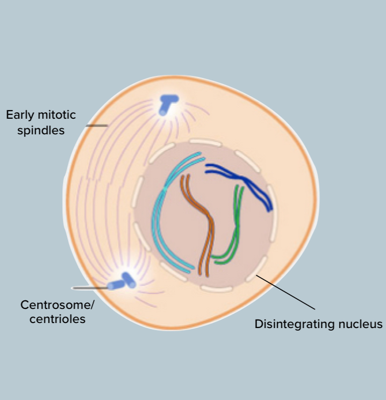
Prophase
Chromatin condenses into chromosomes
Replicated centrioles migrate to opposite poles
Nuclear membrane and nucleolus start to disintegrate

Third phase of cellular division
Prometaphase

Prometaphase
Nuclear membrane breaks down and chromosomes attach to spindle fibers
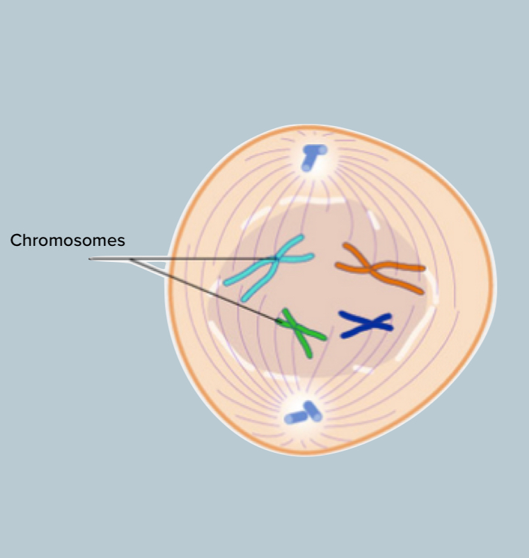
Fourth phase of cell division
Metaphase

Metaphase
Cell’s chromosomes move together, align in the center of cell
Nucleus has dissolved
“Lined up at the 50-50 line”

Fifth phase of cell division
Anaphase
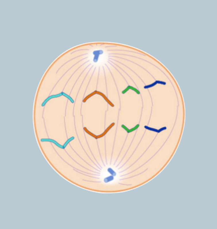
Anaphase
Chromosomes separate and move together opposite ends
Daughter chromosomes being pulled apart

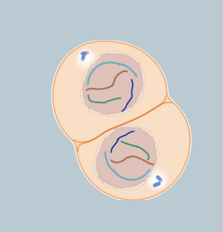
Sixth stage of cell division
Telophase

Telophase
Two nuclear membranes form
Separate the cell into two nuclei
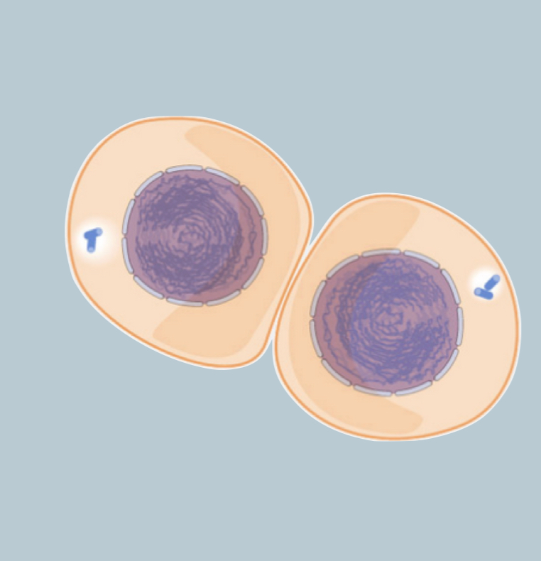
What is the result of cell division
2 daughter cells with 46 chromosomes each
How do cells stick together to form an organ, and further, a system?
Intracellular Junctions
Intracellular Junctions
Helps cells stick together to form an organ and/or system
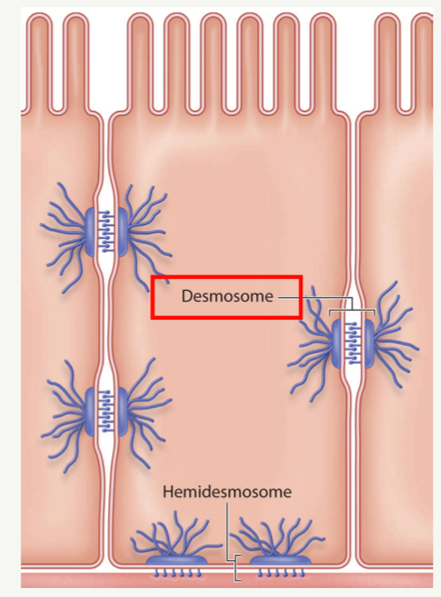
What are the 2 Intracellular Junctions
Desmosomes
Hemidesmosomes
Hemidesmosome
Attachment of a cell to an adjacent noncellular surface like a basement membrane
How are hemidesmosomes involved in the oral cavity
Allows gingival tissue to be secured to the tooth surface by the epithelium attachment
Desmosome
Strong connective junctions between cells
“Glue” to hold cells together
How are desmosomes involved in the oral cavity
In areas exposed to significant stress like the gum line and inner cheek lining where cell turnover is high
Where are desmosomes located in the oral cavity
Gum line
inner cheek lining
Areas where cell turnover is high
Occluding or Tight Junctions
Holds cells tightly together so that space between is impermeable and large molecules cannot enter
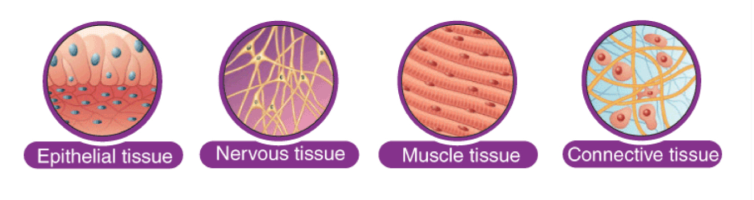
What are the 4 basic tissue types
Epithelial Tissue
Nervous tissue
Muscle tissue
Connective tissue
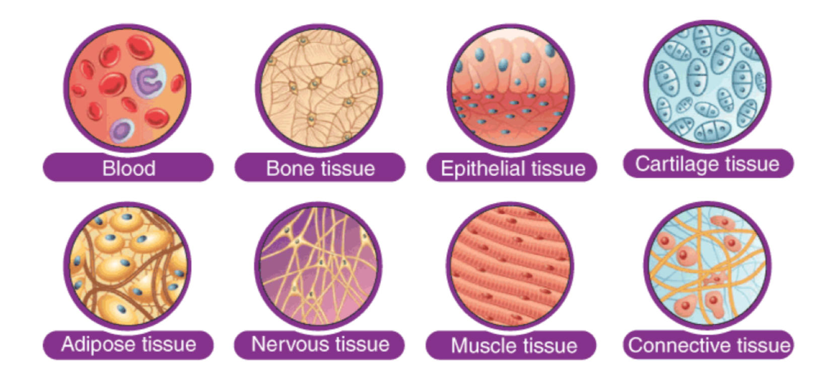
Types of animal/human tissue
Blood
Bone tissue
Epithelial tissue
Cartilage tissue
Adipose tissue
Nervous tissue
Muscle tissue
Connective tissue
Does enamel have regenerative properties
NO
Cell Regeneration
Individual cells die and new ones takes their place
In what age group does cell regeneration occur in more?
Children opposed to older people
How many phases of cell regeneration is there?
4
What are the 4 phases of cell regeneration?
Hemostasis phase
Inflammatory phase
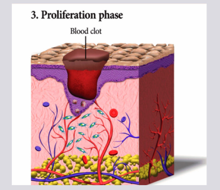
Proliferation phase (blood clot)
Remodeling phase (scab)
What is the first phase of cell regeneration
Hemostasis phase
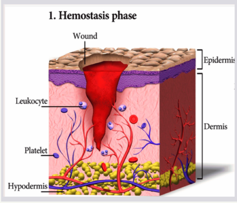
Hemostasis phase
Vasoconstriction; blood vessels constrict to help clotting
Platelets arrive
Leukocytes (WBCs) arrive to fight off infection

What first happens during the hemostasis phase
Vasoconstriction for clotting
What happens secondly during the hemostasis phase
Platelets arrive
What happens thirdly during the hemostasis phase
Leukocytes (WBCs) arrive to fight off infection
What leukocytes are present during the hemostasis phase
Neutrophils and macrophages
What do neutrophils and macrophages do during the hemostasis phase
Cleans the wound site from dead cells, bacteria, and other pathogens or debris
Hemostasis phase basic rundown
Vasoconstriction → Platelets → WBC (leukocytes)
What is the second phase of cell regeneration
Inflammatory phase
Inflammatory phase
Neutrophils arrive first to phagocytose/engulf damaged cells
Clear oozing substance due to damaged blood vessels
Macrophages arrive and phagocytose damaged cells, debris, foreign invaders including the neutrophils
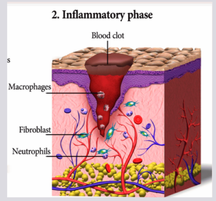
What arrives FIRST during the inflammatory phase
Neutrophils
What do neutrophils do during the inflammatory phase
They release antimicrobial substances
What causes the oozing clear substance mean during the inflammatory phase
Due to damaged blood vessels
What do macrophages do during the inflammatory phase
Phagocytose damaged cells, debris, foreign invaders
Do neutrophils have a short or long lifespan
Short lifespan
How long do neutrophils live
Only a few days
Do macrophages have a short or long lifespan
Long lifespan
How long do macrophages live
Months or even years
Is the first line of defense neutrophils or macrophages
Neutrophils
Is the second line of defense neutrophils or macrophages
Macrophages
What is the third phase of cell regeneration?
Proliferation phase

Proliferation Phase
Granulation tissue is forming and replaces damaged tissue
Rebuilding via growth of blood vessels and the migration of fibroblasts which produce collagen
Macrophages are still cleaning up debris
The wound is typically pink at this point
What is the fourth phase of cell regeneration
Remodeling phase
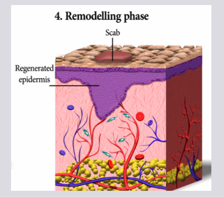
Remodeling phase
Collagen becomes more organized
Vascular regression
Fibroblasts transform into myoflibroblasts which contribute to scar tissue formation

What is granulation tissue
Newly forming connective tissue through proliferation of fibroblasts and capillaries to wound site


What is common in areas with granulation tissue
Commonly sore and bleeds easily

What does the epithelium tissue cover/line
Both the external and internal body surfaces (vessels and small cavities)
What is the epithelium tissue also involved in BESIDES being a protective covering or lining
Involved in tissue absorption, secretion, sensory, other specialized functions
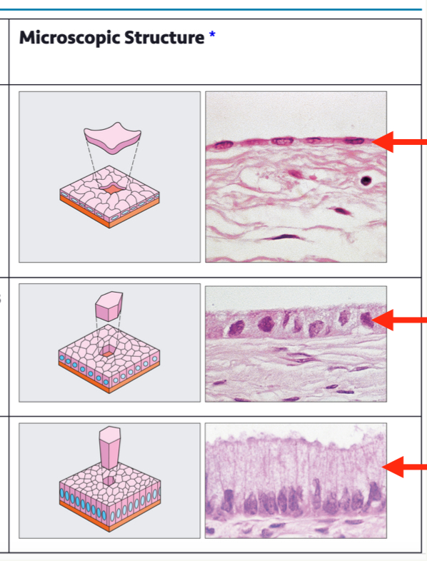
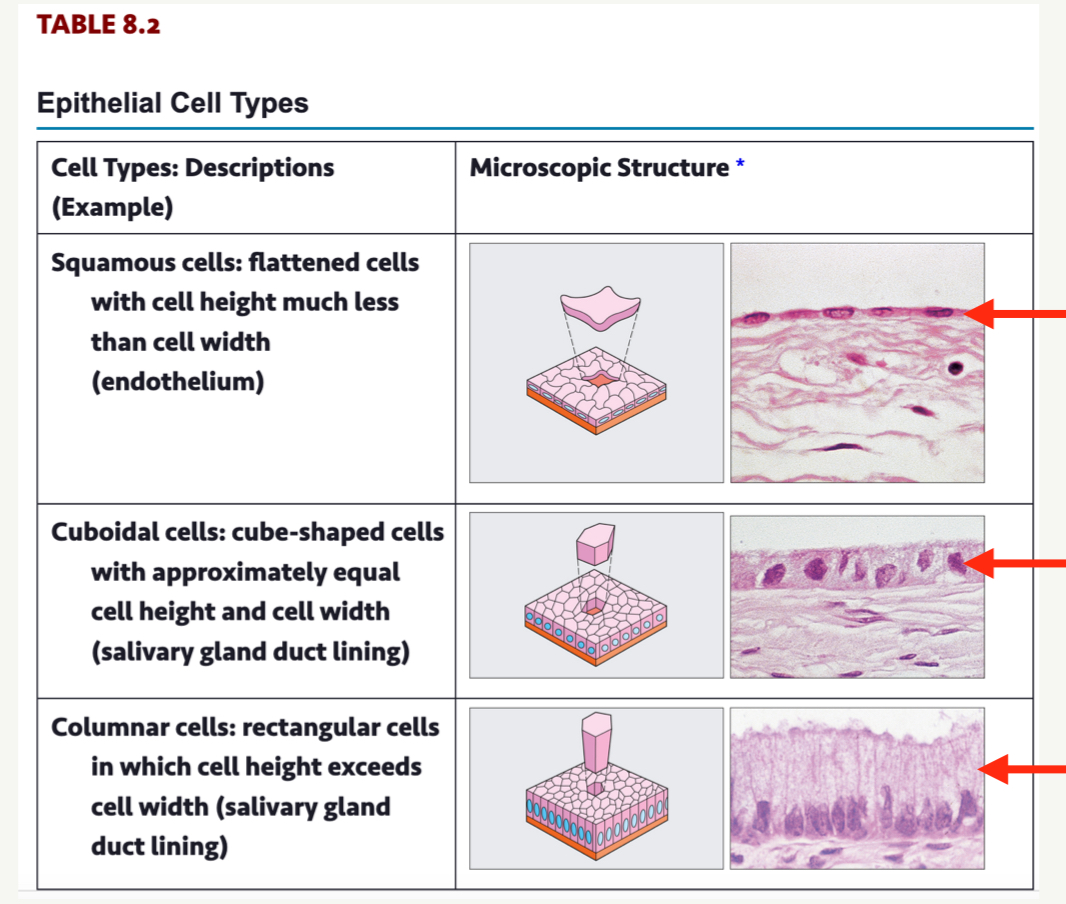
What are three epithelial cell types?
Squamous cells
Cuboidal cells
Columnar cells
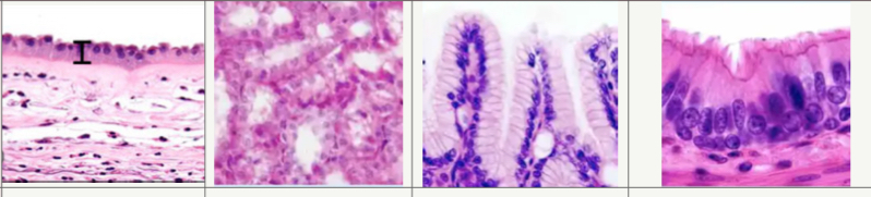
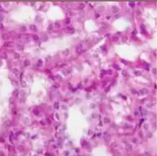
What are 4 types of simple epithelial tissue (non-keratinized)
Simple squamous epithelium
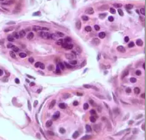
Simple cuboidal epithelium
Simple columnar epithelium
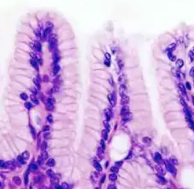
Pseudostratified epithelium
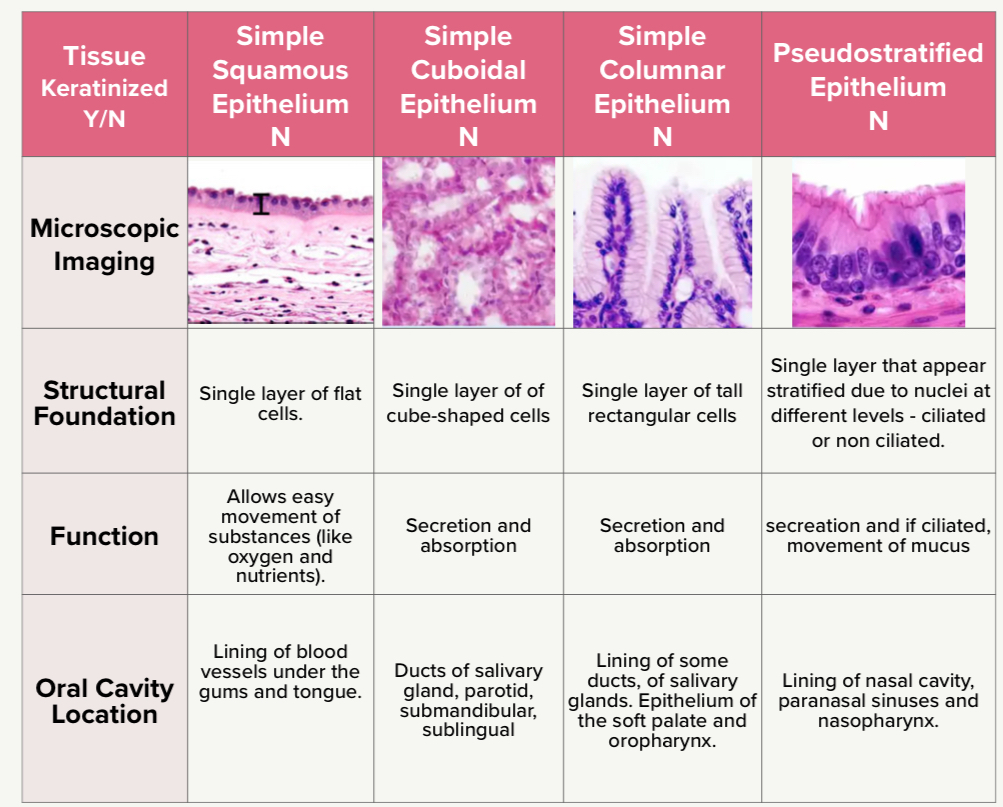

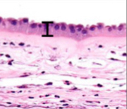
What is the structural foundation of simple squamous epithelium
Single layer of flat cells
What is the structural foundation of simple cuboidal epithelium
Single layer of cube-shaped cells

What is the structural foundation of simple columnar epithelium
Single layer of tall rectangular cells

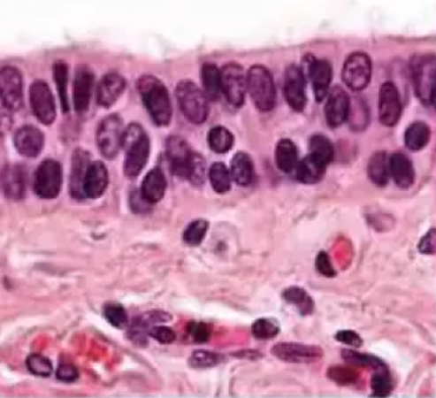
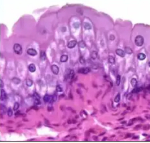
What is the structural foundation of Pseudostratified epithelium
Single layer that appear stratified due to nuclei at different levels - ciliated or non ciliated
Function of simple squamous epithelium
Allows easy movement of substances (like oxygen and nutrients)
Function of simple cuboidal epithelium
Secretion and absorption
Function of simple columnar epithelium
Secretion and adsorption
Function of Pseudostratified epithelium
Secretion and if ciliated, movement of mucus
Oral cavity location of simple squamous epithelium
Lining of blood vessels under the gums and tongue
Oral cavity location of Simple cuboidal epithelium
Ducts of salivary gland, parotid, submandibular, sublingual
Oral cavity location of Simple columnar epithelium
Lining of some ducts of salivary glands. Epithelium of the soft palate and oropharynx
Oral cavity location of Pseudostratified epithelium
Lining of nasal cavity, paranasal sinuses and nasopharynx
What are 4 types of stratified epithelial tissue
Stratified squamous
Stratified cuboidal
Stratified columnar epithelium
Transitional
What is the structural function of ALL stratified epithelial tissue
All multiple layers of cells
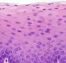
Function of stratified squamous
Provides protection against friction, bacteria, and damage


Function of stratified cuboidal
Provides protection against friction, bacteria, and damage

Function of stratified columnar epithelium
Provides protection against friction, bacteria, and damage
Function of transitional epithelial tissue
Protective barrier/normally in urinary tract

Oral cavity location of stratified squamous
Mucosa buccaneers, labial, floor of mouth, soft palate. Keratinized and non-keratinized
Oral cavity location of stratified cuboidal
Salivary ducts; parotid, submandibular, sublingual
Oral cavity location of stratified columnar epithelium
Covers the tongue, cheeks, gums, roof of mouth to protect against chewing and food abrasion
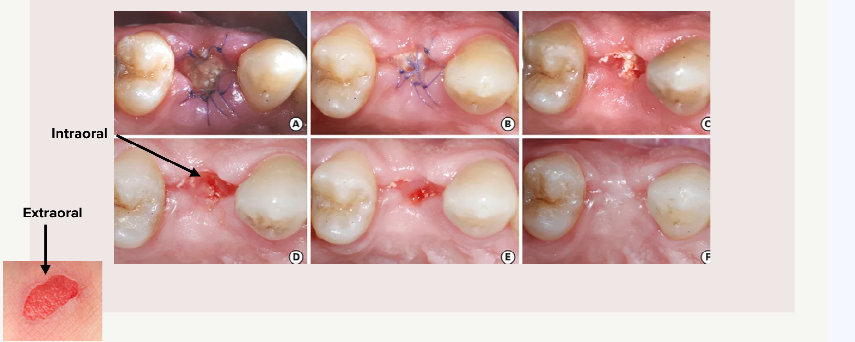
What is going to heal faster? A laceration in the oral cavity, or an abrasion on the knee?
A laceration in the oral cavity
Is cell turnover time high or low in the oral cavity?
High cell turnover time