Brain Anatomy
1/61
There's no tags or description
Looks like no tags are added yet.
Name | Mastery | Learn | Test | Matching | Spaced | Call with Kai |
|---|
No analytics yet
Send a link to your students to track their progress
62 Terms
What structures is the human brain made up of?
Neurones - responsible for electrical signalling
Glial cells - support, protect and nourish neurones
Blood vessels - supply oxygen and nutrients
Which structures protect the brain?
Skull
Hard bony structure
Protects the brain from physical injury
Forms a cavity that houses the brain
Meninges
Three protective membranes:
Dura mater (outer, tough)
Arachnoid mater (middle, web-like)
Pia mater (inner, closely attached to brain tissue)
Cerebrospinal fluid (CSF)
Produced in the choroid plexuses of brain ventricles
Formed by filtration of blood plasma
Cushions the brain, absorbs shock, and helps remove waste
Blood–brain barrier (BBB)
Formed by tight junctions between endothelial cells in brain capillaries
Highly selective
Protects the brain from toxins and pathogens while allowing essential substances through
Which region of the brain is especially important for neurological and psychiatric disorders and drug actions?
The brainstem
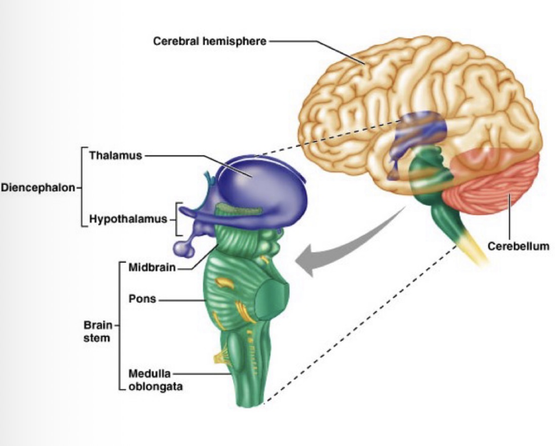
Brainstem
What is the brainstem?
Connects the brain to the spinal cord
3 main parts:
Medulla oblongata
Pons
Midbrain
Brainstem
Medulla oblongata
Lowest part of the brain and brainstem
Connects:
Anteriorly to the pons
Posteriorly to the spinal cord
Merges with the spinal cord at the foramen magnum
Functions:
Controls autonomic nervous system activity:
Breathing (respiration)
Heart rate
Blood pressure
Digestion
Other roles:
Control of movement
Relaying sensory information from internal organs
Regulation of arousal and sleep

Brainstem
Pons
Middle part of the brainstem
Located between the medulla oblongata and the midbrain
Connects to:
Cerebellum
Cerebral cortex
Functions:
Acts as a bridge between different parts of the brain (including the cerebellum and the cerebral cortex)
Gives rise to important cranial nerves:
Trigeminal nerve: allows sensation in the face, controls chewing and swallowing
Facial nerve: controls facial expressions
Contains respiratory nuclei which regulate the depth and frequency of breathing
Contains nuclei which contribute to ‘slow neurotransmitter systems’ which regulates brain activity (such as alertness and mood)

Brainstem
Midbrain
Functions:
Vision and hearing reflexes
Motor control
Sleep–wake cycles
Arousal (alertness)
Temperature regulation
It contains important dopamine-producing nuclei which supply the rest of the brain with dopamine:
Substantia Nigra
Ventral Tegmental Area (VTA)

Brainstem
What is the function of dopamine that is supplied from the midbrain?
Neurotransmitter
Dopamine is essential for:
Movement (basal ganglia function)
Motivation
Habit formation
Brainstem
Why is damage to the brainstem (pons or midbrain) dangerous?
Possible effects of damage to the brainstem?
Damage to the brainstem (pons or midbrain) can be life-threatening because it controls vital functions
Possible effects:
Sleep disorders
Balance and movement problems
Organ failure
Death

Cerebellum
What is the cerebellum?
The cerebellum sits beneath the cerebral cortex and is primarily responsible for motor coordination
Cerebellum
Structure
Two hemispheres - posterior lobe and anterior lobe
A midline structure called the vermis
Cerebellar cortex - tightly folded layer of cells at the outer surface
White matter underneath the cerebellar cortex composed of nerve fibres and cerebellar nuclei
A ventricle at its base

Cerebellum
Key roles
Coordination of movement
Precision and timing
Motor learning
It fine-tunes movements to make them smooth and accurate
It contributes to:
Attention
Language
Emotional regulation (fear and pleasure)
It receives sensory input from:
The spinal cord
Other brain regions
Cerebellum
What can damage to the cerebellum cause?
Tremors
Loss of coordination (ataxia)
Difficulty walking
Dizziness
Slurred or impaired speech
Cerebellum
Cognitive diseases associated with the cerebellum
autism, schizophrenia, and dyslexia
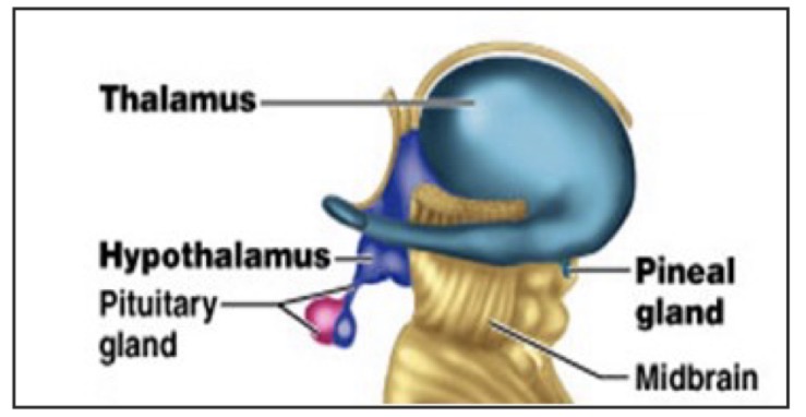
Diencephalon
Where is it located?
What are the 4 parts?
The diencephalon lies between the brainstem and the cerebral cortex.
It includes four major parts:
Thalamus
Subthalamus
Hypothalamus
Epithalamus


Diencephalon
Thalamus
Consists of two large, symmetrical lobes and acts as the brain’s main sensory relay station.
Nearly all sensory information (except smell) passes through the thalamus before reaching the cerebral cortex
Each sensory system (except the olfactory system) has a specific thalamic nucleus that receives signals and sends them to the appropriate primary sensory cortex.
The thalamus has strong feedback connections (2-way communication) with the cerebral cortex which allows it to:
Process sensory info as well as relay it
Play an Important role in consciousness, sleep and wakefulness » strong feedback connections with the cerebral cortex, forms thalamo–cortico–thalamo circuits which are involved in consciousness
Diencephalon
Thalamus
What can damage to the thalamus result in?
Coma
Amnesia
Impaired sensory processing
Movement and posture problems
Pain
Dementia
Excessive sleepiness
Diencephalon
Thalamus
Which cognitive disorders are linked to the thalamus?
Bipolar disorder
ADHD
Autism
Depression
Alzheimer’s disease
Diencephalon
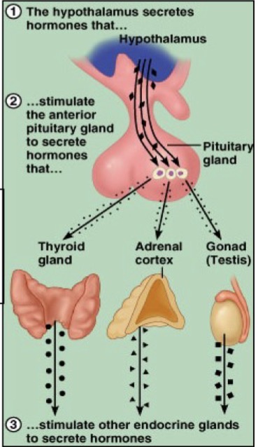
Hypothalamus
Controls many unconscious and hormonal functions of the body:
It regulates the pituitary gland, which means it indirectly controls most hormones
It contains many small nuclei that produce hormones affecting body functions
Its primary role is homeostasis—maintaining stable internal conditions.
Functions include regulation of:
Body temperature
Hunger and thirst
Instinctive behavior
Fatigue and sleep
Circadian rhythms
Stress responses
Diencephalon
Hypothalamus
What controls the release of hormones by the pituitary gland?
The hypothalamus secretes hormones
Which stimulates the pituitary gland to secrete hormones
Which stimulates other endocrine glands to secrete hormones
Diencephalon
Hypothalamus
What can damage to the hypothalamus cause?
Aggression
Hypothermia
Excessive sleep (hypersomnia)
Lethargy
Weight gain or loss
Chronic stress
Diencephalon
Hypothalamus
Which disorders are linked to the hypothalamus?
Depression
Bipolar disorder
Schizophrenia
Hormonal diseases

Diencephalon
Epithalamus
The epithalamus is a dorsal, posterior part of the diencephalon.
It includes several small nuclei and the pineal gland
It is connected to both the limbic system and basal ganglia.
Its primary function is the secretion of melatonin by the pineal gland
Diencephalon
Epithalamus
Function of melatonin
Regulates the circadian rhythm, especially the sleep–wake cycle
Diencephalon
Epithalamus
What can damage to the pineal gland cause?
Disrupted sleep patterns
Disturbed circadian rhythm
Altered pituitary hormone secretion
Diencephalon
Epithalamus
Are any cognitive disorders associated with the epithalamus?
no

Diencephalon
Subthalamus
The subthalamus is a small, ventral anterior part of the diencephalon.
Its main structure is the subthalamic nucleus
It is functionally connected to the basal ganglia.
It plays an important role in motor control
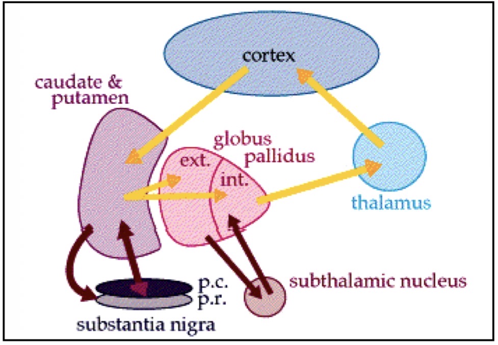
Basal Ganglia
What are the basal ganglia?
A group of interconnected nuclei located deep at the base of the forebrain. They work together to regulate:
Voluntary movement
Procedural learning (learning skills and habits)
Routine behaviors
Eye movements
Cognition and emotion
Rather than directly causing movement, the basal ganglia help select and control appropriate movements.

Basal Ganglia
Basal Ganglia Loop
The basal ganglia form a loop system with the cerebral cortex and thalamus:
Information from the cerebral cortex (especially frontal, prefrontal, and parietal areas) is sent to the striatum.
The basal ganglia process this information.
Signals are sent to the thalamus.
The thalamus sends information back to the cerebral cortex.
This loop helps the brain choose the correct voluntary movement and suppress inappropriate ones
Basal Ganglia
In addition to being part of the basal ganglia loop, what is the striatum also involved in?
The striatum is also involved in processing:
Rewarding stimuli: things that feel good (like food, success, or praise) and supports learning to repeat those behaviours
Aversive (unpleasant) stimuli: negative or uncomfortable experiences (like pain or punishment), helping you learn to avoid them in the future
New or unexpected stimuli: when something is unfamiliar or surprising, helping the brain pay attention and learn from new situations
Basal Ganglia
What can damage to the basal ganglia cause?
Tremors
Involuntary muscle movement
Abnormal increase in muscle tone
Abnormal posture
Difficulty initiating movement
Basal Ganglia
Which movement conditions are linked with the basal ganglia?
Parkinson’s disease
Huntington’s disease
Dyskinesias
Basal Ganglia
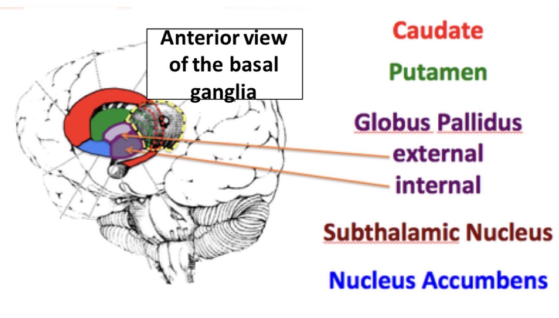
Main components of the basal ganglia
Striatum
Globus Pallidus
Substantia Nigra
Subthalmic Nucleus (STN)
Nucleus Accumbens
Basal Ganglia
Nucleus Accumbens
Plays a key role in the brain’s reward system.
It helps produce feelings of pleasure and motivation
It reinforces behaviors by making them feel rewarding
It is strongly involved in the effects of psychoactive drugs and drugs of abuse
Basal Ganglia
Which cognitive disorders are linked to the basal ganglia?
Damage to which part of the basal ganglia is likely to cause these conditions?
Addiction
Depression
Schizophrenia
» caused by damage to the nucleus accumbens
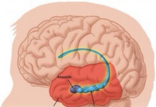
Hippocampus
Small region in the medial temporal lobe of the cerebral cortex
Belongs to the limbic system
Function: involved in the consolidation of information from short-term memory to long-term memory and spatial navigation
Hippocampus
What can damage to the hippocampus cause?
Memory impairment
Disorientation
Hippocampus
Cognitive disorders linked to the hippocampus
Alzheimer’s disease
Depression
Bipolar disorders
Schizophrenia
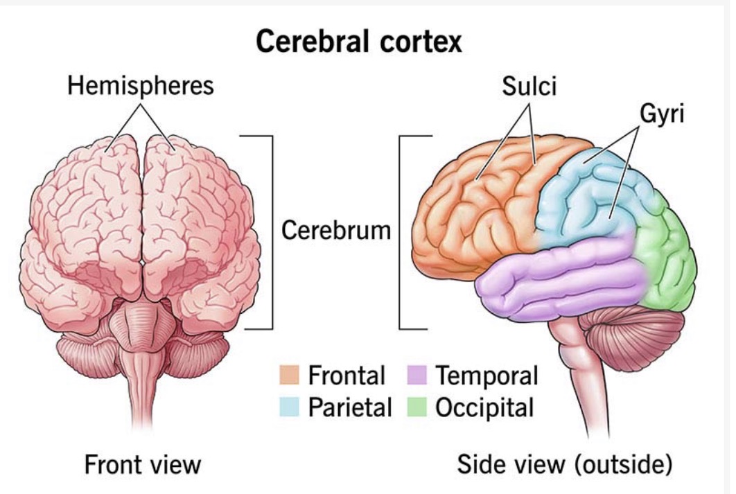
Cerebral Cortex
What is it?
Function?
Which 2 parts is it divided into?
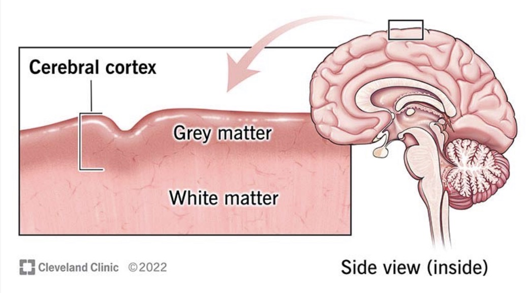
The cerebral cortex is the outer layer of the brain where complex information processing occurs
It is responsible for perception, thought, memory, language, and voluntary movement
The cerebral cortex is divided into two main parts:
Neocortex – the largest part
Allocortex – a smaller, older part

Cerebral Cortex
Which areas of the brain are the primary sites of seizures and epilepsy?
Neocortex and Allocortex in the cerebral cortex
Cerebral Cortex
Neocortex
Contains six distinct layers of neurons
Develops later and is fully formed in the mature human brain
Divided into lobes, each with specific functions and connected to other brain regions
Responsible for conscious experience, complex thinking, planning, and memory
Cerebral Cortex
Allocortex
Has fewer than six layers of neurons
Includes structures such as:
Olfactory cortex
Hippocampus
Involved in smell and memory
Cerebral Cortex
Name the 4 lobes of the neocortex
Frontal lobe
Parietal lobe
Temporal lobe
Occipital lobe


Cerebral Cortex
Frontal lobe
Where is it located?
Main regions and their functions
What can damage to the frontal lobe cause?
Disorders linked to the frontal lobe
Located at the front of the cerebral cortex
Main regions:
Prefrontal Cortex – personality, decision-making, planning complex behaviours
Premotor Area – planning movements
Motor Area – executing voluntary movements
Damage to the frontal lobe can cause:
Impaired movement and motor learning
Speech difficulties
Distorted body image
Associated disorders:
Autism
ADHD
Depression
Bipolar disorder
Some forms of schizophrenia

Cerebral Cortex
Parietal lobe
Functions
Which area does it contain?
What can damage to the parietal lobe cause?
Disorders linked to the parietal lobe
Functions:
Processes sensory information from: Skin, Muscles, Joints, Internal organs
It also plays a role in:
Spatial awareness
Visuospatial processing
It contains the Somatosensory Area, which receives information from the somatosensory thalamus.
Damage to the parietal lobe may result in:
Difficulty recognizing or locating objects
Problems identifying body parts or events
Associated disorders:
ADHD
Alzheimer’s disease
Schizophrenia
Cerebral Cortex
Temporal lobe
Functions
Which area does it contain?
What can damage to the temporal lobe cause?
Disorders linked to the temporal lobe
Functions:
The temporal lobe is involved in: Hearing, Language, Learning, Memory
It contains the Auditory Area, which processes sound information received from the auditory thalamus (originating from the ear).
Damage to the temporal lobe can affect:
Recognition
Language comprehension
Memory formation
Associated disorders:
Schizophrenia
Early Alzheimer’s disease
Autism

Cerebral Cortex
Occipital lobe
Functions
Where does it receive input from?
What can damage to the occipital lobe cause?
Function: Primarily responsible for vision.
It contains the Visual Cortex which receives visual input from the visual thalamus, which gets information from the retina of the eye
Damage to the occipital lobe can cause:
Hallucinations
Blindness
Inability to distinguish colours
Associated disorders:
Schizophrenia
Bipolar disorders
Autism
Depression
Cerebral Cortex
What are the Association Areas of the cerebral cortex?
Regions that are not primary sensory regions (do not receive direct input from the sensory thalamus) and are not primary motor regions (do not directly control muscle movement)
Instead, they integrate and coordinate information from many areas of the cortex and other brain regions
Cerebral Cortex
Examples of functions that the association areas coordinate and which lobes these functions originate from
Frontal lobe → Motivation
Premotor cortex (part of the frontal lobe) → Planning movements before carrying them out
Parietal lobe → Understanding the relationship between our body and the world around us
Temporal lobe → Formation of episodic memories (memories about events and places)
Cerebral Cortex
What is cerebral white matter?
Allows communication between different parts of the brain
This communication occurs via myelinated axon fibres bundled up into:
Tracts - connect cortex with brainstem and spinal cord
Comissures - connects left and right hemispheres
Association fibers - connects areas within the same hemisphere
Projection fibers - connects the cortex with the lower brain regions vertically
Cerebral Cortex
Cerebral White Matter
Example of a comissures
Corpus callosum - connects left and right hemispheres

Cerebral Cortex
Cerebral Grey Matter
Made up of:
Cerebral cortex
Basal ganglia nuclei
Base forebrain nuclei
Other nuclei (clusters of neuronal cell bodies)
Limbic System
The limbic system is a network of interconnected brain regions that are especially important for emotions and feelings
These interconnected brain regions include:
Cingulate gyrus (part of the cortex)
Amygdala
Hippocampus
Nucleus accumbens
How is movement controlled in the brain?
Movement is controlled by a network of brain regions, not a single area:
Motor cortex (precentral gyrus) – initiates voluntary movement
Basal ganglia – selects and regulates movements
Cerebellum – coordinates and fine-tunes movements
These regions send signals to:
Motoneurons in the spinal cord → move limbs and trunk
Motor nuclei in the brainstem → control head, neck, eye movements, speech, and swallowing
Motoneurons then activate muscle contraction
What allows fast and precise voluntary movements?
The corticospinal tract
Corticospinal Tract
A pathway carrying motor commands from the cortex to the spinal cord
It passes through the pyramidal tracts in the brainstem
Just before entering the spinal cord, most fibers cross over (decussate):
Left motor cortex controls the right side of body
Right motor cortex controls the left side of body
Allows fast, precise movements to occur
What is the fastest descending motor pathway?
The corticospinal (pyramidal) tract
What 2 main classes of neurones does the cerebral cortex contain?
Projection neurons
Send signals to distant brain regions
E.g. pyramidal neurons
Mostly excitatory
Use glutamate (glutamatergic cells)
Interneurons
Usually act locally to influence nearby neurons
Some interneurones have extensive axonal projections so can control the activity of larger groups of neurones = strong synchronisation of activity
E.g. basket cells
Mostly inhibitory
Use GABA (GABAergic cells)
Which classes of glial cells does the cerebral cortex contain?
Glia support and protect neurones:
Astrocytes
Regulate the chemical environment of the neurone
Oligodendrocytes
Form myelin around axons to speed up conduction of action potentials
Microglia
Primary immune cells of the CNS