Human Anatomy and Physiology Overview
1/145
There's no tags or description
Looks like no tags are added yet.
Name | Mastery | Learn | Test | Matching | Spaced | Call with Kai |
|---|
No analytics yet
Send a link to your students to track their progress
146 Terms
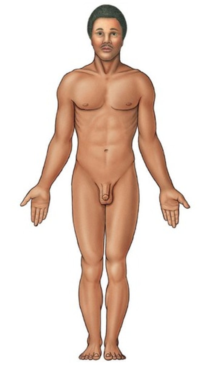
What is the anatomical position used for?
It serves as a reference point when referring to specific areas of the human body.
Describe the anatomical position.
The body is erect, with the head and toes pointing forward, and arms hanging downwards with palms facing forward.
What does surface anatomy refer to?
It refers to visible surface landmarks that can be used for the study of the human body and for unambiguous location.
What are the two divisions of surface anatomy?
Axial areas (head, neck, trunk) and appendicular areas (limbs and their attachments).
How can body landmarks be further categorized?
They can be categorized into anterior and posterior landmarks.
Why is terminology important in anatomy?
Understanding terminology is crucial for accurately describing body structures and locations.
What does the term 'anterior' refer to in surface anatomy?
Anterior refers to the front surface of the body.
What does the term 'posterior' refer to in surface anatomy?
Posterior refers to the back surface of the body.
What does the suffix '-al' or '-ar' indicate in anatomical terms?
It indicates that the term belongs to a specific region.
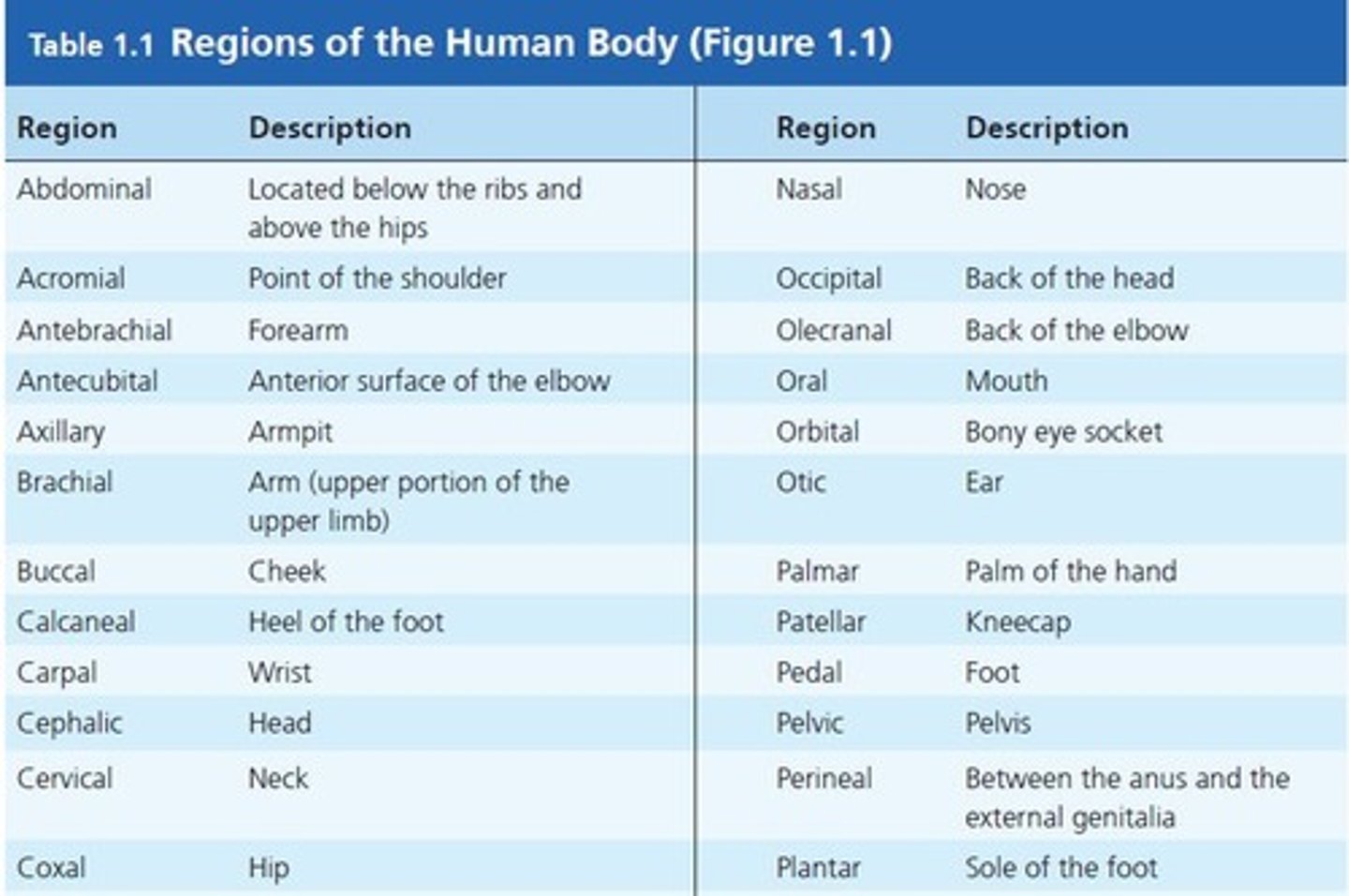
What is the difference between 'pedal' and 'plantar'?
Pedal refers to the whole foot, while plantar refers specifically to the bottom of the foot.
What does 'thoracic' refer to?
Thoracic refers to the whole chest area.
What are the pairs of directional terminology used to locate body structures?
Superior vs Inferior, Cranial vs Caudal, Anterior vs Posterior, Ventral vs Dorsal, Lateral vs Medial, Proximal vs Distal, Superficial vs Deep.

What does 'superior' mean in directional terminology?
It means towards the top of the body.
What does 'inferior' mean in directional terminology?
It means towards the bottom of the body.
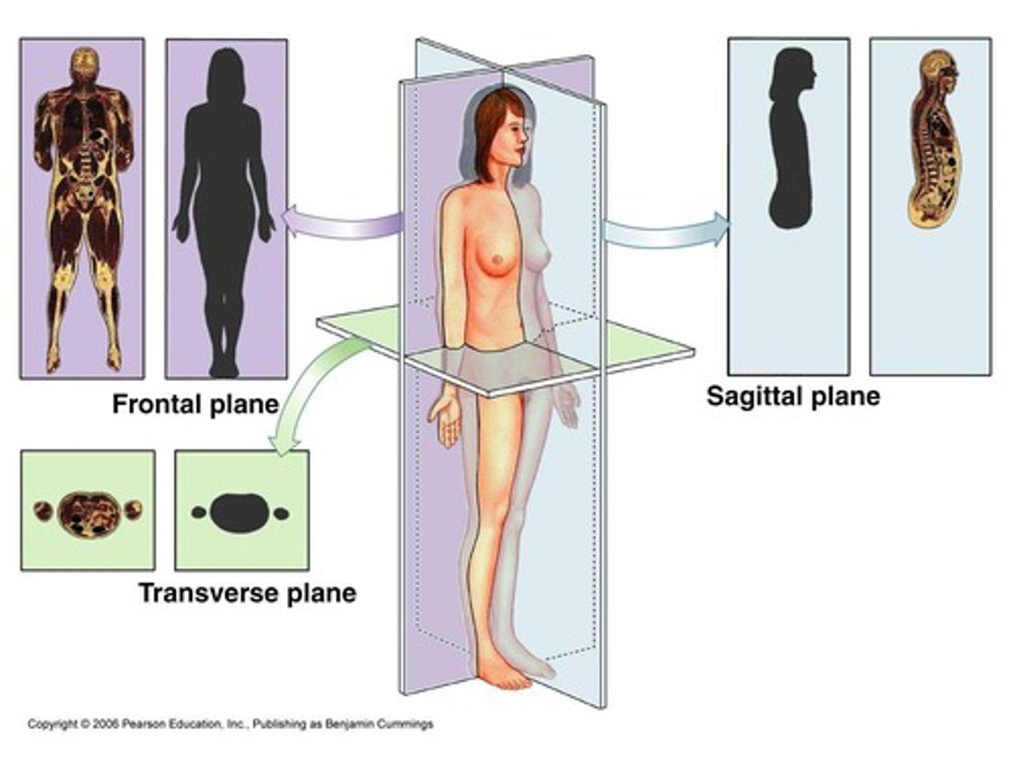
What is the sagittal plane?
A longitudinal section that divides the body into left and right sides.
What is the midsagittal plane?
It is the median plane that divides the body into equal mirror images.
What is the frontal (coronal) plane?
A longitudinal plane that divides the body into anterior and posterior parts.
What is the transverse plane?
A horizontal plane that divides the body into superior and inferior parts.
What does an oblique cut refer to?
A cut made at an angle.
What should you keep in mind when viewing histological slides or imaging scans?
Objects can look odd when viewed in section.
What is the significance of understanding body sections in anatomy?
It helps in visualizing and understanding the spatial relationships between different body structures.
What is the difference between medial and median?
Medial refers to being closer to the midline of the body, while median refers to the middle value or position.
How do you describe the pollex in relation to the pinky when in anatomical position?
The pollex is lateral to the pinky.
How many digits are there in fingers II to V?
There are four digits (II, III, IV, V) in fingers II to V.
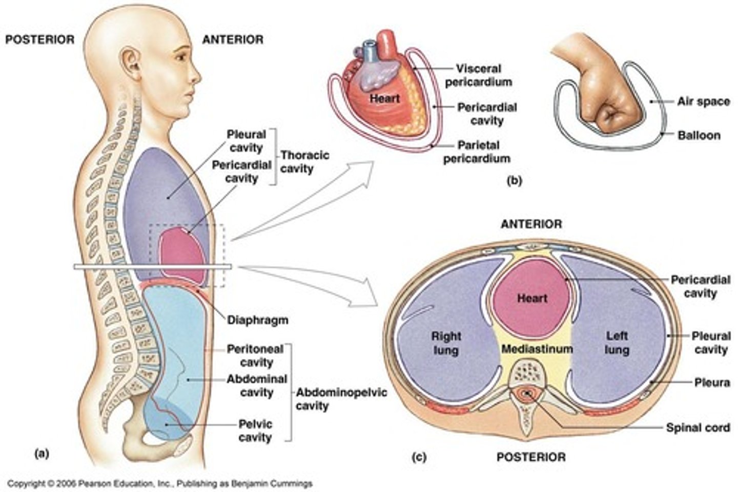
What are the two main body cavities in the axial body portion?
Dorsal body cavity and Ventral body cavity.
What does the dorsal body cavity consist of?
Cranial cavity and Spinal cavity.
What are the components of the ventral body cavity?
Thoracic cavity (above diaphragm) and Abdominopelvic cavity (below diaphragm), which includes the Abdominal cavity and Pelvic cavity.
What organs are contained within the cranial cavity?
The brain.
What organs are found in the thoracic cavity?
The heart and lungs.
What does the spinal cavity contain?
The spinal cord.
What is the function of the diaphragm in relation to body cavities?
It separates the thoracic cavity from the abdominopelvic cavity.
What are serosa membranes?
Thin double-layered membranes that cover the internal walls of the ventral body cavity and the organs within it.
What are the two layers of serosa membranes called?
Visceral serosa layer (touching the organs) and parietal serosa layer (not touching the organs).
What is found between the visceral and parietal serosa layers?
Serosa fluid.
What is the name of the serosa membrane around the heart?
Pericardium.
What is the name of the serosa membrane around the lungs?
Pleura.
What is the name of the serosa lining the abdominal cavity?
Peritoneum.
What are mucosa membranes?
Membranes lining the inside openings of organs that are not considered body cavities.
What is a lumen?
A generic name for an opening of a tube-like structure.
How are the abdominopelvic area subdivided for medical purposes?
Into quadrants and regions.
What are the four quadrants of the abdominopelvic area?
Right upper quadrant, left upper quadrant, right lower quadrant, and left lower quadrant.
How many regions are used to subdivide the abdominopelvic area?
Nine regions.
What is the epigastric region?
The region located above the stomach.
What organs are located in the left hypochondriac region?
Spleen and part of the stomach.
What is the hypogastric region?
The region located below the stomach.
What are some examples of smaller body cavities?
Oral cavity, nasal cavities, orbital cavities, sinuses, middle ear cavities, and synovial (joint) cavities.
What is the function of synovial cavities?
They are found in joints and contain synovial fluid to reduce friction.
What are body membranes also known as?
Body system membranes or tissue membranes.
What is the primary function of body membranes?
To provide protective sheets around organs.
What are the two main types of body membranes?
Epithelial membranes and connective tissue membranes.
What are the three kinds of epithelial membranes?
Mucous membranes, serous membranes, and cutaneous membranes.
What do mucous membranes line?
The lumen of internal organs that have an opening to the outside of the body.
What type of connective tissue is typically found in mucous membranes?
Areolar connective tissue, referred to as the lamina propria.
What is the function of mucous membranes?
To stop pathogens and dirt from entering the body and to prevent bodily tissues from becoming dehydrated.
What do serous membranes line?
The outside of internal organs.
What type of epithelial tissue is found in serous membranes?
Simple squamous epithelium or mesothelium.
What are the two layers of serous membranes?
Visceral layer (touches the organ) and parietal layer (does not touch the organ).
What fills the space between the visceral and parietal layers of serous membranes?
Serous fluid that prevents friction.
What is the pericardium?
A two-layered serous membrane sac that surrounds the heart.
What is the pleura?
The serous membrane that surrounds the lungs in the pleural cavity.
What is the peritoneum?
The serous membrane that surrounds several organs in the abdominopelvic cavity.
What is the tunica vaginalis?
The serous membrane that surrounds the male gonad, the testis.
What is the cutaneous membrane?
The skin, consisting of the epidermis and dermis.
What type of epithelium is found in the epidermis of the cutaneous membrane?
Keratinized stratified squamous epithelium.
What type of connective tissue is primarily found in the dermis of the cutaneous membrane?
Dense irregular connective tissue, with the top region being areolar connective tissue.
What are synovial membranes?
Membranes that line the cavities where moveable joints between two bones are formed.
What is the function of synovial membranes?
To provide a smooth surface and secrete a fluid that cushions impact.
What are meninges?
Layers of connective tissue that surround the brain and spinal cord.
What is the Integumentary System?
The Integumentary System is the skin and its associated structures.
What are the two major regions of the Integument?
The two major regions are the Epidermis and the Dermis.
What is the Hypodermis, and how is it related to the skin?
The Hypodermis, also called superficial fascia, is the deeper region located below the dermis and is not part of the skin but is always discussed with it.
What does the prefix 'Epi-' in Epidermis mean?
The prefix 'Epi-' means 'on top', indicating that the epidermis is the layer on top of the dermis.
What type of epithelium composes the Epidermis?
The Epidermis is composed of keratinized stratified squamous epithelium.
What are the four distinct cell types found in the Epidermis?
The four distinct cell types are keratinocytes, melanocytes, Merkel cells, and Langerhans cells.
What is the primary function of the Epidermis?
The primary function of the Epidermis is protection, as it is exposed to the external environment.
What is the Stratum Basale?
The Stratum Basale is the deepest epidermal layer, firmly attached to the basal lamina and dermis, consisting of a single row of the youngest keratinocytes.
What occurs in the Stratum Basale?
Cells undergo rapid division, pushing upwards to form new cells.
What is the significance of damage to the Stratum Basale?
Damage to the Stratum Basale interferes with the production of new epidermal layers.
What characterizes the Stratum Spinosum?
The Stratum Spinosum contains cells with a weblike system of intermediate filaments attached to desmosomes, and is abundant in melanin granules and Langerhans' cells.
What happens in the Stratum Granulosum?
In the Stratum Granulosum, drastic changes in keratinocyte appearance occur, with kerato-hyaline and lamellated granules accumulating to waterproof the skin.
What is the Stratum Lucidum, and where is it found?
The Stratum Lucidum is a thin, transparent band present only in thick skin, consisting of a few rows of flat, dead keratinocytes.
What is the Stratum Corneum?
The Stratum Corneum is the outermost layer of keratinized cells, accounting for three quarters of the epidermal thickness.
What are the functions of the Stratum Corneum?
The functions include waterproofing, protection from abrasion and penetration, and rendering the body relatively insensitive to biological, chemical, and physical assaults.
What is the Dermis?
The Dermis is the second major skin region containing strong, flexible connective tissue.
What cell types are found in the Dermis?
Cell types in the Dermis include fibroblasts, macrophages, and occasionally mast cells and white blood cells.
What are the two layers of the Dermis?
The two layers of the Dermis are the papillary layer and the reticular layer.
What is the papillary layer of the dermis?
The area of the dermis that makes contact with the epidermis, containing mostly areolar connective tissue with collagen and elastic fibers.
What structures are found in the dermal papillae?
Capillary loops, Meissner's corpuscles (tactile corpuscles), and free nerve endings (pain receptors).
What is the function of the epidermal ridges?
They increase the surface area of contact and provide stronger attachment between the epidermis and dermis.
What do friction ridges in thick skin represent?
They are produced by the surface of the skin following epidermal ridges, which are genetically determined (e.g., fingerprints).
What percentage of the skin's thickness does the reticular layer account for?
Approximately 80%.
What type of connective tissue primarily makes up the reticular layer of the dermis?
Dense irregular connective tissue.
What is the role of collagen fibers in the reticular layer?
They add strength and resiliency to the skin.
What do elastin fibers provide in the reticular layer?
Stretch-recoil properties.
What structures are located in the reticular layer of the dermis?
Blood vessels, sweat and sebaceous glands, and deep pressure receptors (Pacinian corpuscles).
What is the cutaneous plexus?
A deep network of arteries located along the reticular layer.
What is a contusion?
A bruise caused by damage to blood vessels in the dermis.
What are free nerve endings in the skin sensitive to?
Painful stimuli, hot and cold temperatures, and light touch.
Where are Merkel discs located and what do they sense?
Located in the stratum basale of the epidermis, they sense light touch and help translate surface features into perception.
What is the function of Meissner's corpuscles?
They are sensitive to touch and vibrations but adapt quickly.