Full Biopsychology Notes
1/119
Earn XP
Description and Tags
https://pmt.physicsandmathstutor.com/download/Psychology/A-level/Notes/AQA/9-Biopsychology/Detailed%20Notes.pdf
Name | Mastery | Learn | Test | Matching | Spaced | Call with Kai | Chat |
|---|
No analytics yet
Send a link to your students to track their progress
120 Terms
What are a part of the nervous system?
Brain
Spinal Cord
What is the peripheral nervous system?
The part of the nervous system that includes all the nerves outside the brain and spinal cord. It connects the central nervous system to the limbs and organs.
What is the central nervous system?
The part of the nervous system consisting of the brain and spinal cord, responsible for processing information and coordinating bodily functions.
What does the peripheral nervous system do?
It transmits messages from the environment to the central nervous system (CNS) via sensory neurons and from the CNS to effectors via motor neurons.
What does the peripheral nervous system consist of?
Autonomic nervous system
Somatic nervous system
What does the autonomic nervous system do?
It controls involuntary functions like heart and breathing rate
What does the somatic nervous system do?
It receives sensory information and stimulates effectors via motor neurons
What branches does the autonomic system have?
Sympathetic
Parasympathetic
What does the sympathetic nervous system do?
It prepares the body for 'fight or flight' responses, increasing heart rate and redirecting blood to muscles.
What is an example of the role of the sympathetic nervous system?
It increases heart rate, breathing rate, causes vasoconstriction and pupil dilation
What does the parasympathetic nervous system do?
It conserves energy by slowing heart rate and promoting 'rest and digest' functions.
What is an example of the role of the parasympathetic nervous system?
It decreases heart rate, breathing rate, causes vasodilation and pupil constriction
What does the endocrine system do?
It uses hormones secreted into the bloodstream from glands, transported towards target cells with complementary receptors
What is the pituitary gland or ‘master’ gland do?
It controls hormone release from other glands
What does the thyroid gland do?
It releases thyroxine, increasing heart rate and growth rate
What does the adrenal gland do?
It releases adrenaline, creating physiological arousal for the fight or flight response by increasing activity in the sympathetic nervous system
What are the steps of the fight or flight response?
The steps of the fight or flight response include:
The identification of a threat,
Activation of the hypothalamus
Stimulation of the adrenal glands to release adrenaline
This prepares the body for a quick response by increasing heart rate, blood flow, and energy levels.
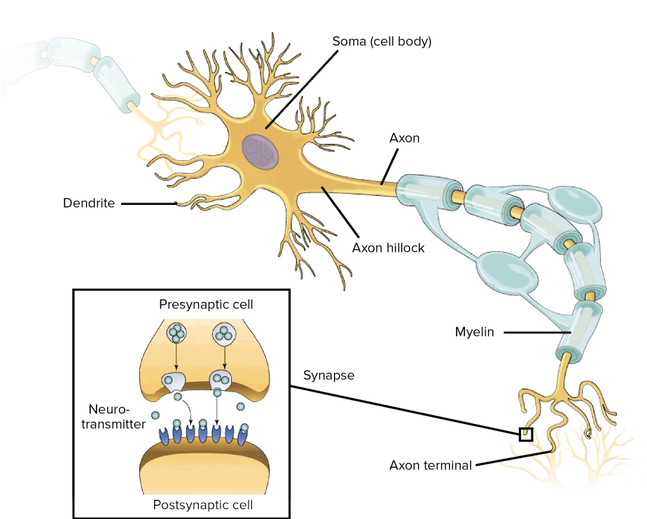
What does this image show?
Nerve and Synapse

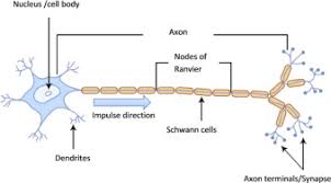
What does this image show?
Motor neuron
What does this image show?
Sensory neuron
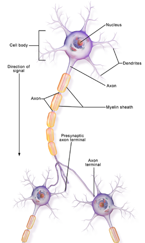
What does this image show?
Interneuron
What is a synaptic transmission?
It is how neurons communicate, relaying information to the CNS via sensory neurons and carrying out responses dictated by the brain through sending information to effectors via motor neurons
What is the process of a synaptic transmission?
An action potential arrives at the presynaptic membrane, causing depolarisation through the opening of voltage-dependent calcium ion channels and the influx of calcium ions.
The increased calcium ion concentration causes vesicles containing neurotransmitters to fuse with the presynaptic membrane and release their contents into the synaptic cleft through exocytosis.
The neurotransmitters diffuse across the synaptic cleft, down a concentration gradient, and bind to complementary receptors on the postsynaptic membrane. This can result in inhibitory or excitatory effects.
The resulting action potential is transmitted along the axon of the following neuron, creating a cascade of neurotransmission.
What are types of neurotransmitters?
Inhibitory neurotransmitters
Excitatory neurotransmitters
What do inhibitory neurotransmitters do?
They reduce the potential difference across the postsynaptic membrane by closing voltage-dependent sodium ion channels, reducing the likelihood of an action potential.
What are examples of an inhibitory neurotransmitter?
Gamma-aminobutyric acid (GABA) and serotonin
What do excitatory neurotransmitters do?
They increase the potential difference across the postsynaptic membrane by opening more voltage-dependent sodium ion channels, increasing the likelihood of an action potential.
What are examples of excitatory neurotransmitters?
Glutamate and dopamine.
What is the localisation theory?
Specific brain areas are responsible for particular processes, behaviours, and activities
What is the motor area?
Found in the frontal lobe, separated from the auditory area by the central sulcus. Regulates and coordinates movement. Lesions result in an inability to control voluntary fine motor movements.
What is the auditory area?
Located in the temporal lobe on the superior temporal gyrus. Processes auditory information and speech. Lesions cause hearing loss, while damage to specific parts (Wernicke’s area) results in Wernicke’s aphasia
What is the visual area?
Located in the occipital lobe. Processes visual information
What is the somatosensory area?
Located in the parietal lobe. Processes information associated with the senses (touch, heat, pressure, etc.). Receives neuronal input from the thalamus that corresponds with the handling of sensation along the lines of touch, pain, temperature and limb position. Lesions result in a loss of ability to denote sensitivity to particular bodily areas.
What is the Wernicke’s area?
Responsible for speech comprehension, located in the temporal lobe (usually the left). Lesions result in Wernicke’s aphasia, characterised by nonsensical words (syllogisms), no awareness of incorrect word usage, but no issues with pronunciation and intonation.
What is the Broca’s area?
Responsible for speech production, located in the frontal lobe, usually in the left hemisphere. Lesions result in Broca’s aphasia, characterised by difficulty forming complete sentences and understanding sentences, failing to understand word order and who they are directed towards (I, you, we, he, me, etc.).
What is the left hemisphere know for?
It is associated with language production and comprehension; therefore, language is a cognitive ability that is both localised and lateralised (to the left hemisphere).
Who supports localisation of brain function?
Tulving et al
Petersen et al (1988)
What did Tulving et al demonstrate?
Using PET scans that semantic memories were recalled from the left prefrontal cortex, while episodic memories were recalled from the right prefrontal cortex. This shows that different brain areas are responsible for different functions, as predicted by localisation theory.
What did Petersen et al (1988) find out?
Wernicke’s area activation is required for listening tasks, whereas Broca’s area is required for reading tasks. This confirms that Wernicke’s area is involved in speech comprehension, while Broca's area is responsible for speech production.
Which case study supports localisation of function?
Phineas Gage
What happened to Phineas Gage?
He was injured by a blasting rod that damaged his prefrontal cortex. This damage caused a defect in rational decision-making and the processing of emotion, supporting the idea that specific brain areas are responsible for specific functions. However, the subjectivity of conclusions drawn, the unusual sample, and a lack of control over confounding and extraneous variables must be considered
What theory contradicts localisation of function?
A holistic view of brain function suggests that each function requires several brain areas to be activated, and these functions are not restricted to these areas
Why does the holistic view of brain function go against localisation of function?
After removing 20-50% of the cortices belonging to rats, researchers found that no specific brain area or lesion was associated with learning how to traverse through a maze. This suggests that intelligence, or even learning, is too complex and advanced a cognitive ability to be restricted to certain areas of the brain. Therefore, this suggests that localisation theory may provide a better explanation for ‘simple’, rather than complex, brain functions
What evidence supports the link between certain brain areas and symptoms of OCD?
Dougherty et al. (2002) studied 44 OCD sufferers who’d undergone lesioning of the cingulate gyrus (cingulotomy) in order to control their symptoms. The researchers found that, after a mean follow-up of 32 months after one or more cingulotomies, 32% met criteria for treatment response, and 14% were partial responders. This suggests that not only are certain brain areas responsible for symptoms of OCD, but that an improved understanding of localisation of brain function has practical applications in the development of more advanced treatments for serious mental disorders
What is plasticity?
The brain’s ability to physically and functionally adapt and change in response to trauma, new experiences, and learning. Neuroplasticity was demonstrated by Maguire et al. (2006).
What idea does plasticity opose?
The idea of a ‘critical window’ for synaptic and neuronal connection formation during the first 3 years of life, after which no new neuronal connections would be formed (Gopnik et al.)
What do we control?
The strength and number of neuronal connections in our brains through synaptic pruning
What is synaptic pruning?
The process by which extra neurons and synaptic connections are eliminated in order to increase the efficiency of neuronal transmissions.
What did Maguire et al find?
A larger grey matter volume in the mid-posterior hippocampi (and a lower volume in the anterior hippocampi) of London taxi drivers' brains, alongside a positive correlation between increasing grey matter volume and how long the individuals had been taxi drivers. They concluded that a complex spatial representation, which facilitates expert navigation and is associated with greater posterior hippocampal grey matter volume, might come at a cost to new spatial memories and grey matter volume in the anterior hippocampus. This may be because the hippocampus is associated with spatial awareness, an ability which taxi drivers must have when they complete The Knowledge test.
What is functional recovery?
The ability of the brain to transfer the functions of damaged areas to other healthy parts of the brain, allowing normal functioning to continue
What enables functional recovery?
The law of equipotentiality (secondary neural circuits surrounding the damaged area become activated).
Axonal sprouting (formation of new synapses and strengthening of axonal connections between damaged and healthy areas).
Reformation of blood vessels (as part of the haemodynamic response, where activated areas experience a higher blood deoxygenation level).
Recruiting homologous areas on the opposite side of the brain.
Who are examples of functional recovery?
Ramachandran
Jodi Miller
What did Ramachandran do?
His research into phantom limb syndrome: caused by sensory input from the face skin ‘invading’ and activating deafferented hand zones in the cortex and thalamus… There appears to be tremendous latent plasticity even in the adult brain. This demonstrates negative plasticity because the neuroplasticity results in painful or negative consequences.
What happened to Jodi Miller?
Her entire right hemisphere was removed to control epileptic seizures. However, through neuroplasticity, she could still control the right side of her body. This demonstrates positive plasticity, because the neuroplasticity results in desirable or positive consequences.
Who demonstrated the negative effects of plasticity and how?
Ramachandran et al. demonstrated negative plasticity through providing an explanation for phantom limb syndrome in terms of cortical reorganisation in the cortex and thalamus (particularly, the somatosensory area).
Who demonstrated positive effects of plasticity and how?
Positive plasticity has been demonstrated by the case study of Jodi Miller, who has shown the power of recruiting homologous areas on the opposite side of the brain, axonal sprouting and the reformation of blood vessels. Therefore, there is evidence supporting not only the existence of, but also the uses of plasticity.
Who found that neuroplasticity happens in animals?
Hubel and Weisel (1970)
What did Hubel and Weisel (1970) find?
They sutured the right eye of kittens, who are blind from birth, for 6 months, opening the eyes and several points and monitoring brain activity in the visual cortex. The researchers found that, although the right eye was closed, there was still activity in the left visual cortex, corresponding to the development of ocular dominance columns. This was demonstrated by how, during the period of high susceptibility in the fourth and fifth weeks, eye closure for as little as 3-4 days leads to a sharp decline in the number of cells that can be driven from both eyes. This therefore supports the idea that areas of the brain receiving no input can take over the function of highly stimulated areas, despite originally having different functions.
How might cognitive reserve increase the rate of functional recovery?
The level of education a person has attained and how long they have been in education. Research suggests that an increased cognitive reserve increases the likelihood of making a disability-free recovery (DFR) after trauma, due to increased rates of neuroplasticity. For example, Schneider et al (2014) found that of the 769 patients studied, 214 achieved DFR after 1 year. Of those, 50.7% had between 12 and 15 years of previous education, and 25.2% had more than 16 years. This suggests that individuals who have been in education for a longer time may have developed the ability to form neuronal connections at a high rate, and therefore experience high levels of functional recovery, demonstrating positive plasticity.
What limits are their to spontaneous and functional recovery?
After trauma, the brain activates secondary neural circuits that contribute toward reinstating normal function (law of equipotentiality). The brain can only ‘repair’ itself up to a specific point, after which motor therapy or electrical stimulation is needed to increase recovery rates. For example, Lieperta et al (1998) found that after constraint-induced movement therapy, the motor performance of stroke patients improved significantly. Therefore, this suggests that functional recovery cannot be relied upon to reinstate normal function.
What is hemispheric laterisation?
Each hemisphere (half) of the brain is mainly responsible for certain behaviours, processes, and activities. This contrasts with the holistic theory of brain function, which suggests that function is distributed across the whole brain (i.e., is global).
What does the right hemisphere control?
The left side of the body, and vice versa
What is an example of the control of the right hemisphere?
Information which we receive from the left visual field is processed by the right hemisphere, which then coordinates a response to affect the left side of the body.
Who did split brain research?
Sperry and Gazzaniga (1968)
What happened in Sperry and Gazzaniga’s split brain research?
It was conducted on 11 epileptic patients who had undergone surgical lesioning of the corpus callosum (cerebral commissurotomy) to control seizures. This procedure prevents information processed by one hemisphere from being relayed to the other, allowing researchers to expose a single hemisphere to certain stimuli and infer the functions of each hemisphere.
What was the set up for the split brain research?
Patients had one eye covered, and stimuli were flashed on a screen for one-tenth of a second to prevent both visual fields from being exposed to the information. This research was conducted under strictly controlled conditions using a laboratory experiment.
What were the procedure steps for the split-brain research?
Describing what you see
Matching words or faces
Words presented simultaneously
Recognising objects placed into the right hands
What happened when participants described what they saw?
If the stimulus word was exposed to the right visual field (processed by the left hemisphere), the patient would say the word because the left hemisphere contains the ‘language centres’ of the brain. However, if the same stimulus word was exposed to the left visual field (processed by the right hemisphere), the patient would write the word using their left hand because the right hemisphere contains the visuo-spatial centres of the brain and allows for the physical act of writing. The patient would not be able to give a verbal description of the word because the right hemisphere contains no language centres.
What happened when participants matched words or faces?
The right hemisphere appeared to dominate the ability to match a list of faces to a given stimulus due to the right hemisphere containing the brain’s visuo-spatial centres, thus allowing for the visual identification and processing of the faces.
What happened when participants were presented with words simultaneously?
If two words were presented at the same time, each to one of the visual fields, the patient would say the word presented to the right visual field (processed by the left hemisphere with language centres) and write down the word presented to the left visual field (processed by the right hemisphere and containing visuo-spatial centres).
What happened when participants had to recognise objects placed into their hands?
If an object was placed into the patient’s right hand, they would be unable to identify that it is there because the information is processed by the left hemisphere, which only has language centres and no visuo-spatial centres. Therefore, if an object was placed into the patient’s left hand, they would be able to identify the object and choose a similar one from a hidden bag due to the action of the visuo-spatial centres.
Why was there a lack of control with the sample selection?
The epileptic patients had been taking anti-epilepsy medications for extended and different periods of time, which may have affected their ability to recognise objects and match words due to causing cerebral neuronal changes. Secondly, although all patients had undergone a commissurotomy, there may have been differences in the exact procedures, e.g. differing extent of the lesioning of the corpus callosum. This would have affected the degree to which the two hemispheres could relay information between themselves. Therefore, these two confounding variables had not been controlled, meaning that the lateralised functions may be examples of unreliable causal conclusions.
How is there clearly demonstrated lateralisation of function?
Split-brain research was pivotal in establishing the differences in functions between the two hemispheres and so opposing the holistic theory of brain function. The left hemisphere was demonstrated as being dominant for language tasks due to containing language centres, whereas the right hemisphere was demonstrated as being dominant for visuo-spatial tasks. Therefore, this suggests that the left hemisphere is the analyser, whereas the right hemisphere is the synthesiser, and so there are marked differences between the two.
What is the contribution to discussions about lateralisation theories?
Such evidence strongly supported the idea of a ‘dual mind’ where the two hemispheres represent two sides of the mind.
What is the difference why function may not be so clear cut?
With evidence making the drastic distinctions that the left hemisphere is responsible for language (analyser) whilst the right is responsible for visual-spatial tasks (synthesiser), this has given the public the false impression that the two hemispheres are ‘opposite’ in function and that they can receive such labels. However, as suggested by Pucetti (1980), there have been cases of split-brain patients who are left-handed but produce and comprehend speech in the right hemisphere, which opposes the predictions made by lateralisation theory. Therefore, it is important not to jump to conclusions and to appreciate that, through the recruitment of homologous areas on the opposite side of the brain, each hemisphere is not restricted to specific functions.
What does the brain’s electrophysiological respond to?
Specific events (sensory, motor or cognitive event) are isolated by doing a statistical analysis of EEG data
What does the statistical averaging technique remove?
It removes all extraneous brain activity from the original EEG recording by filtering it out, leaving only those responses that relate to the presentation of a specific stimulus or performance of a specific task
What is left in event-related potentials?
Brainwaves that are triggered by particular events
What are the different types of ERP?
Attention
Perception
Memory
Motor response
What are the advantages of the Brain’s Electrophysiological?
Excellent Temporal Resolution- Neural processes are measured more specifically than in an EEG.
Widely used in the measurement of cognitive deficits and functions.
What are disadvantages of the Brain’s Electrophysiological?
Lack of standardisation in ERP methodology in different research studies.
Background noise and extraneous material can be an obstacle.
What are fMRI scans?
Rely on the haemodynamic response. Areas of the brain with high levels of activity have a larger requirement for oxygenated blood, leading to a higher rate of blood deoxygenation. As measured through the bold response, the deoxyhaemoglobin in the blood in these highly active areas absorbs the signal produced by the scan, so such areas appear brightly coloured on the scan.
What are advantages of fMRI scans?
High spatial resolution as up to 4 images can be produced per second.
It can be used while a patient is carrying out a task.
Does not use ionising radiation, and so is safer.
What is a disadvantage of fMRI scans?
Poor temporal resolution because there is approximately a 5-second difference between neuronal activity and the produced image.
What EEG scans?
Use electrodes attached to the scalp to measure and amplify the electric activity across the whole brain (action potentials being transmitted across the axons of neurons).
What are advantages of EEG?
Useful in investigating the characteristics of the different stages of sleep.
Much higher temporal resolution than fMRI scans, more appropriate for monitoring ongoing cerebral states and activity.
Useful in the diagnosis of epilepsy.
What are disadvantages of EEGs?
Lower spatial resolution compared to fMRI scans, with particular difficulty in differentiating activity between adjacent areas.
What are post-mortem examinations?
They involve a comparison of the patient’s brain with that of a healthy, neurotypical brain. Any differences (e.g., lesions, damage, abnormally large or small areas) are assumed to have caused the neurological problem the patient faced in their lifetime.
What are advantages of Post-mortem examinations?
Particularly useful for advancing medical knowledge and being the basis of further research into certain areas of the brain. Led to the identification of Broca’s area and further localisation theory research.
What are disadvantages of post-mortem examinations?
Incorrectly assumes that differences compared with the neurotypical brain must be the explanation for neurological or cognitive deficits. Prolonged drug use, stress, and genetic factors may be other plausible explanations.
Ethical issues: informed consent cannot always be obtained before the patient dies or from the family.
What are biological rhythms?
Periodic biological fluctuations in an organism that correspond to, and are in response to, periodic environmental change. Biological rhythms can be endogenous (controlled by internal clocks, e.g. the suprachiasmatic gyrus) or exogenous (controlled by external, environmental factors, e.g. exposure to sunlight). The three types of biological rhythms are circadian, infradian and ultradian.
What are exogenous zeitgebers?
External changes in the environment which affect or ‘entrain’ our biological rhythms.
What are circadian rhythms?
A type of biological rhythm which completes one full cycle every 24 hours, e.g. the sleep-wake cycle. Like other biological rhythms, it is affected by both endogenous pacemakers and exogenous zeitgebers.
What is the main example of an exogenous zeitgeber?
Light as it changes in light exposure can trigger desynchronisation of a ‘pre-set’ sleep-wake cycle.
What happened in Siffre’s cave study?
Siffre descended into a cave completely devoid of natural light. He finished his experiment on September 14th, believing it to be August 20th! This demonstrates that prolonged exposure to a strong exogenous zeitgeber, such as light, disrupts the sleep-wake cycle becomes disrupted and there is a disconnection between psychological time and the clock. His sleep-wake cycle did not conform to a cyclical 24-hour period but was around 24 hours and 30 minutes, with Siffre himself determining when to sleep and when to eat. Therefore, this demonstrates that “there was an internal clock independent of the natural terrestrial day/night cycle”. This describes a ‘free-running’ circadian rhythm, i.e. one which is not affected by exogenous zeitgebers.
What did Aschoff and Wever do in 1967?
They deprived 55 participants of natural light while spending 4 weeks in an underground bunker. The researchers found that “all subjects showed free-running circadian rhythms, with the average periods of wakefulness and sleep ranging from 23.9 to 50.0 hours. 36 subjects remained internally synchronised during the whole experiment”.
What were the findings of Aschoff and Wever’s research?
The findings demonstrate that although the free-running circadian rhythm is more than 24 hours long, as a society, we have specific exogenous zeitgebers which entrain the rhythm to conform to a 24-hour cycle.
What are individual differences in circadian rhythms?
Circadian rhythms may not and do not always have to conform to cyclical 24-hour periods. Therefore, this is a real-life example of how the circadian rhythms of teenagers specifically are not always in line with those of adults, and so an appreciation of this can improve educational attainment.
What are the confounding effects of artificial light?
As demonstrated by Czeisler et al (1999), artificial lighting can create shifts in circadian rhythms by up to 6 hours. Siffre’s research was conducted at a time when researchers believed that artificial lighting had no effect on biological rhythms. The use of artificial light meant that over 2 months, Siffre could have entrained his own circadian rhythm through signalling sleeping and waking times by using the light, meaning that the conclusions made about his ‘free-running’ circadian rhythm may not be entirely accurate.
What are the detrimental impacts on health in shift workers?
Shift work was associated with obesity, high triglycerides, and low concentrations of HDL cholesterol, which the researchers suggest may demonstrate a link between the desynchronisation of circadian sleep-wake cycles in shift workers and the consequent disruptions in the biological control of metabolism (and therefore core body temperature). This suggests that there may be practical uses in an improved understanding of the effects of desynchronisation. This in turn has economic implications, in terms of companies that employ shift-workers making the effort to revise their policies in order to reduce days taken off sick.