all on
1/218
There's no tags or description
Looks like no tags are added yet.
Name | Mastery | Learn | Test | Matching | Spaced | Call with Kai |
|---|
No analytics yet
Send a link to your students to track their progress
219 Terms
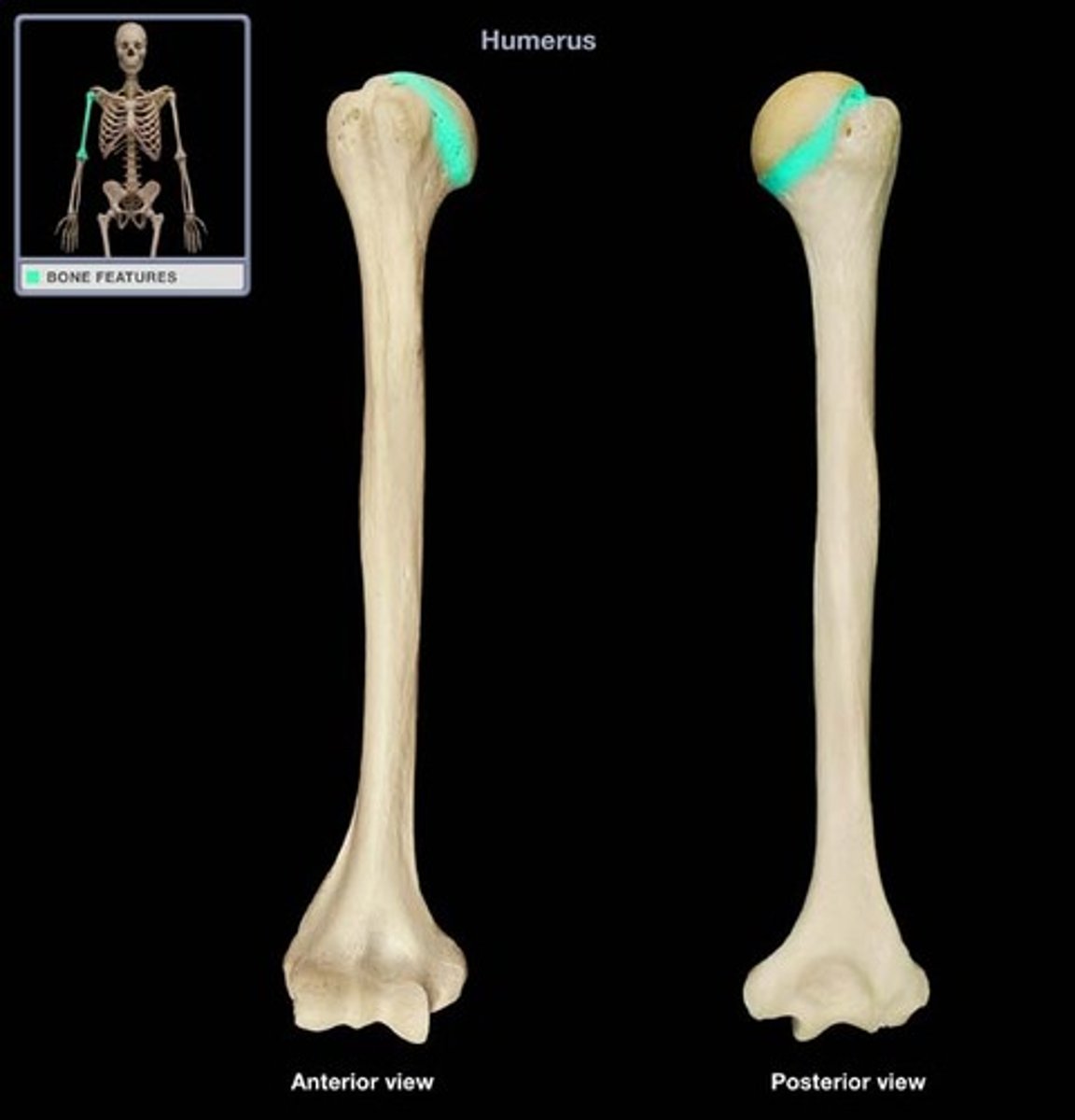
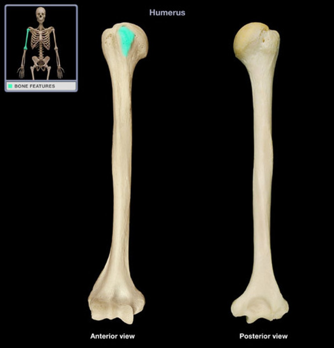
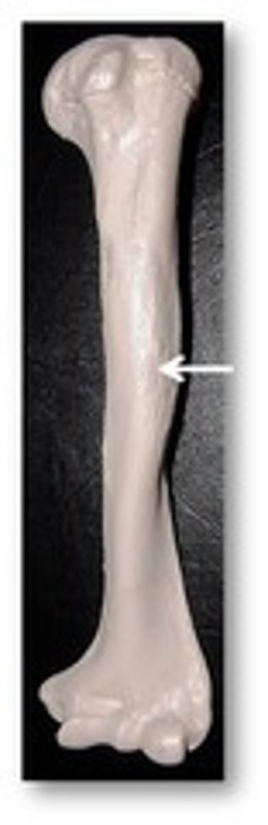
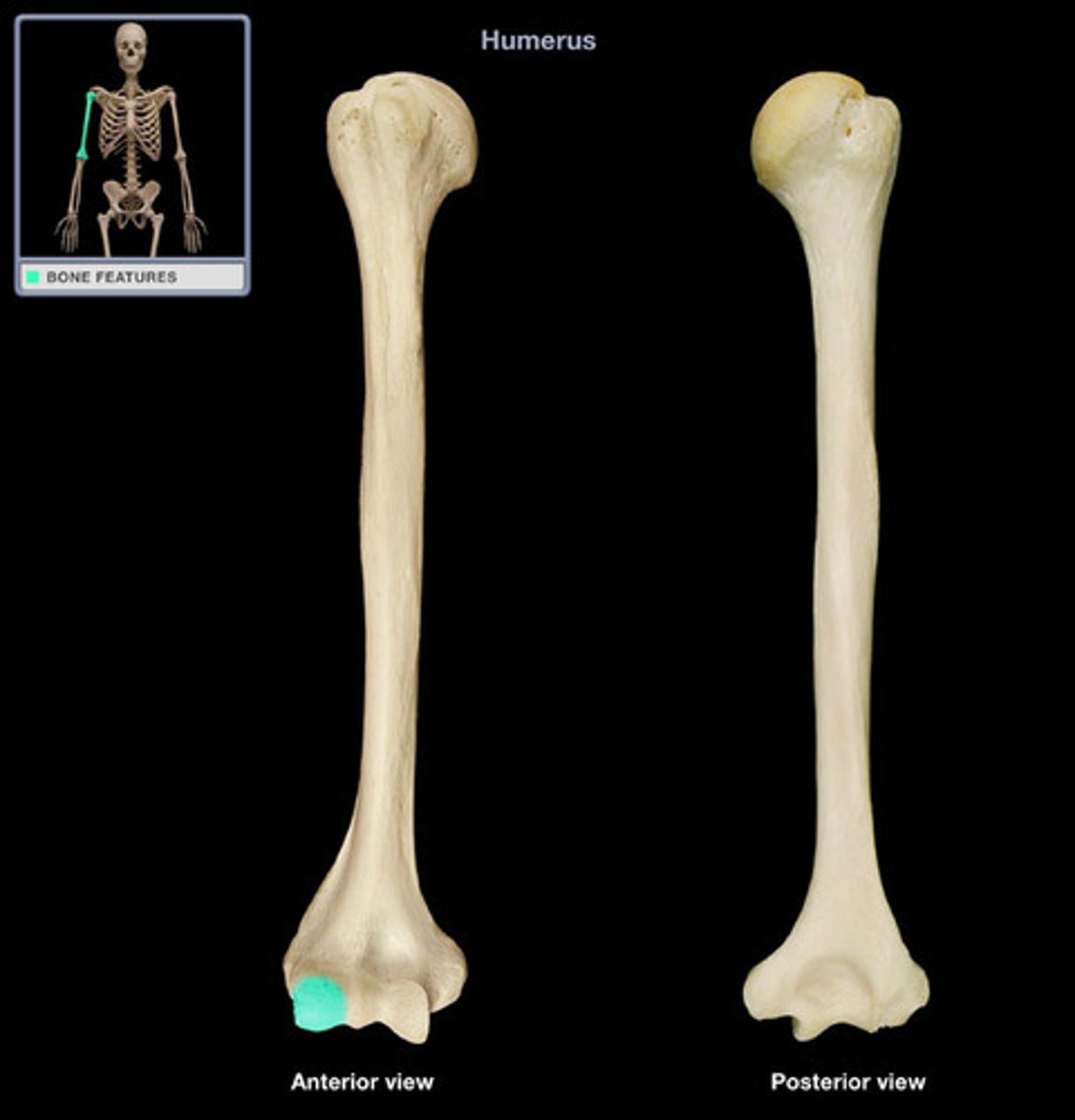
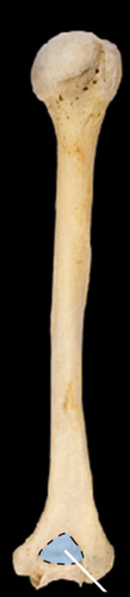
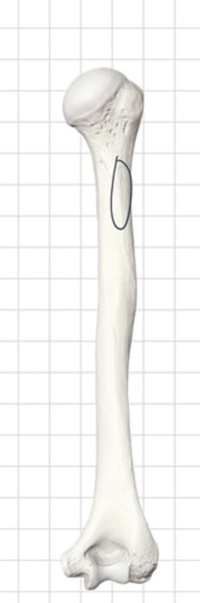
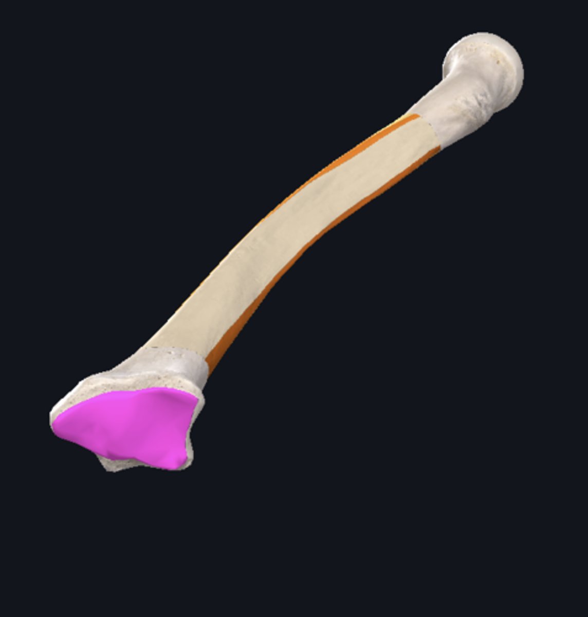
Humerus
longest bone in the arm
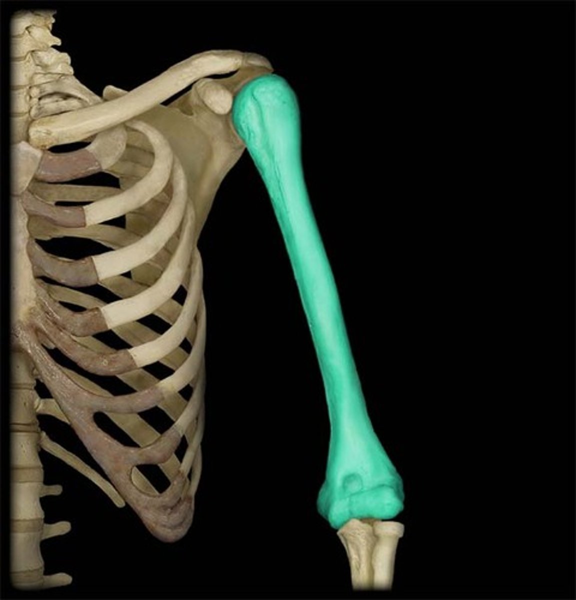
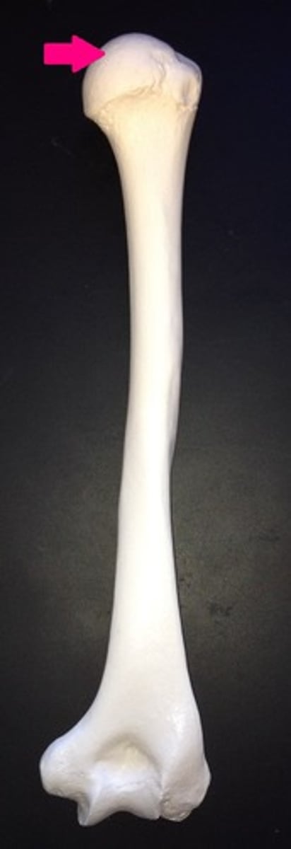
Humeral Head
rounded, medially oriented, proximal end that articulates with the glenoid fossa of the scapula
Humeral Shaft
long, tubular portion of bone
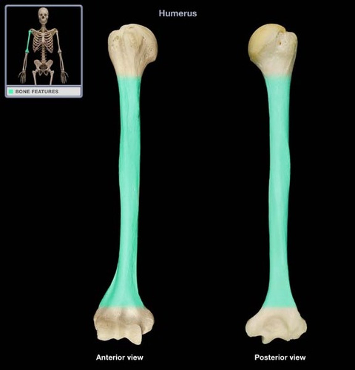
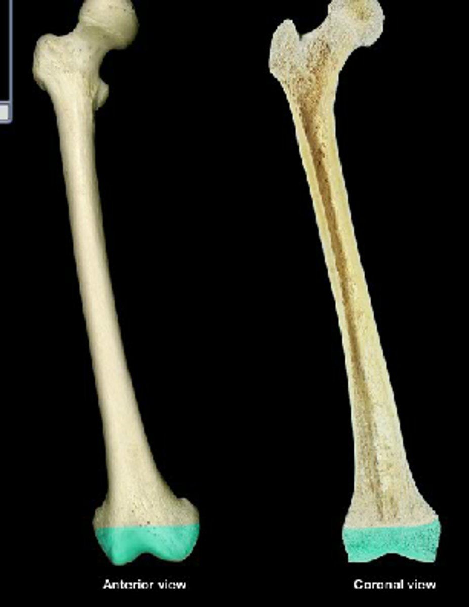
Distal End of Humerus
articulations for ulna and radius
Anatomical Neck of Humerus
groove that encircles the articular surface of the head
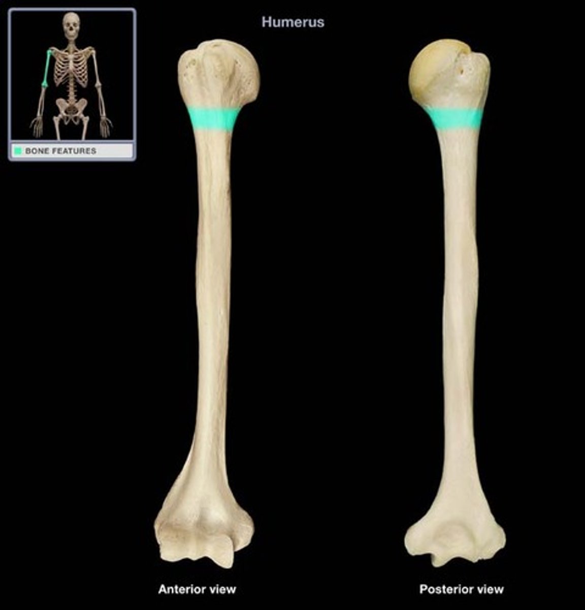
Surgical Neck of Humerus
link between the head and shaft
Greater Tubercle of Humerus (Anterior View, Proximal Shaft)
lateral
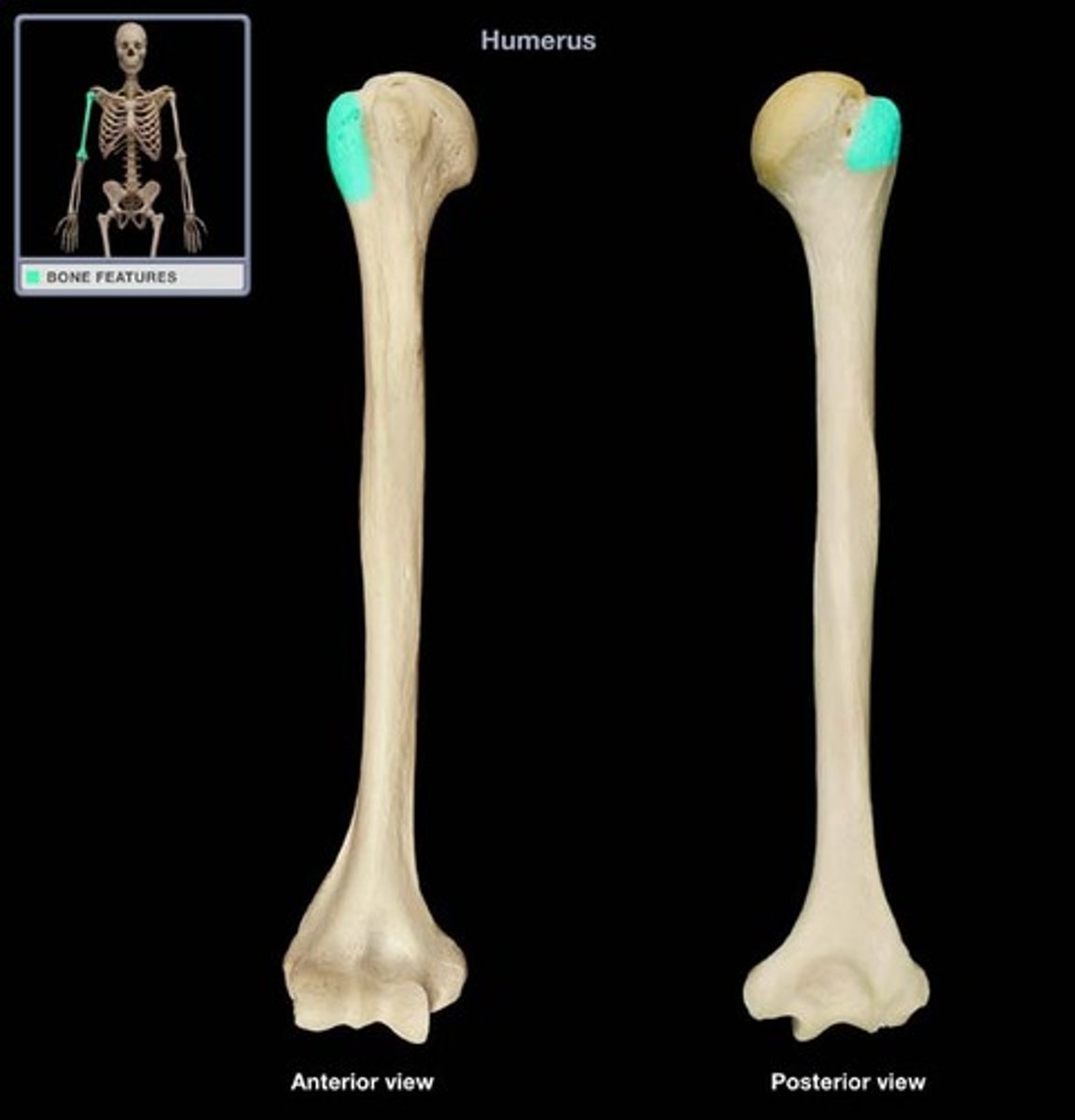
Lesser Tubercle of Humerus (Anterior View, Proximal Shaft)
medial
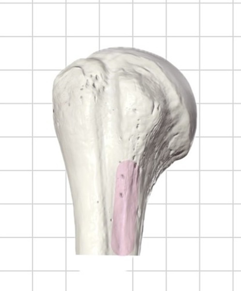
Crest of the Greater Tubercle
runs distally from the
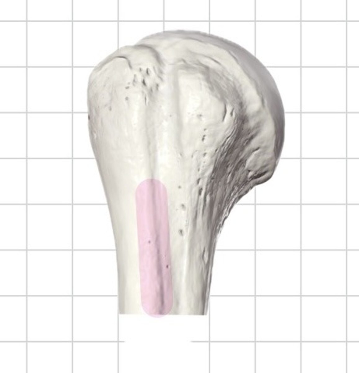
Crest of the Lesser Tubercle
prominent, longitudinal bony ridge running inferiorly from the lesser tubercle on the proximal, medial aspect of the humerus
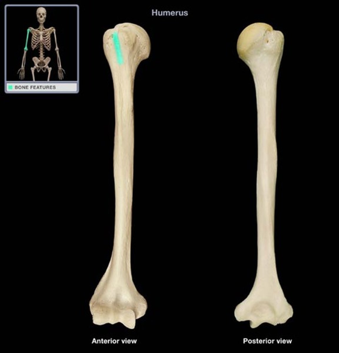
Intertubercular Groove of Humerus
houses the tendon of the long head of the biceps brachii muscle
Deltoid Tuberosity of Humerus
on the lateral surface of the shaft, an insertion for the deltoid muscle
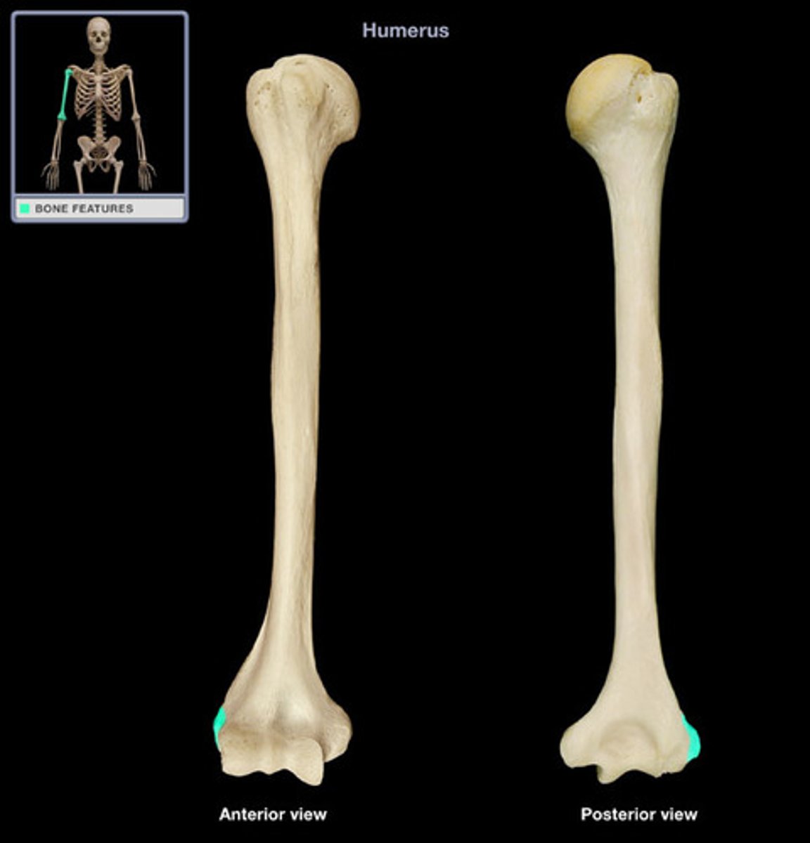
Medial Epicondyle of Humerus (Anterior View, Distal Shaft)
prominent, bony bump on the inner side of the distal humerus
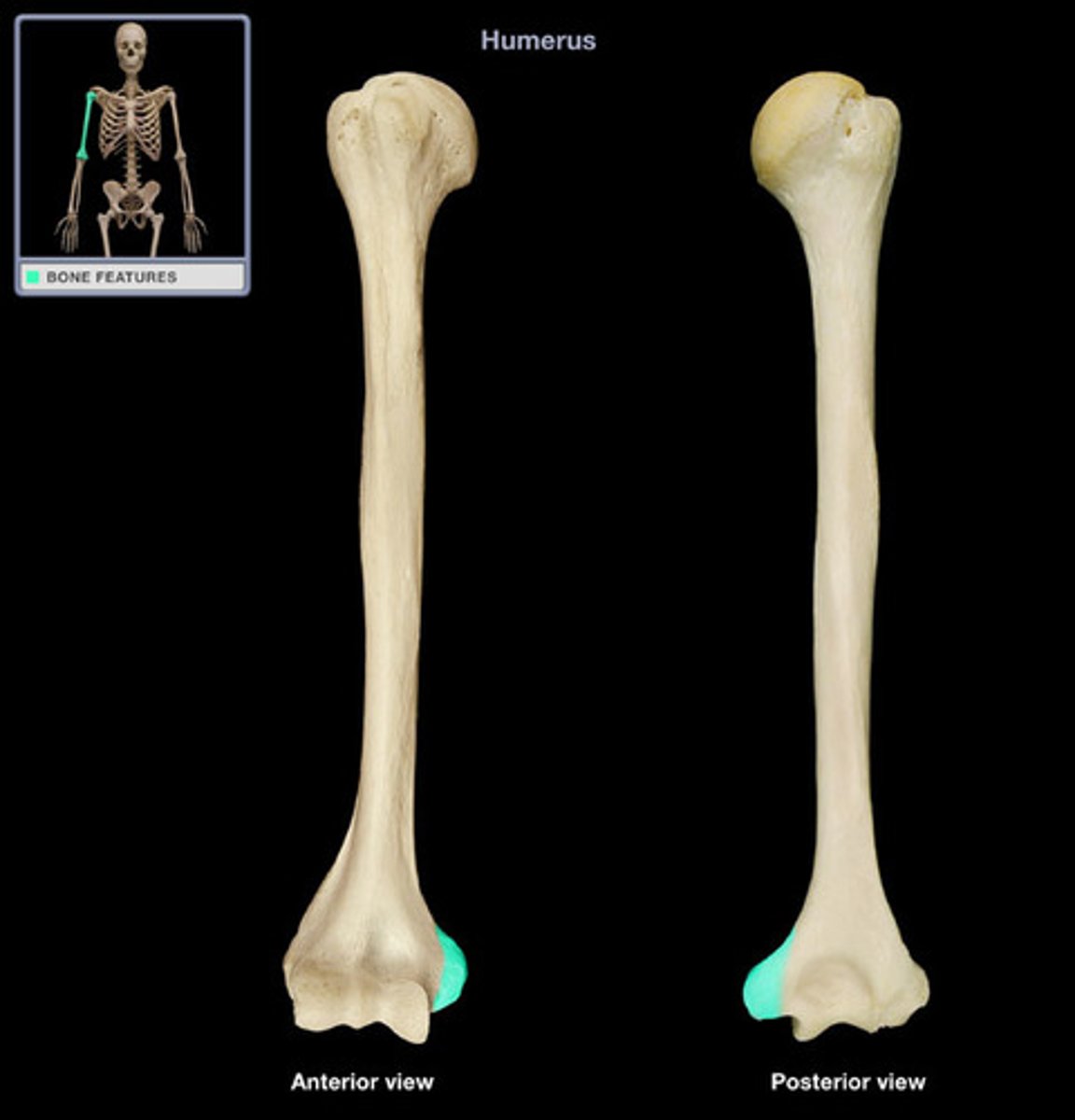
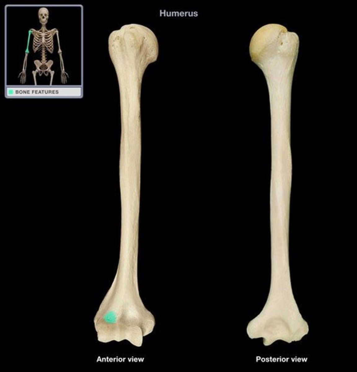
Lateral Epicondyle of Humerus (Anterior View, Distal Shaft)
bony prominence on the outer side of the distal humerus
Trochlea of Humerus
articulation with the ulna, medial
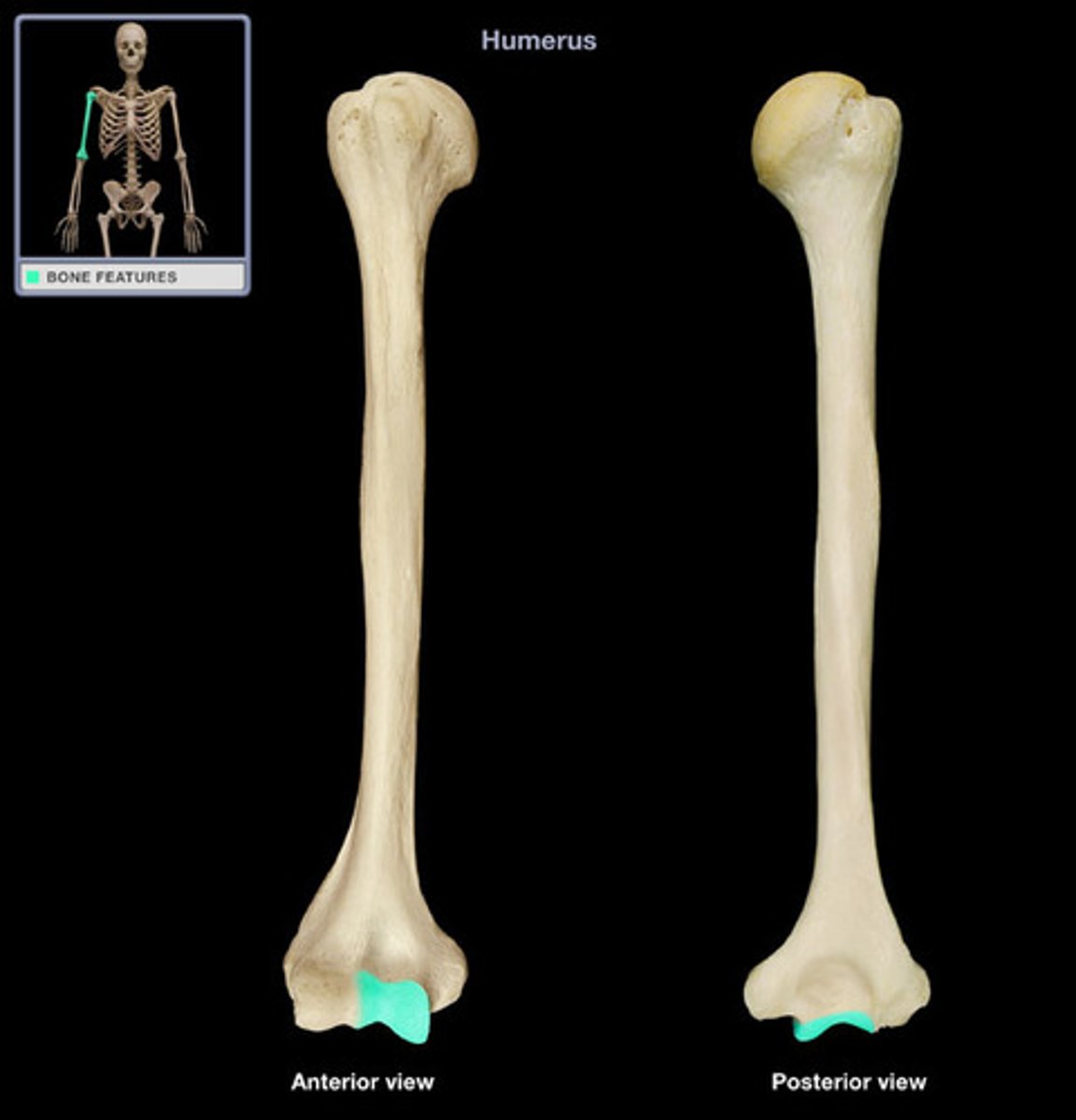
Capitulum of Humerus
articulation with the radius, lateral
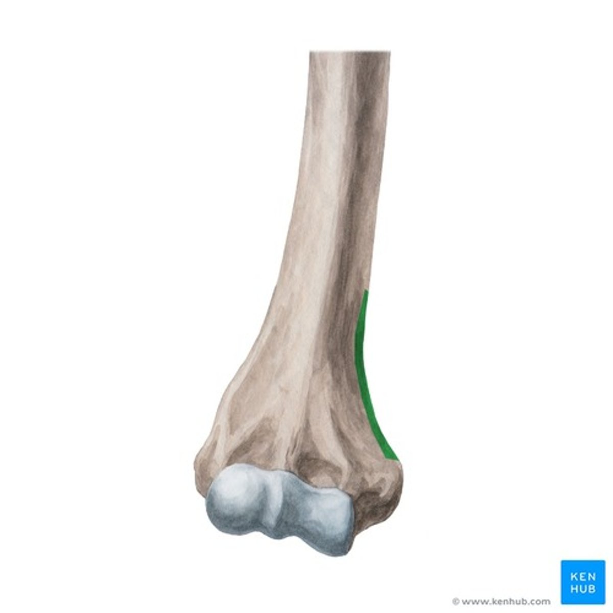
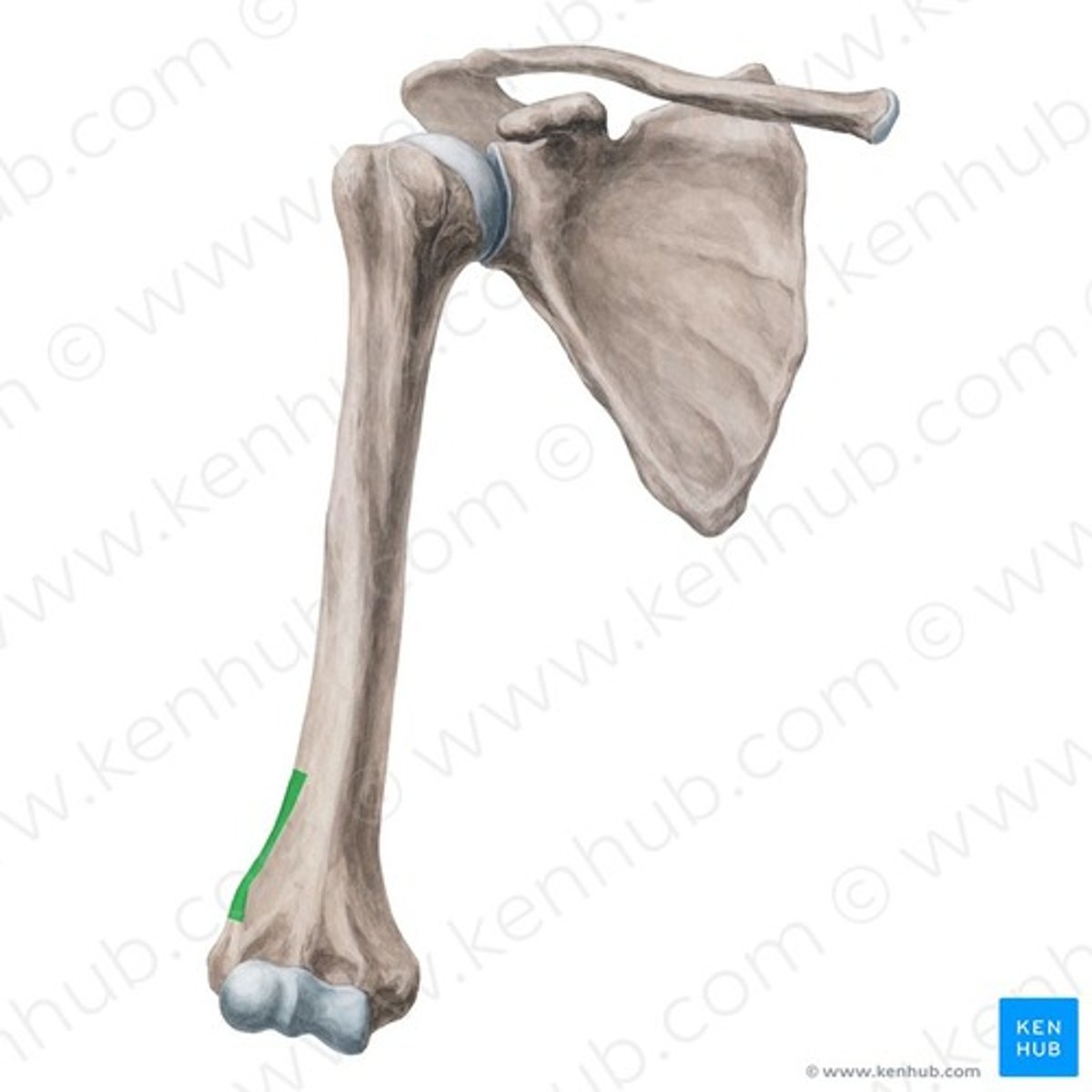
Medial Supracondylar Crest of Humerus
sharp, prominent bony margin located on the distal-medial aspect of the humerus, extending superiorly from the medial epicondyle
Lateral Supracondylar Crest of Humerus
prominent, rough bony margin on the distal, outer side of the humerus
Coronoid Fossa of Humerus
receives the coronoid process of the ulna during flexion of the forearm

Radial Fossa of Humerus
receives head of the radius during flexion of the forearm
Olecranon Fossa of Humerus (Posterior View)
receives the olecranon fossa if the ulna in extension of the forearm
Crest for the Triceps Brachii
runs from the surgical neck to the deltoid tuberosity
Siding for Humerus
- head is proximal with the articular surface oriented medially
- capitulum is lateral, the trochlea is medial
- medial epicondyle is larger/ projects at a more acute angle than the lateral
- the greater tubercle is lateral and anterior
- deltoid tuberosity is lateral and curves from superior/ posterior to inferior/ anterior
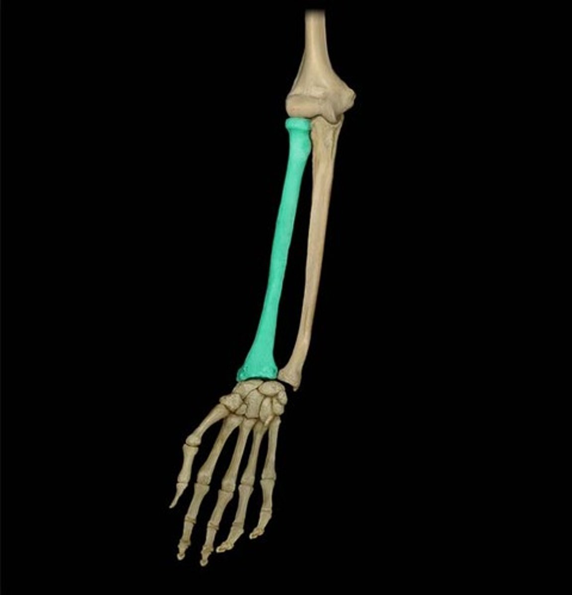
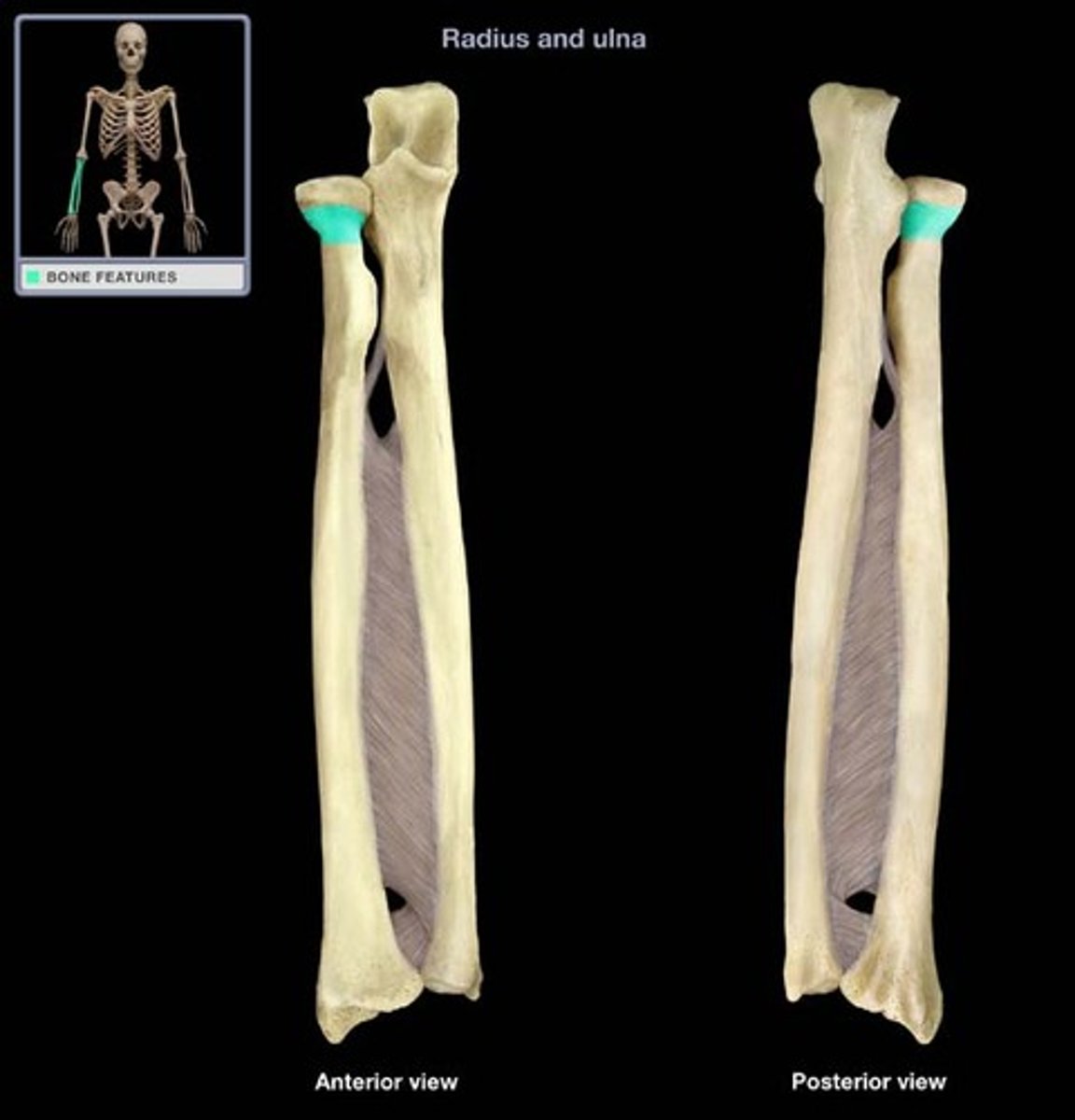
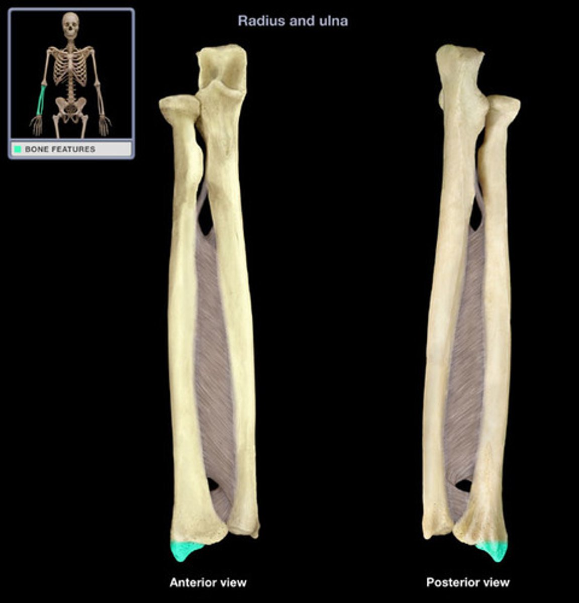
Radius
shortest bone in the arm, the 'thumb side'
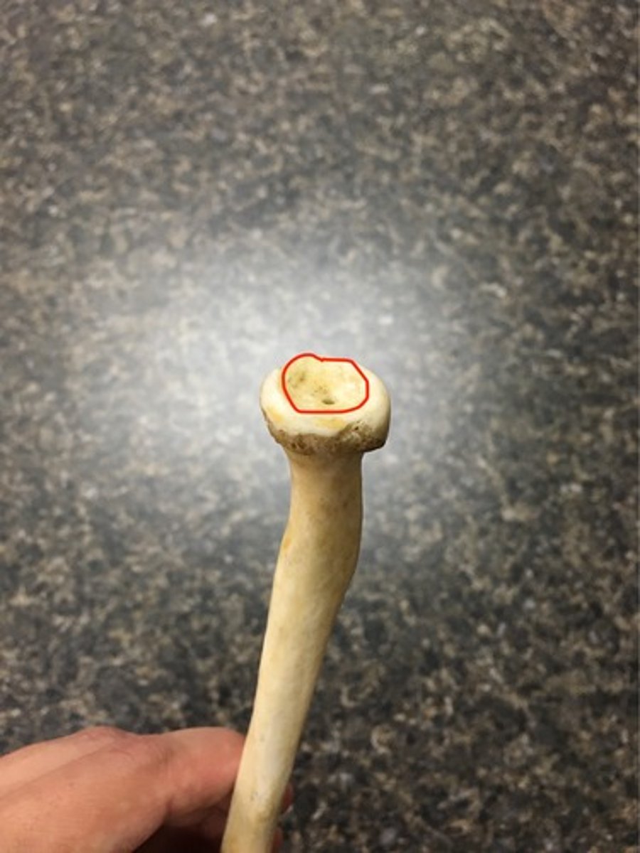
Head of Radius
has two articulations:
- articular facet
- articular circumference
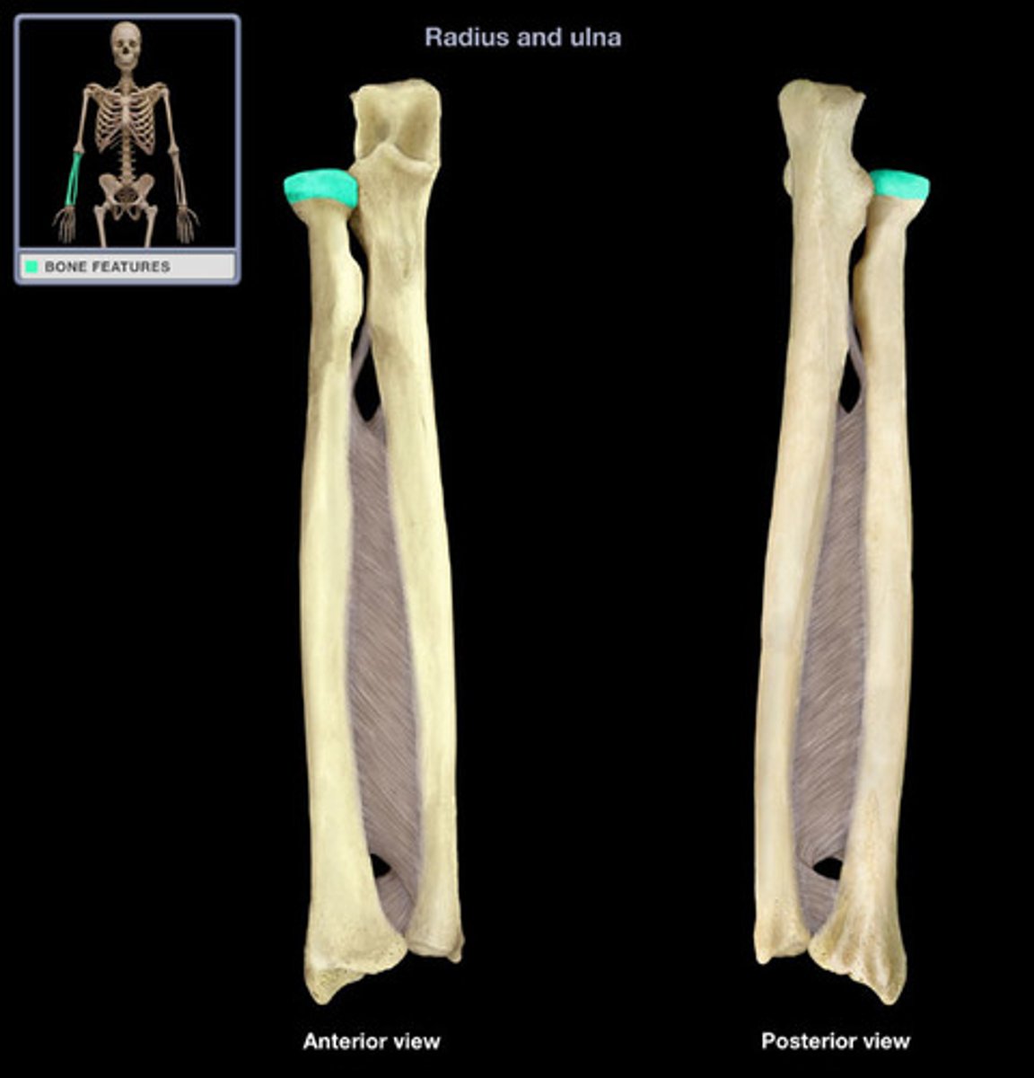
Articular Facet of Radius
for the capitulum of the humerus
Articular Circumference of Radius
for the radial notch of the ulna
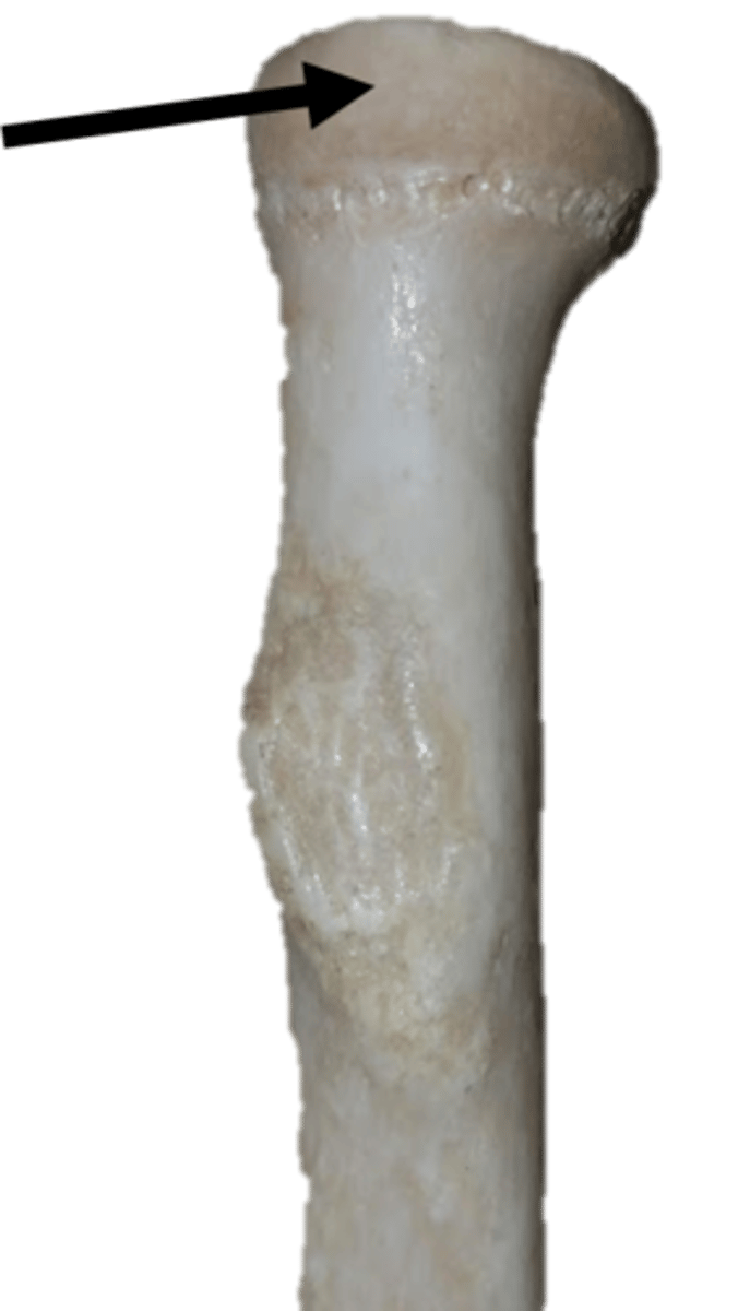
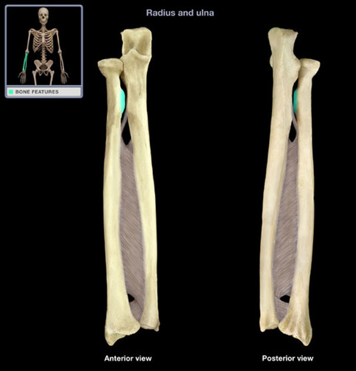
Neck of Radius
narrowed region immediately distal to the head of the radius
Radial Tuberosity
medial, insertion of the biceps brachii
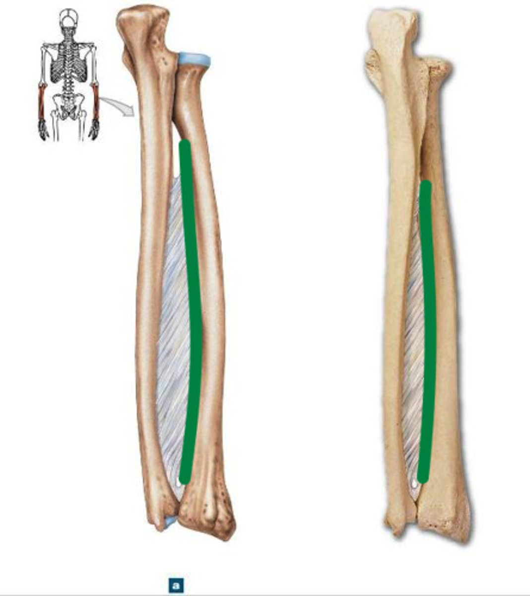
Interosseous Crest of Radius (Anterior View, Distal End)
sharp, medial attachment site for the interosseous membrane
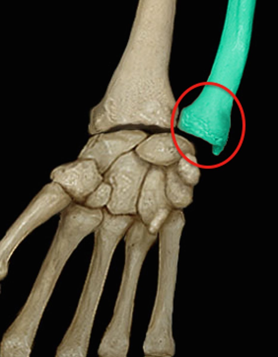
Styloid Process of Radius (Anterior View, Distal End)
most distally projecting point, on lateral side of bone
Dorsal Surface of Radius (Posterior View)
side that the dorsal tubercle resides on
Dorsal Tubercle
large bump on the posterior surface of the distal end, radius
Ulnar Notch of Radius (Anterior View)
for articulation with the distal end of the ulna
Carpal Articular Surface of Radius (Anterior View)
articulates with the lunate (medial) and the scaphoid (lateral)
Radius Siding
- head is proximal, dorsal tubercle is posterior
- ulnar notch is medial
- interosseous crest is medial
- when viewed posteriorly, the styloid process extends inferiorly on the side the bone is from
- when viewed anteriorly, radial tuberosity is found on the side the bone is from
- anterior side is slightly concave, posterior side is rounded
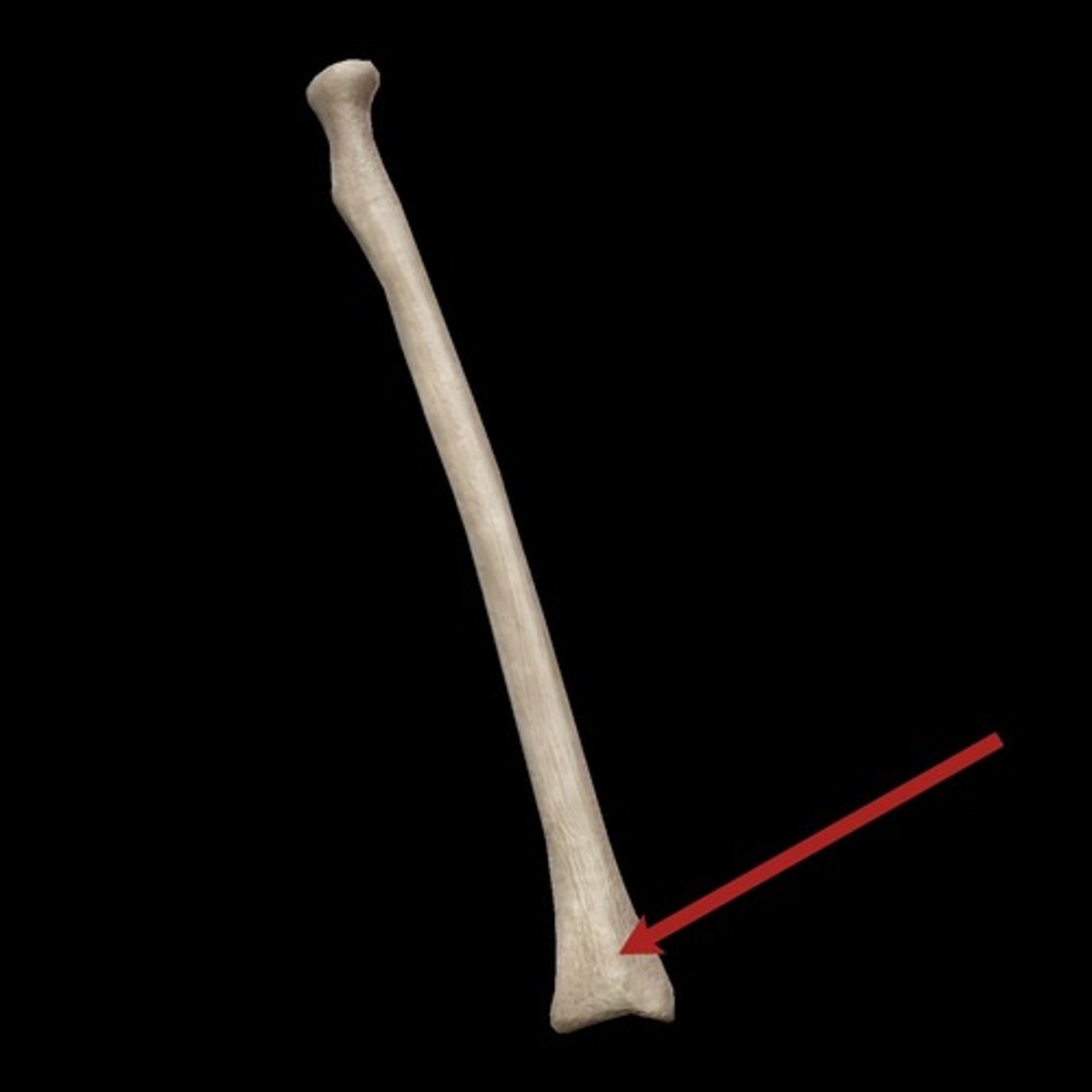
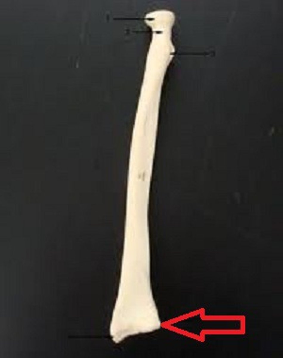
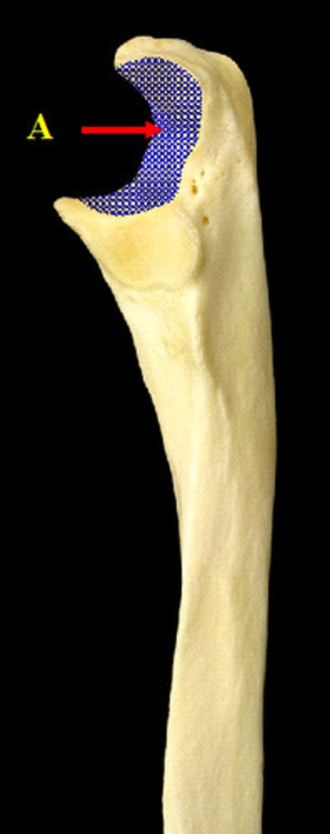
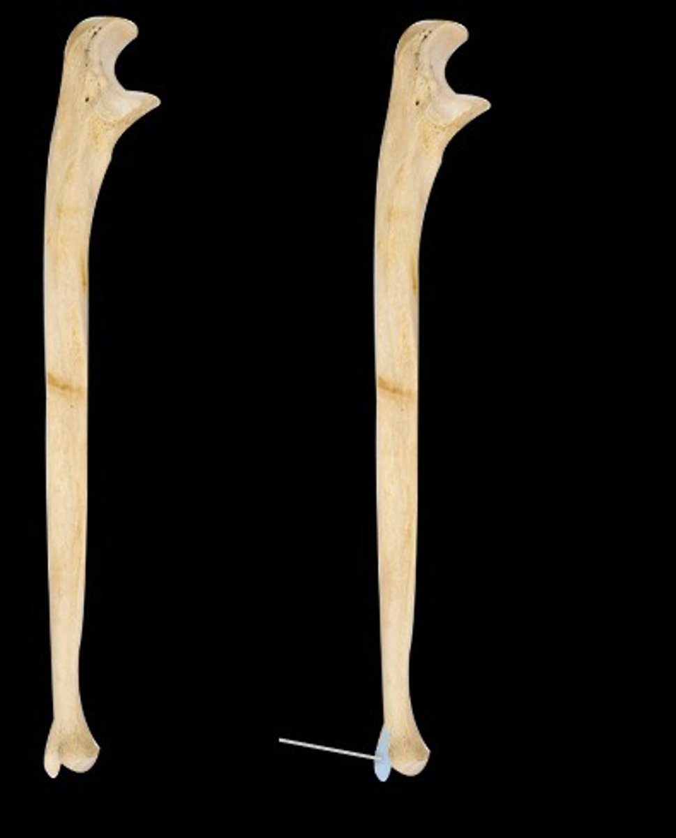
Ulna
- pinky side, medial bone when put in anatomical position

Trochlear Notch of Ulna (Anterior View, Proximal End)
articulates with the humerus
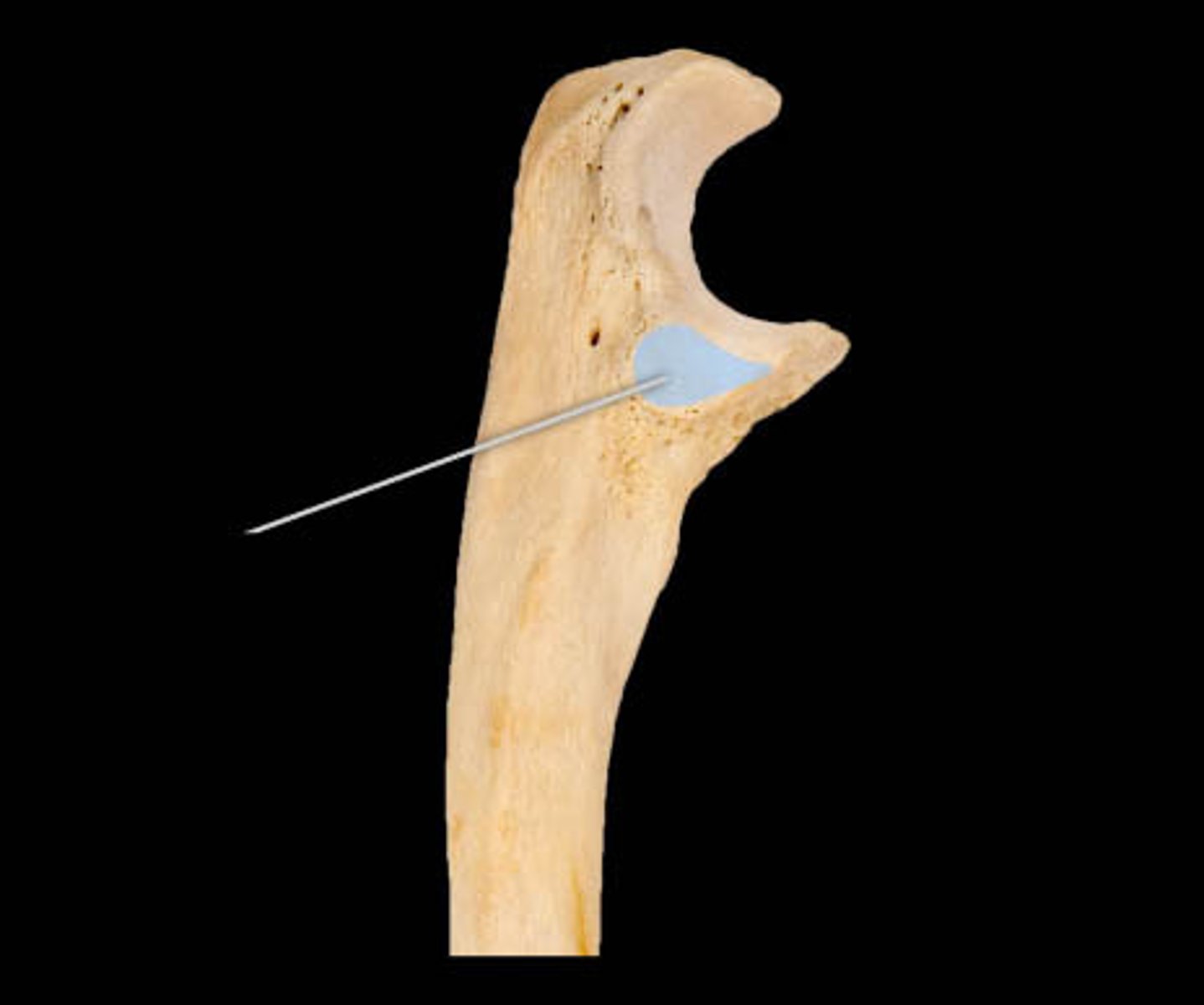
Radial Notch of Ulna (Anterior View, Proximal End)
for the radius
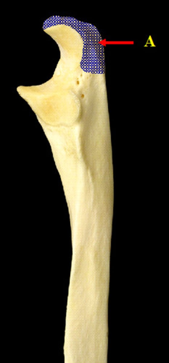
Olecranon of Ulna
the 'elbow'
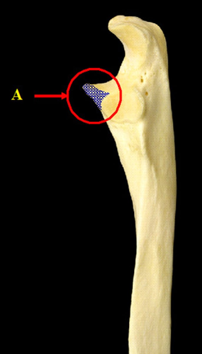
Coronoid Process of Ulna
triangular, bony prominence on the proximal, anterior surface of the ulna
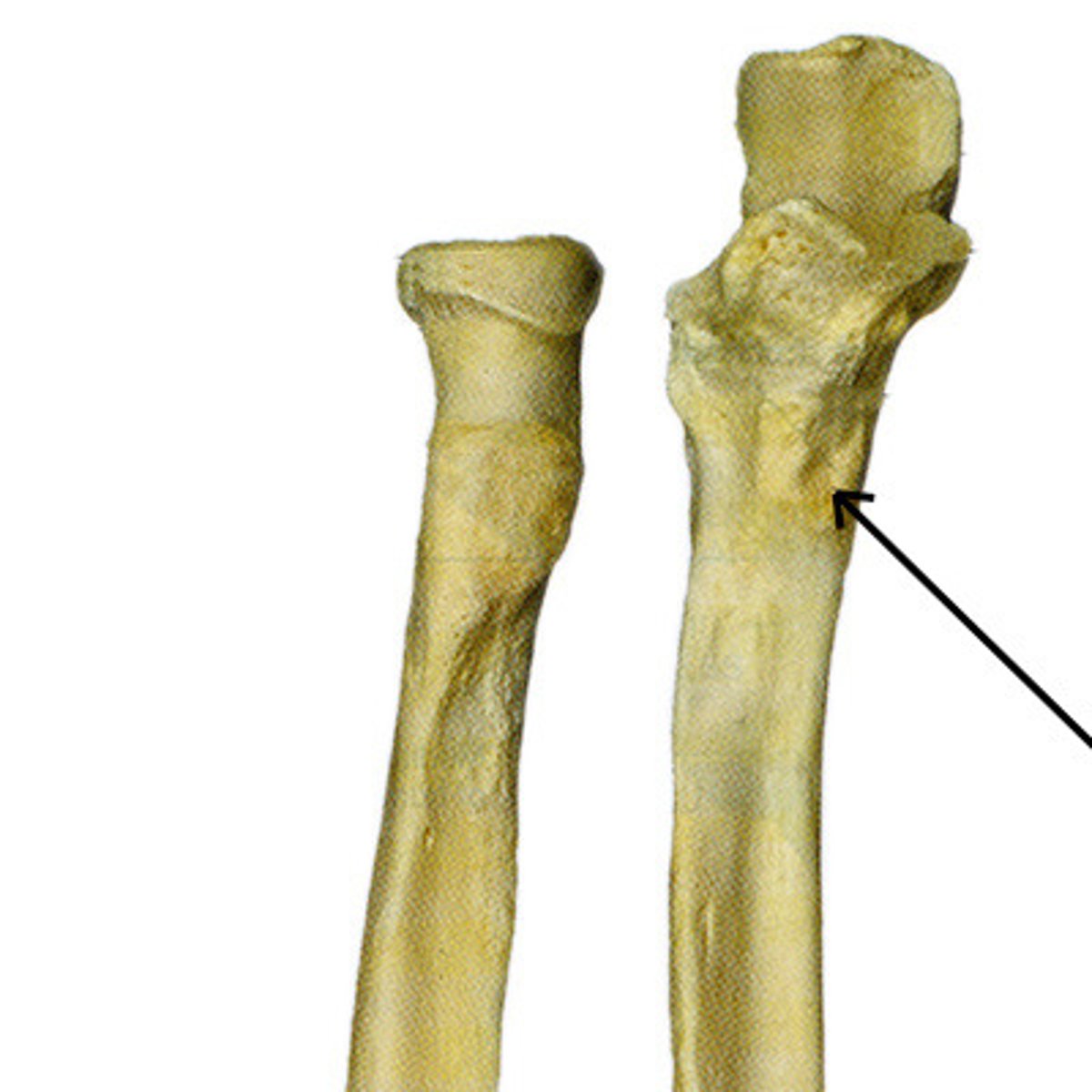
Ulnar Tuberosity
roughened area located on the anterior, proximal ulna inferior to the coronoid process
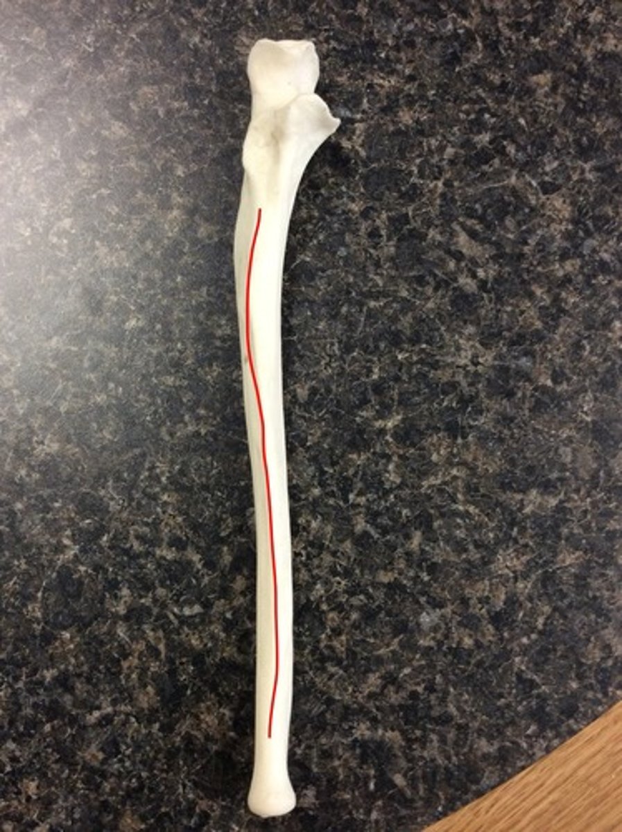
Interosseous Crest of Ulna
ridge along the lateral portion of the ulnar shaft that points towards the radius, sharp, lateral attachment site for the interosseous membrane
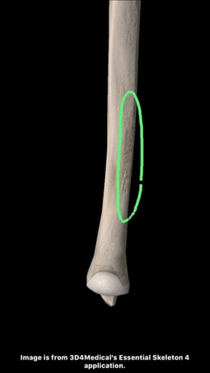
Pronator Ridge of Ulna (Anterior View, Distal End)
distal, medial
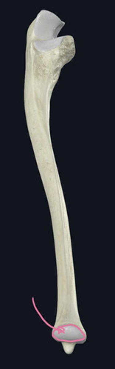
Head of Ulna (Anterior View, Distal End)
distal end
Styloid Process of Ulna (Anterior View, Distal End)
sharp, distal-most projection on the posteromedial corner
Articular Circumference of Ulna (Anterior View, Distal End)
for the ulnar notch of the radius
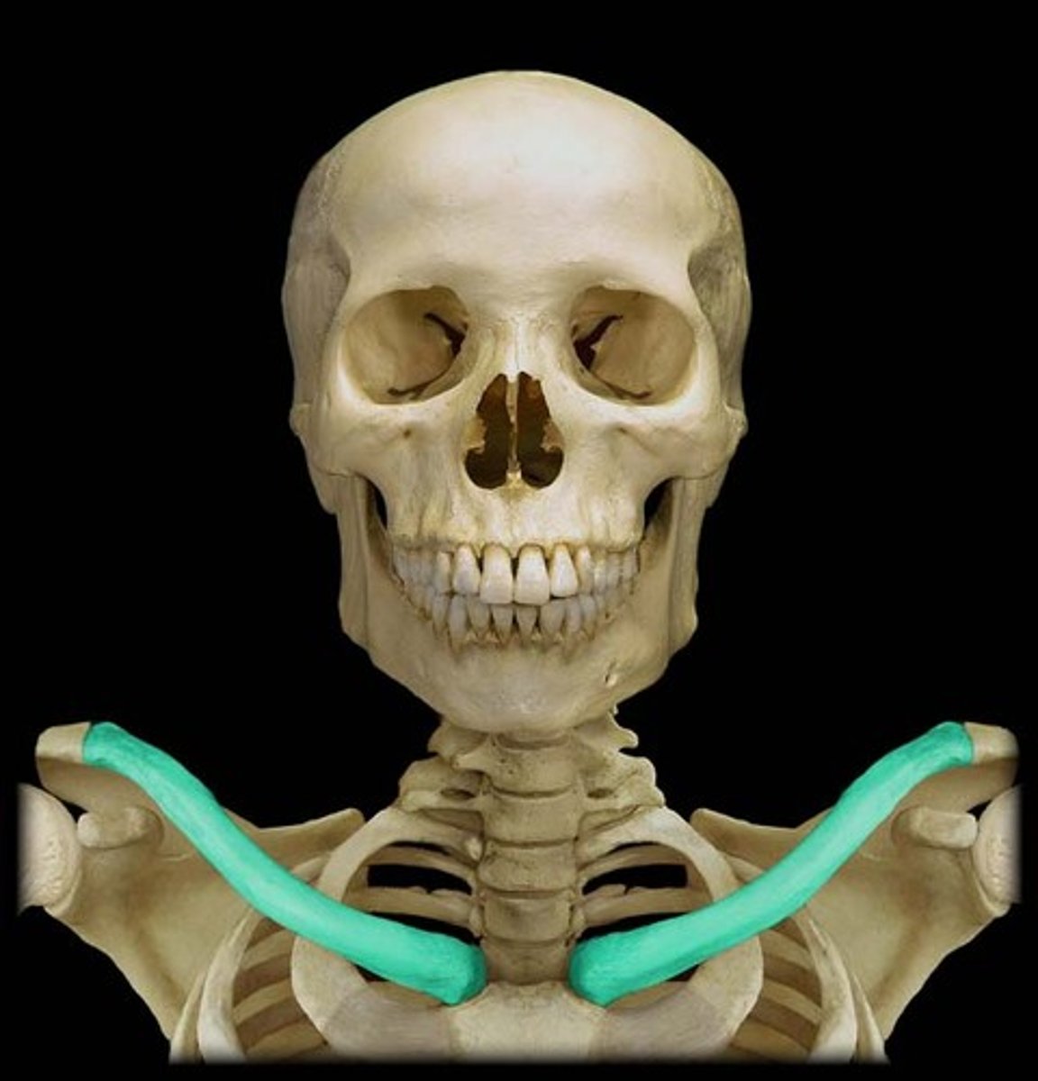
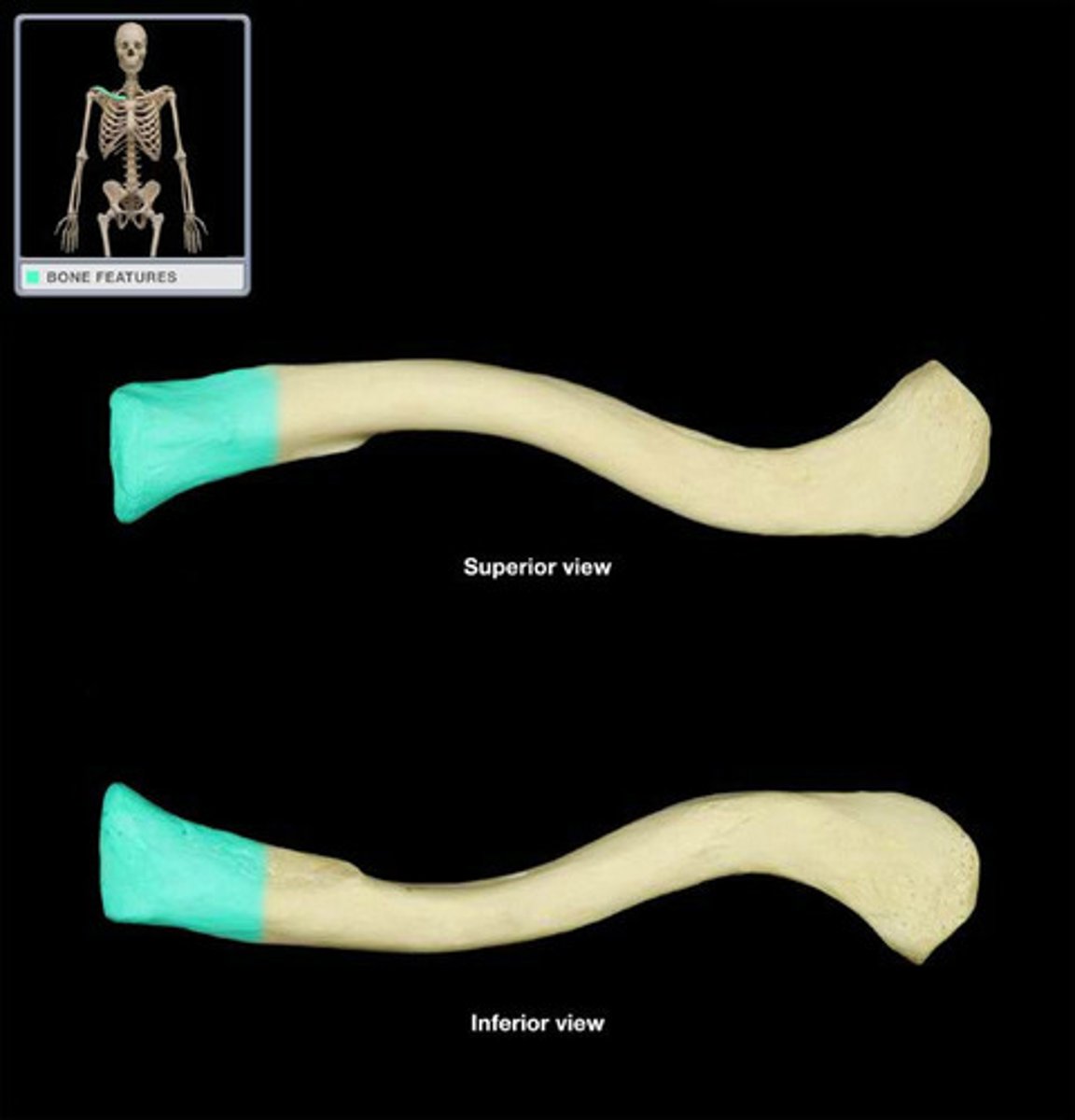
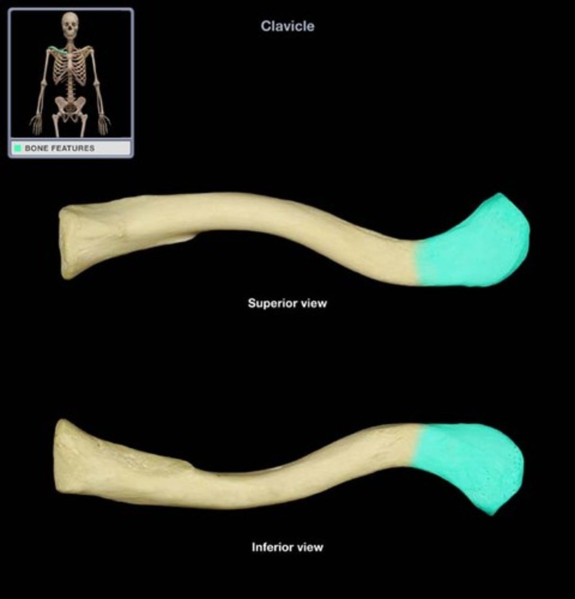
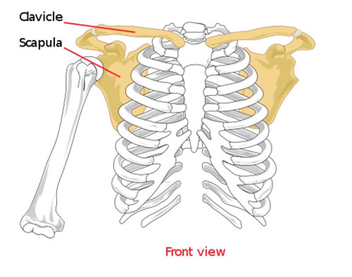
Clavicle
- articulates with the manubrium of the sternum medially and with the scapula laterally
- smooth on the superior surface, majority of features found on inferior surface
Sternal End
- medial
- articular facet for the first costal cartilage and the manubrium
Acromial End
- lateral end
- acromial facet for articulation
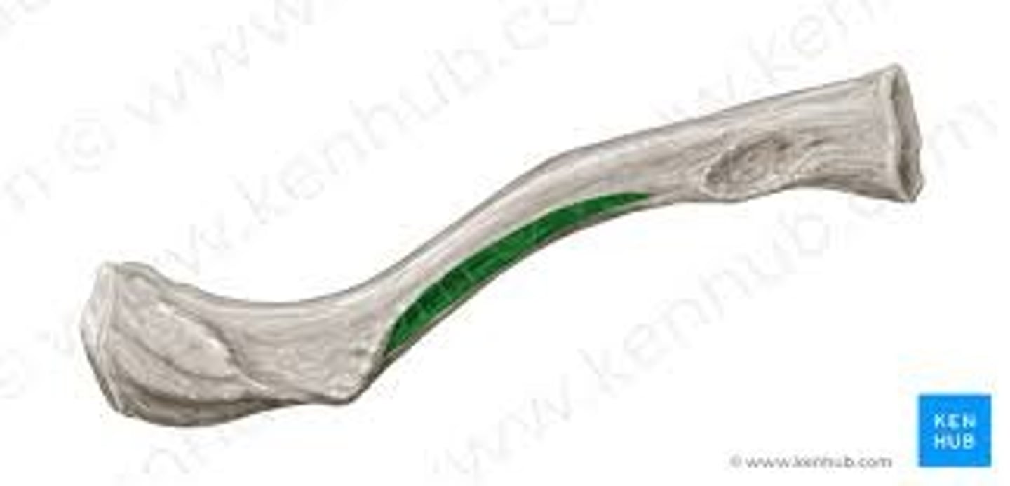
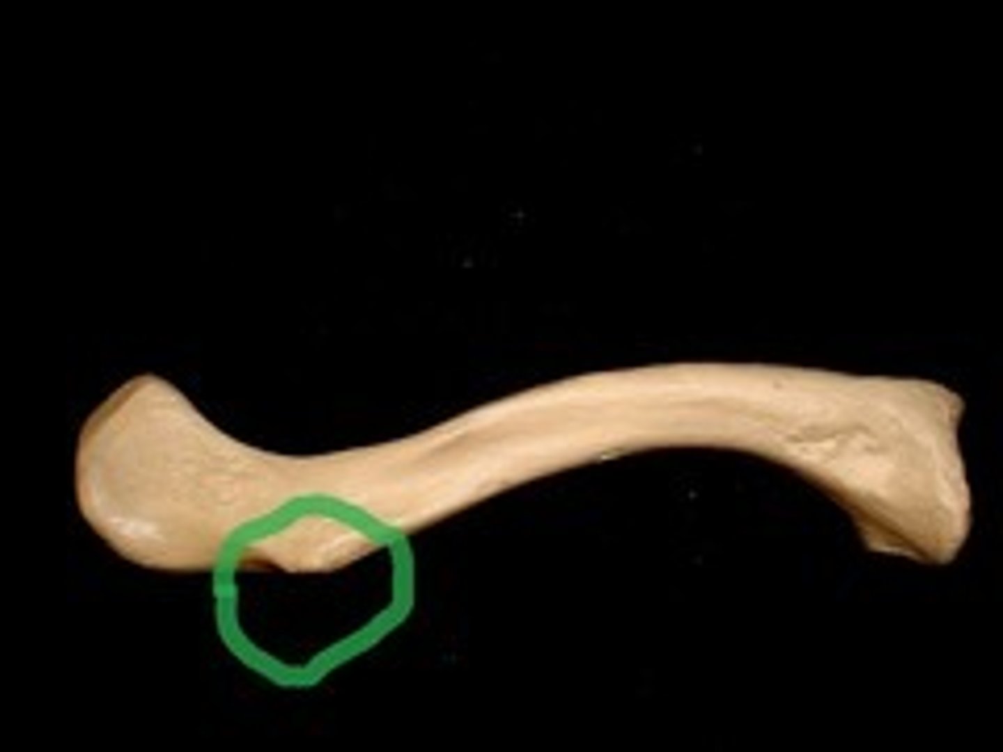
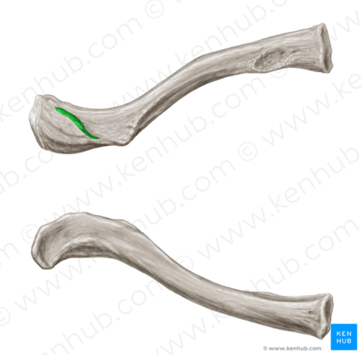
Subclavian Groove/ Sulcus
shallow depression on the middle of the inferior surface of the clavicle
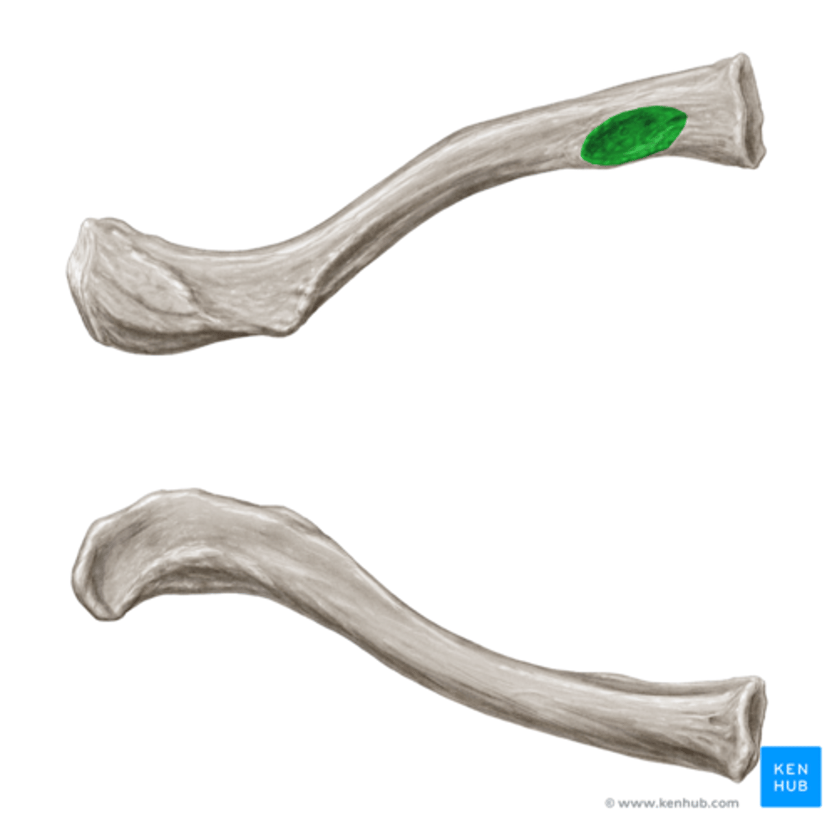
Costal Tuberosity (Costoclavicular Impression)
raised, roughened surface on the inferior sternal end, anchors the costoclavicular ligament
Conoid Tubercle
prominence on inferior surface of the lateral end of clavicle
Trapezoid Line
extends from the conoid tubercle and is the attachment for the trapezoid ligament
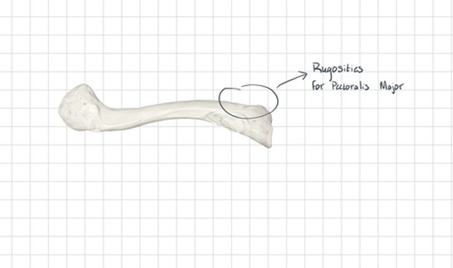
Rugosities for Pectoralis Major
the attachment site for the Pectoralis Major muscle
Siding of Clavicle
- medial end is rounded, lateral end is flattened
- superior surface is smooth, inferior surface is rough
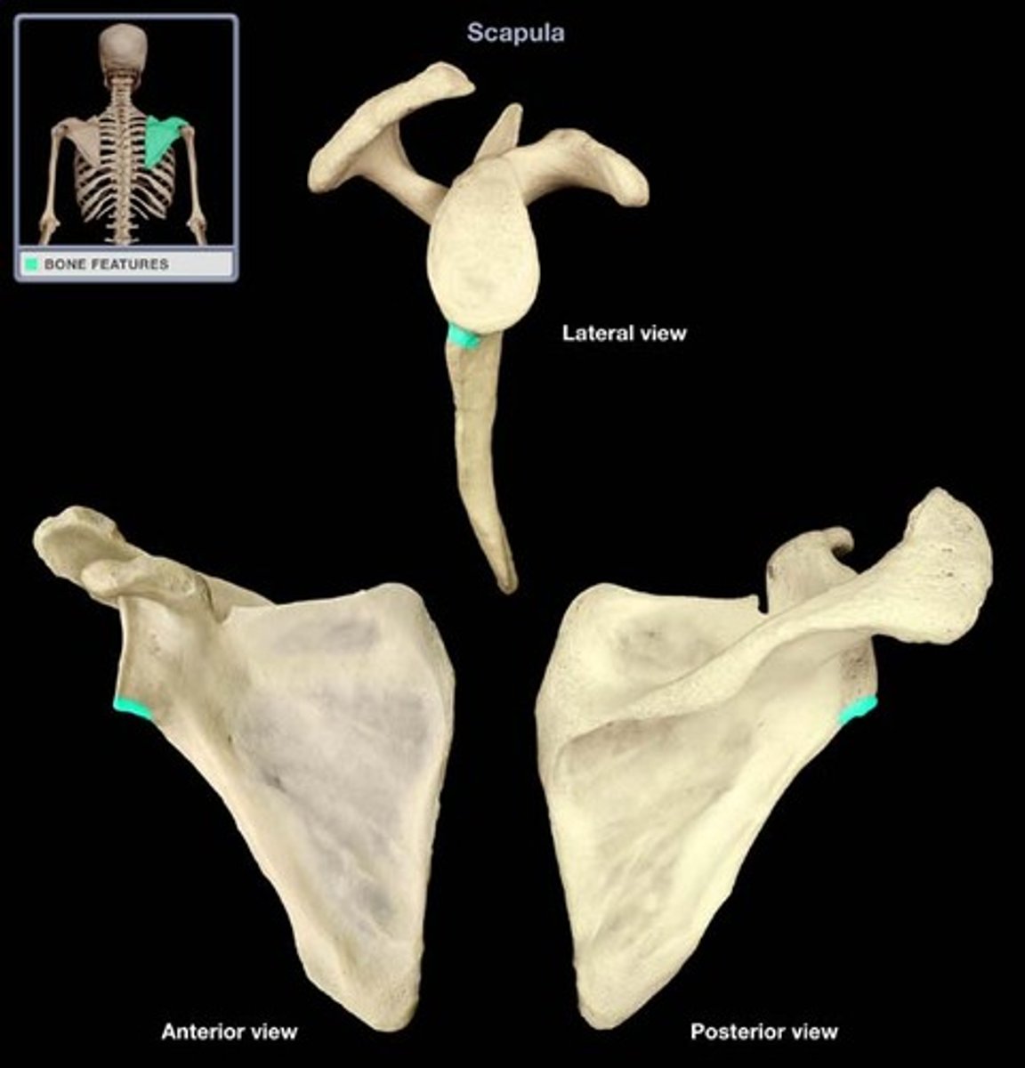
Scapula
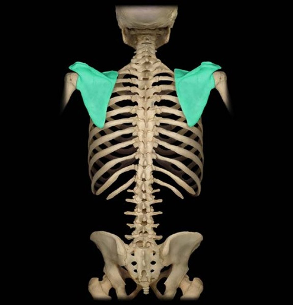
Cranial Border of Scapula
superior, shortest, and most irregular
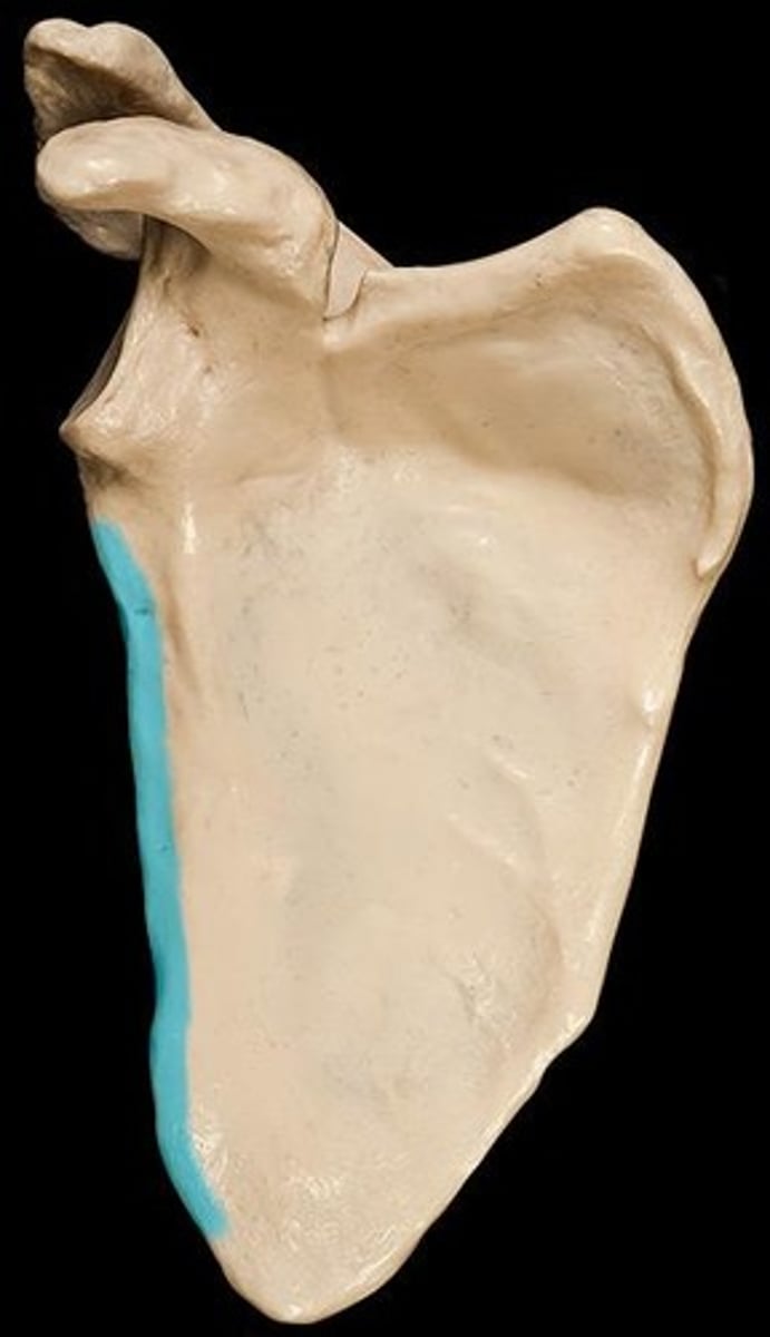
Axillary Border of Scapula
lateral, thickest, slightly concave
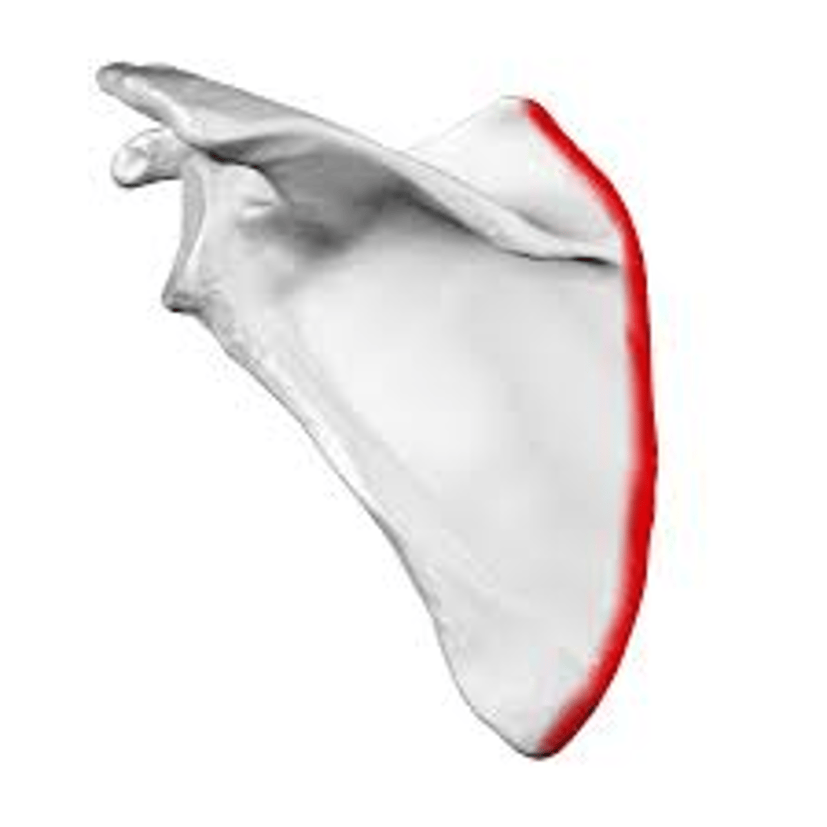
Vertebral Border of Scapula
medial, straightest, long and thin
Inferior Angle
the intersection of the axillary and vertebral borders

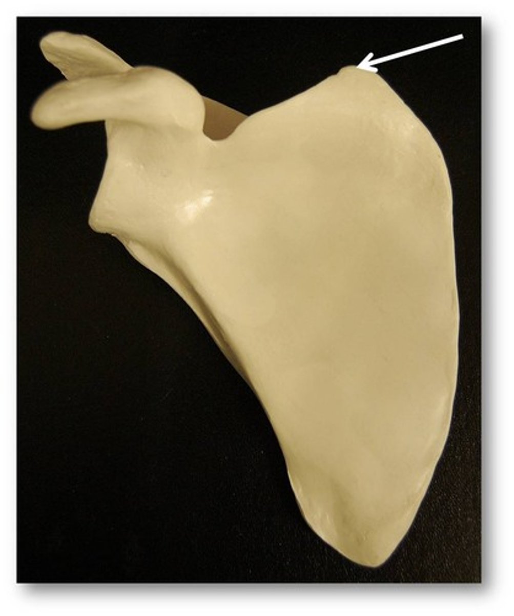
Superior Angle
intersection of the cranial and vertebral
Scapular Spine
raised projection of the posterior surface of the scapula
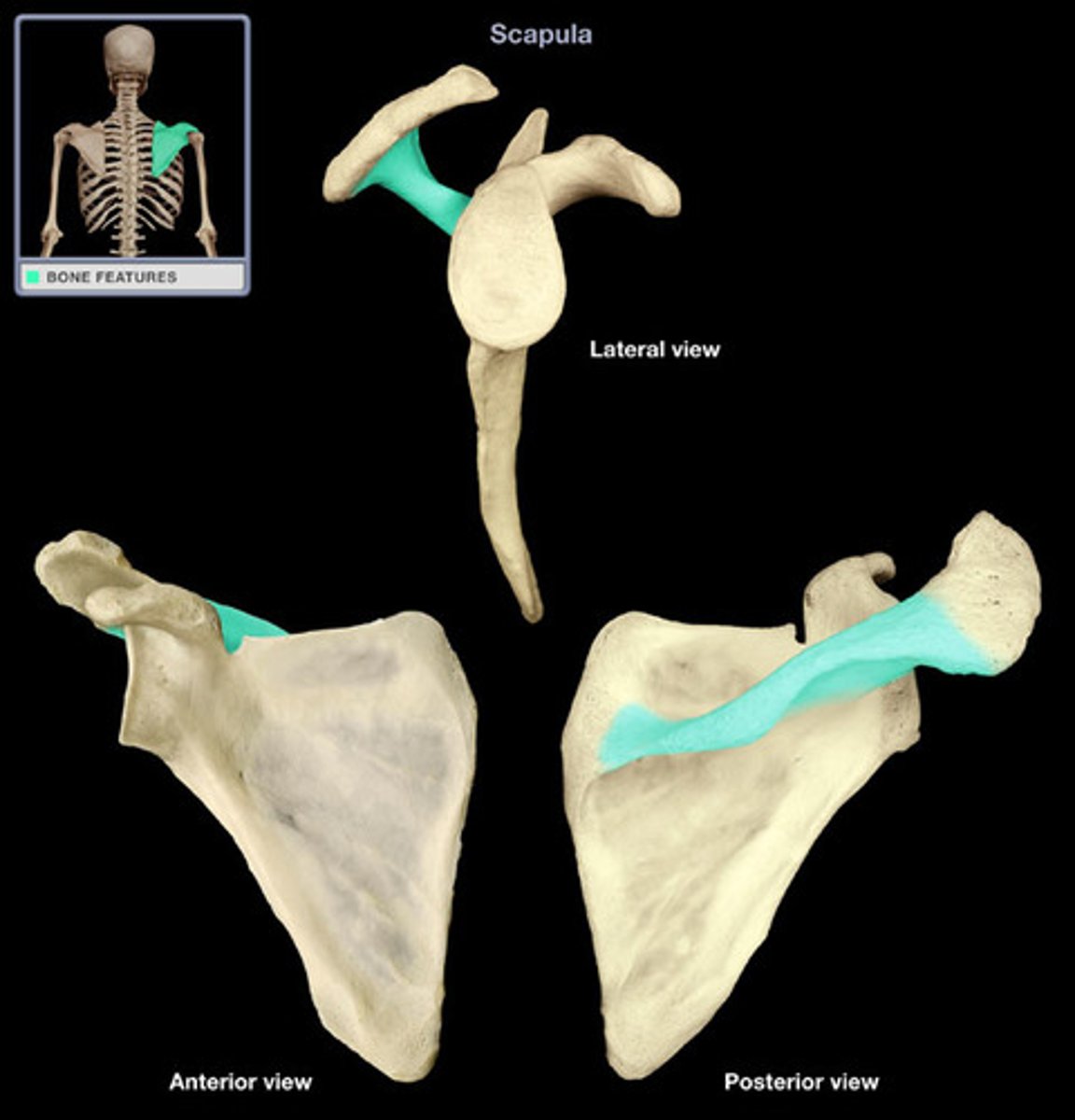
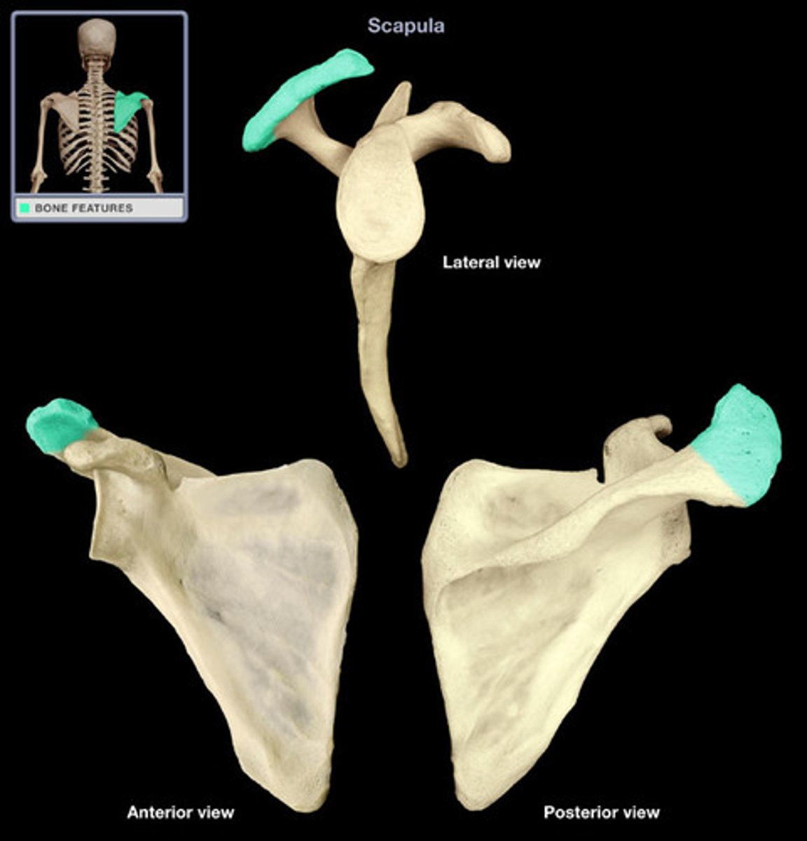
Acromion
the lateral projection of the scapular spine, bears the clavicular facet for articulation with the clavicle
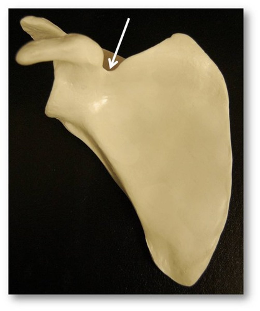
Scapular Notch
depression in superior border of scapula
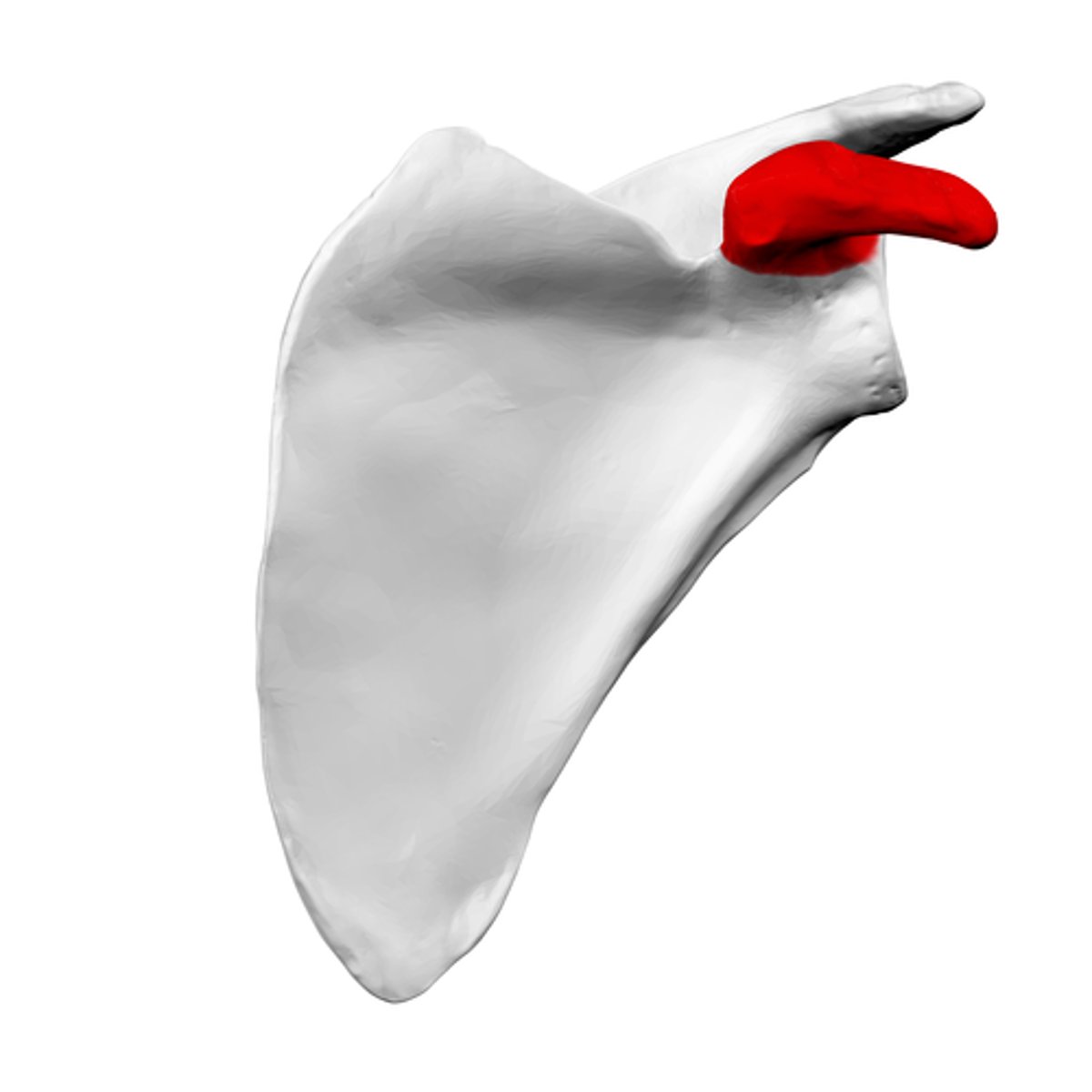
Coracoid Process
projects anterior and laterally from the cranial border
Supraspinous Fossa
superior to the spine
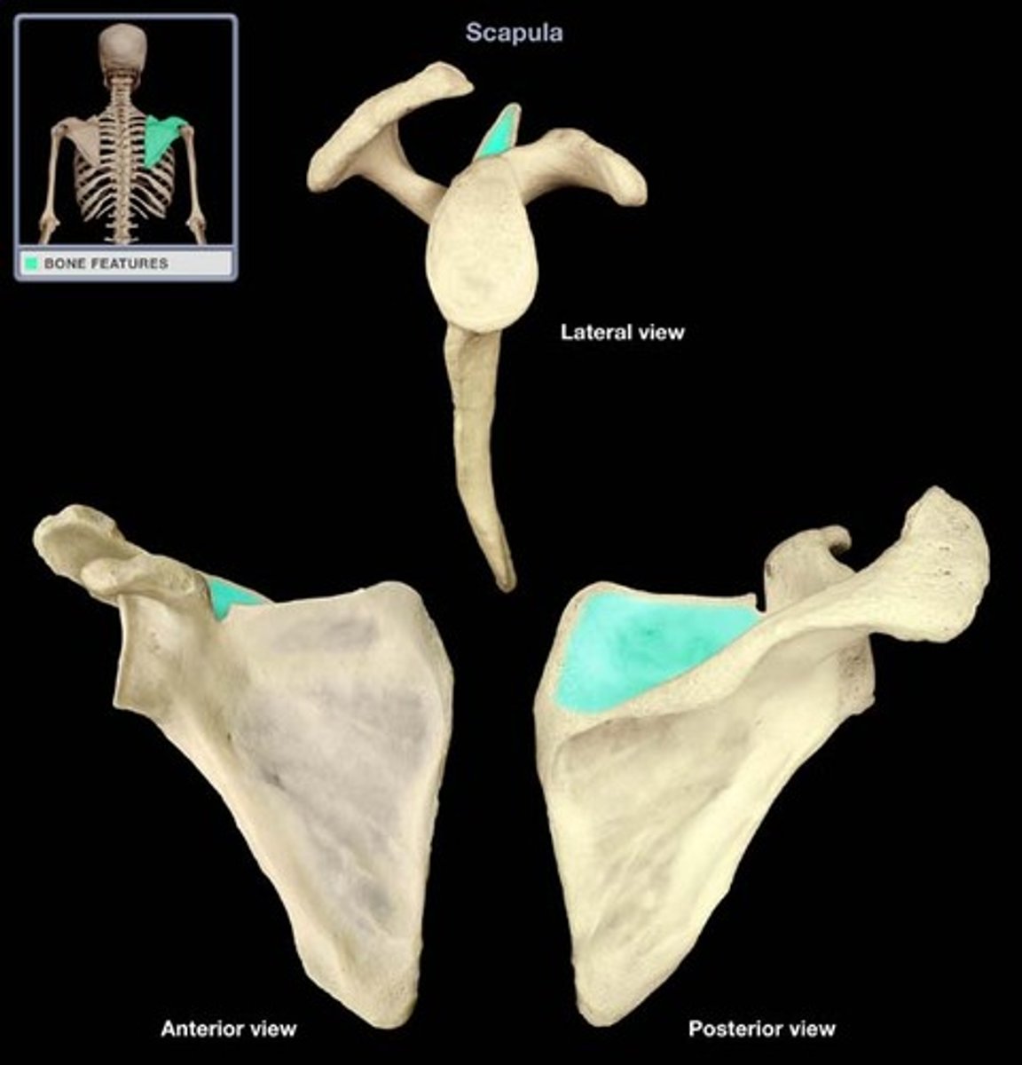
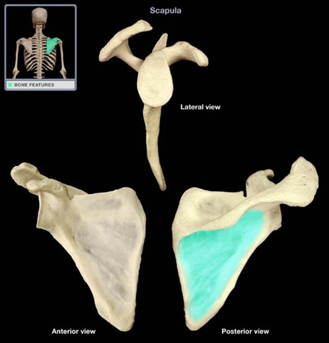
Infraspinous Fossa
inferior to the spine
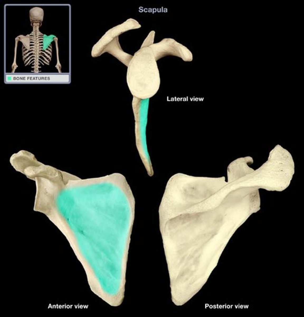
Subscapular Fossa (Anterior Feature)
concave surface of the anterior scapula
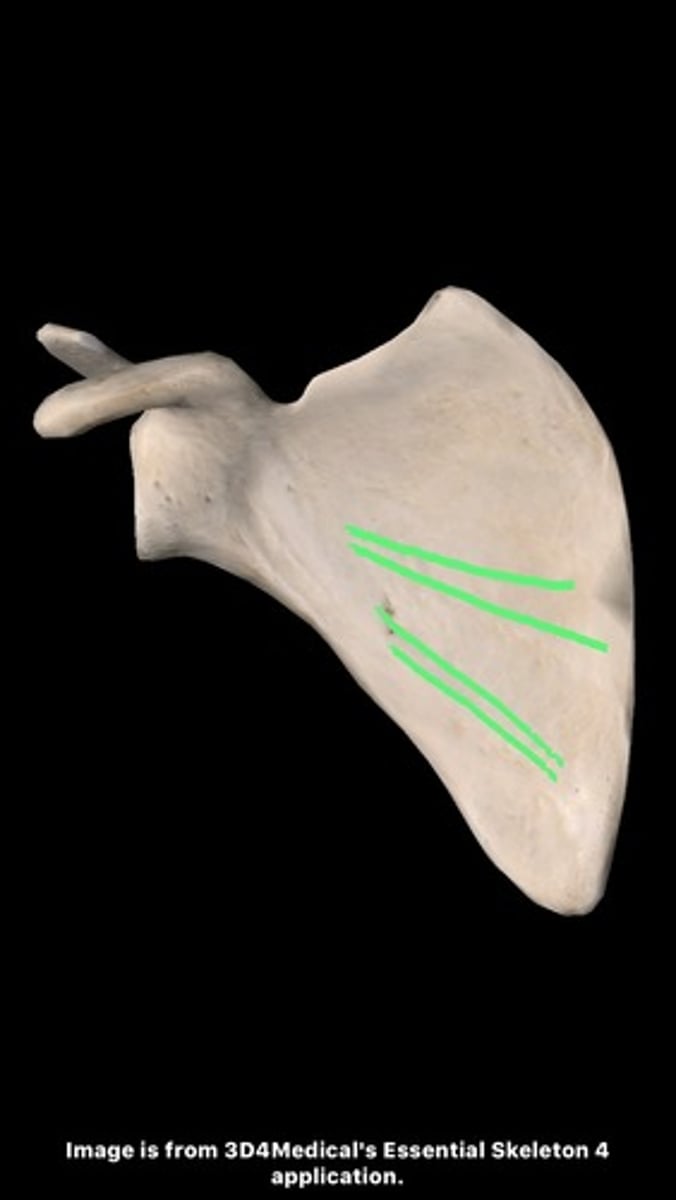
Oblique Ridges (Anterior Feature)
run superolateral to the inferomedial, are muscle attachments
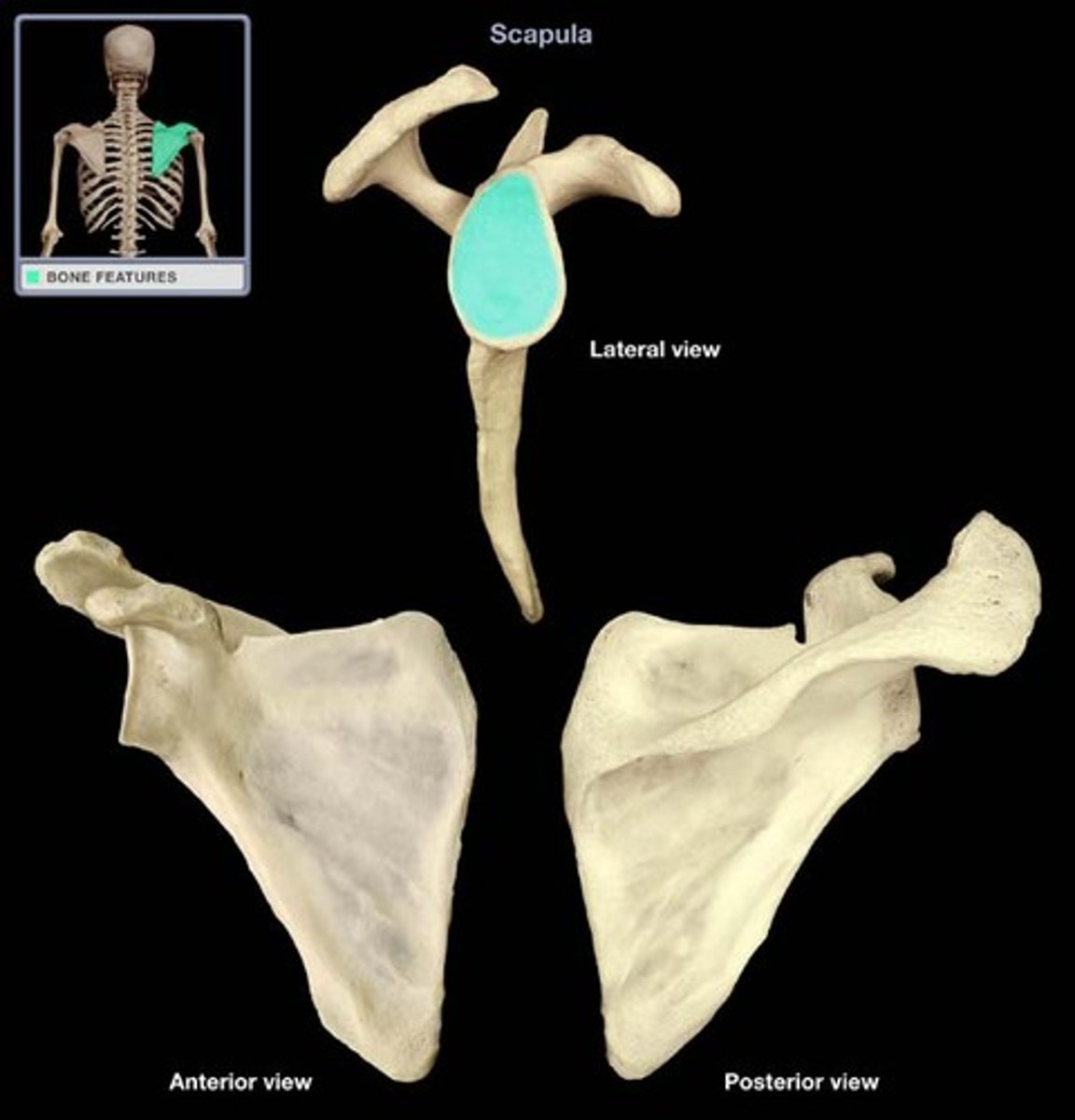
Glenoid Fossa (Lateral Feature)
the articular surface for the head of the humerus
Supraglenoid Tubercle (Lateral Feature)
anchors the long head of the biceps brachii muscle
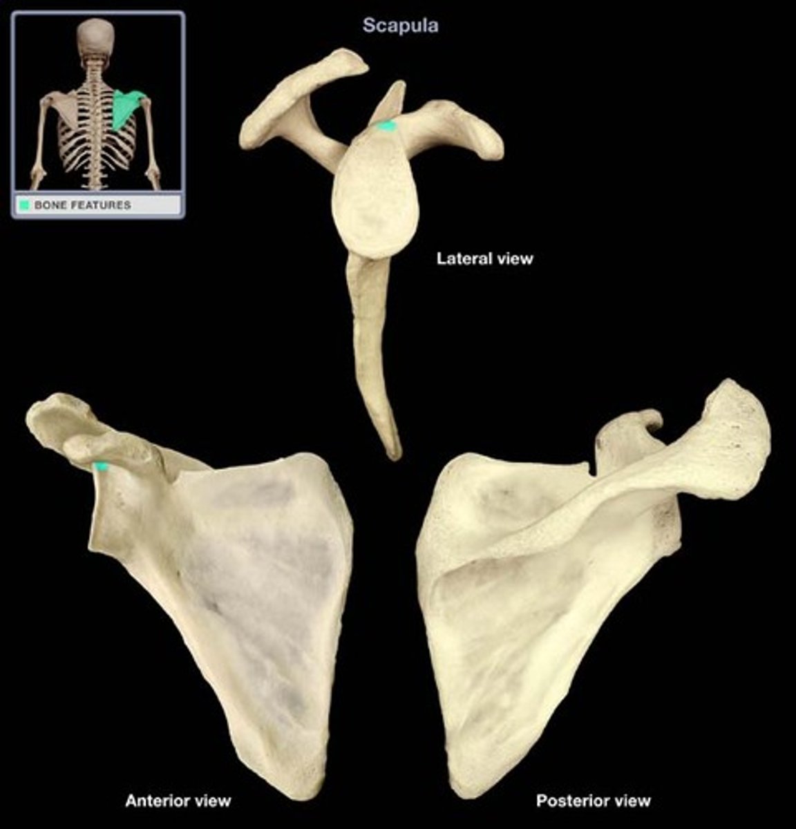
Infraglenoid Tubercle (Lateral Feature)
anchors the long head of the triceps brachii muscle
Shoulder Girdle
- composed of the clavicle and scapula (articulate with each other at the acromioclavicular joint)
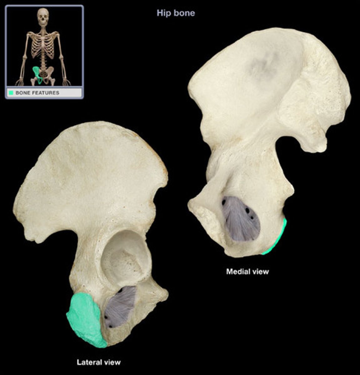
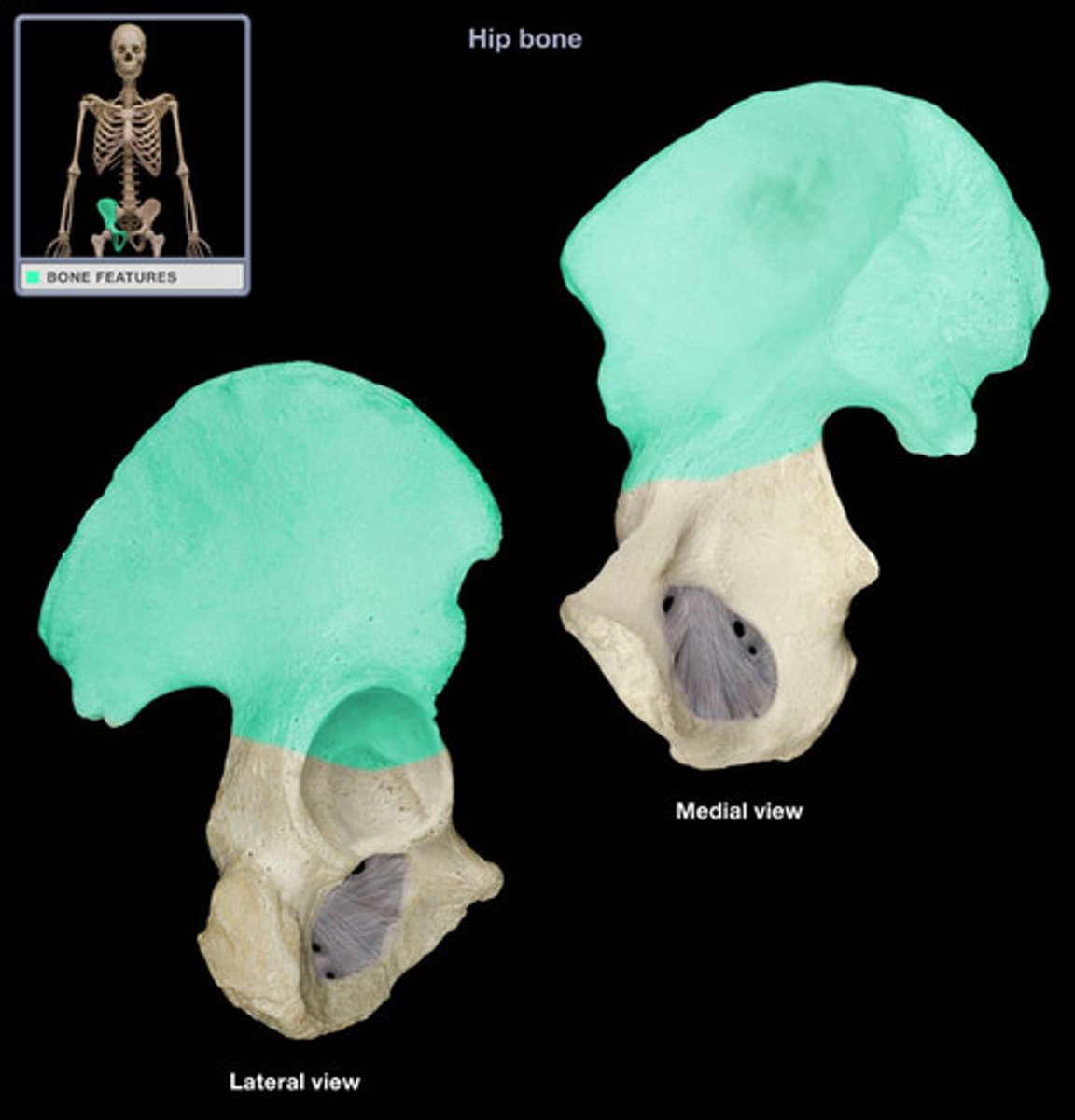
Os Coxa
hip bone; composed of the ilium, ischium, and pubis
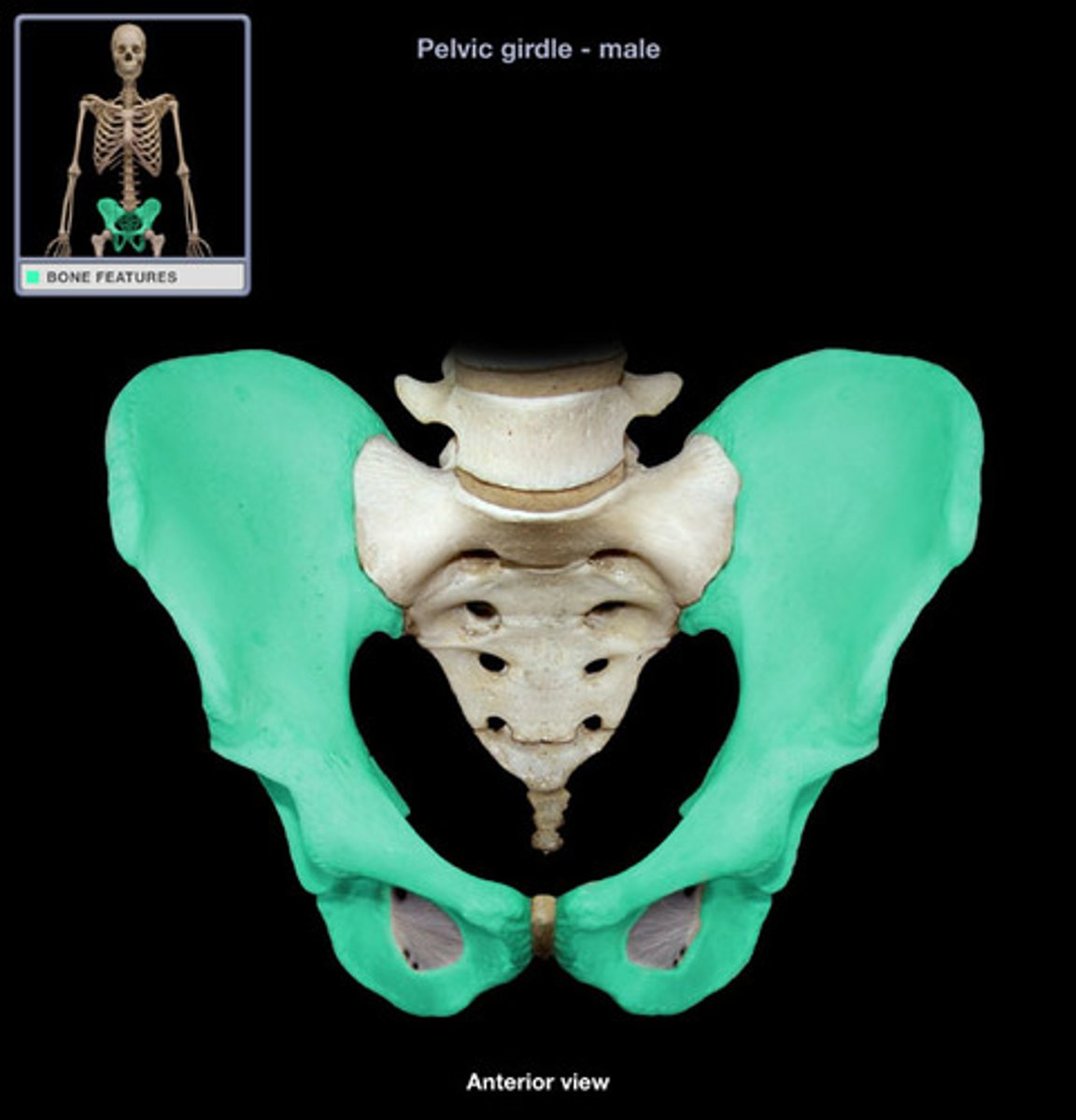
Ilium
large broad bone forming the upper part of each half of the pelvis
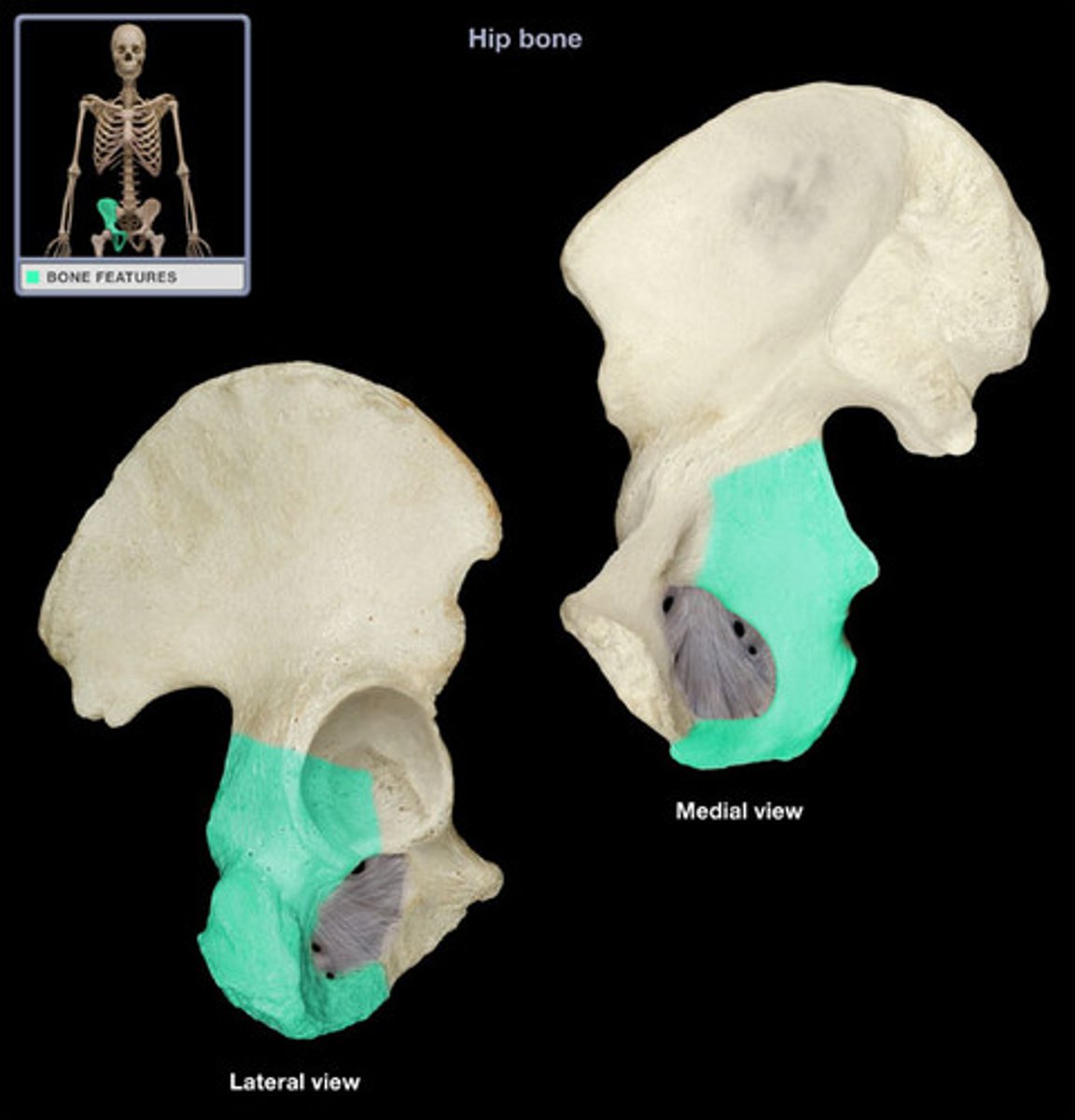
Ischium
the lower, posterior portions of the pelvis
Pubis
the medial anterior portion of the pelvis
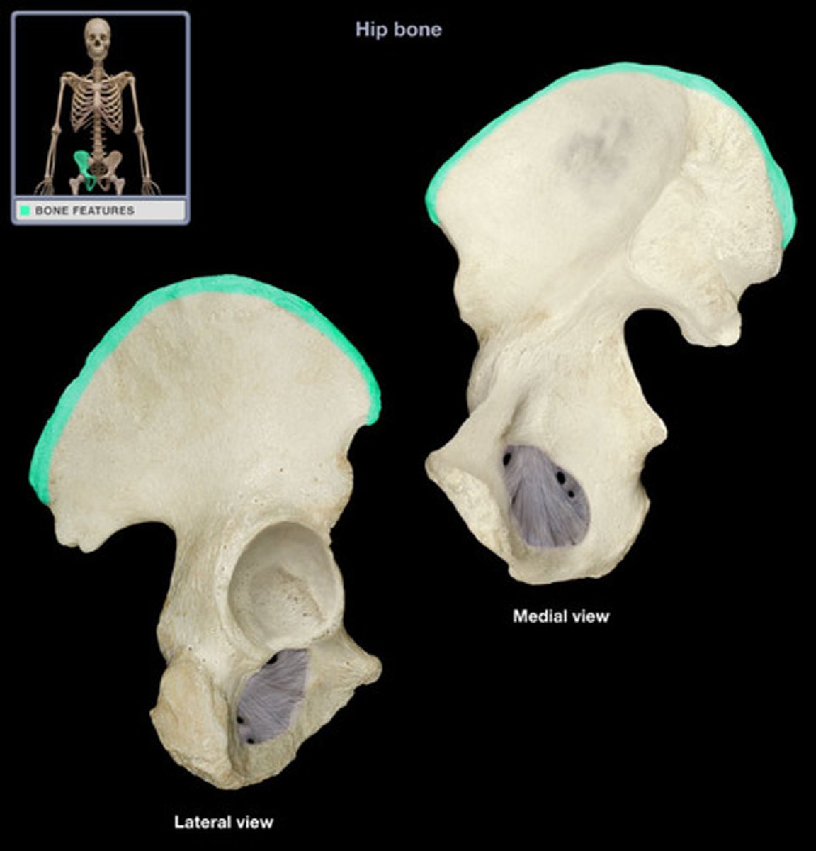
Iliac Crest (Posterior Ilium)
found on the top of the hip bone.
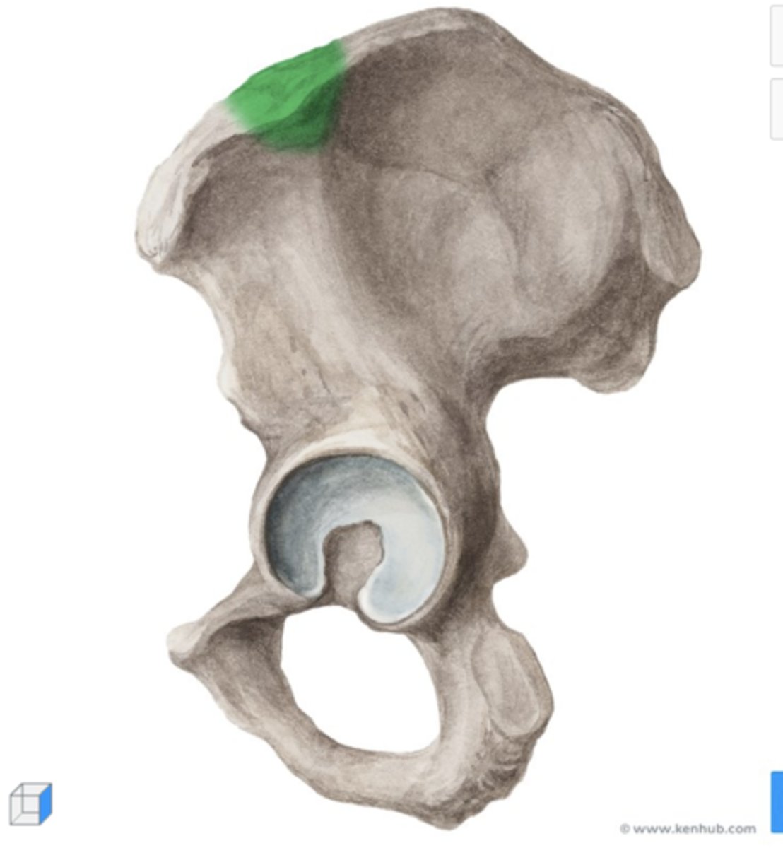
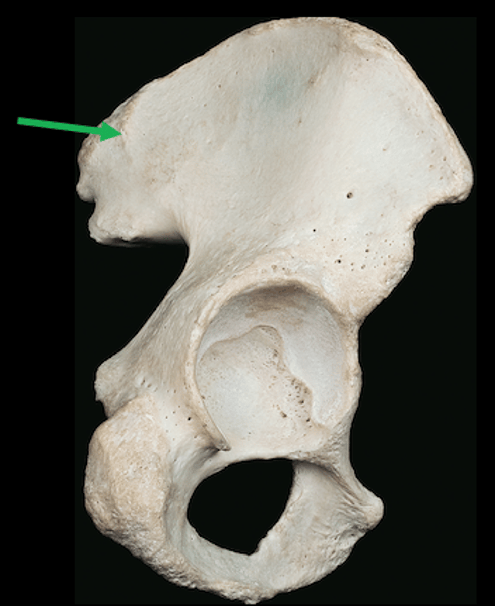
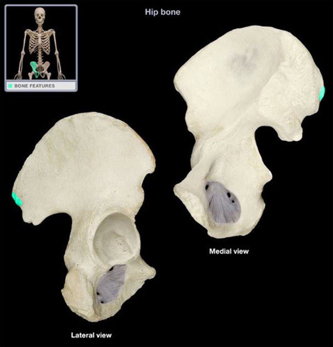
Iliac Tubercle (Posterior Ilium)
prominence found along the iliac crest
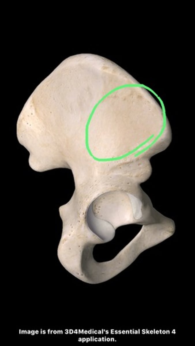
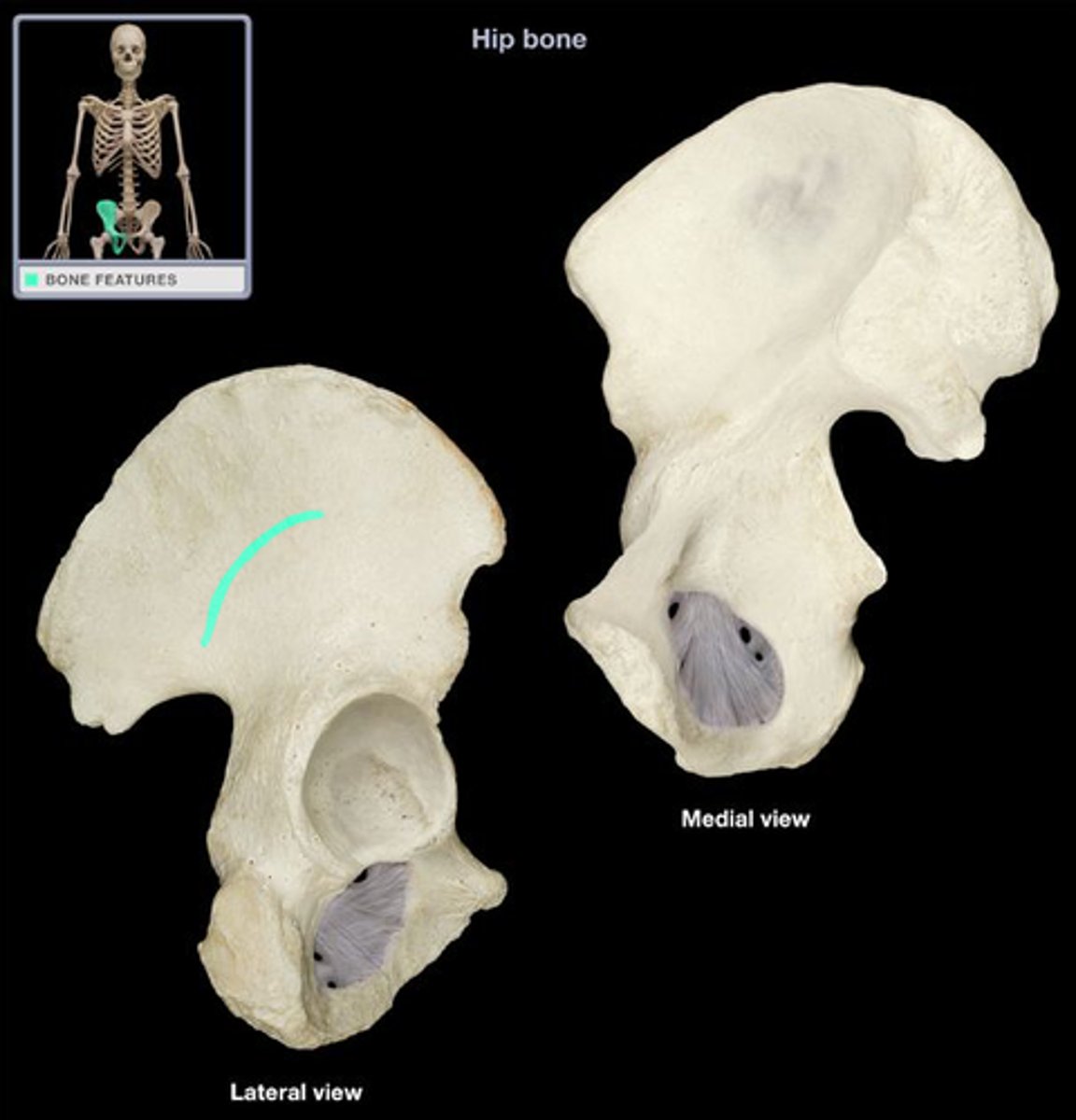
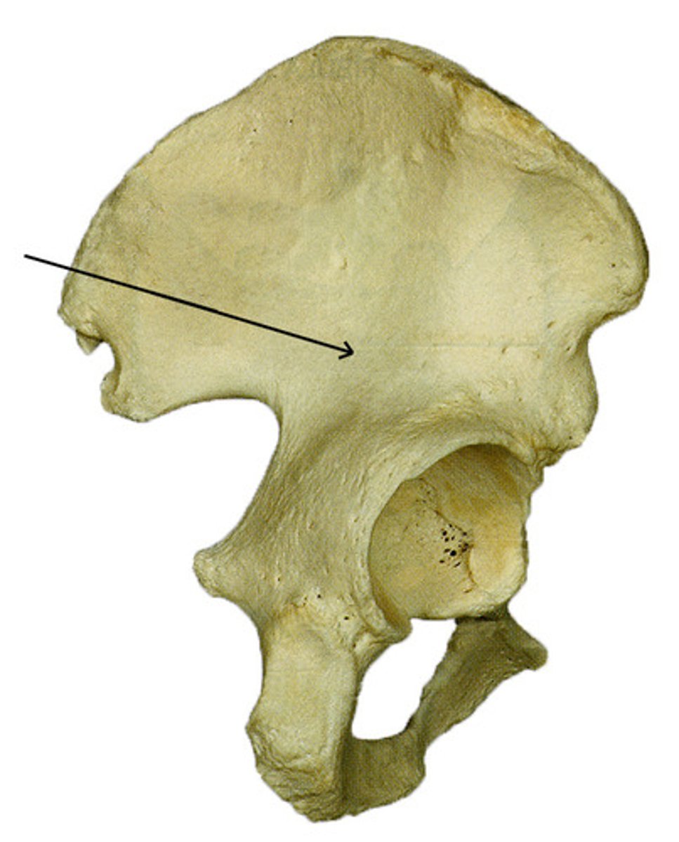
Iliac Pillar (Posterior Ilium)
dense, thick structural column of bone within the ilium of the pelvis
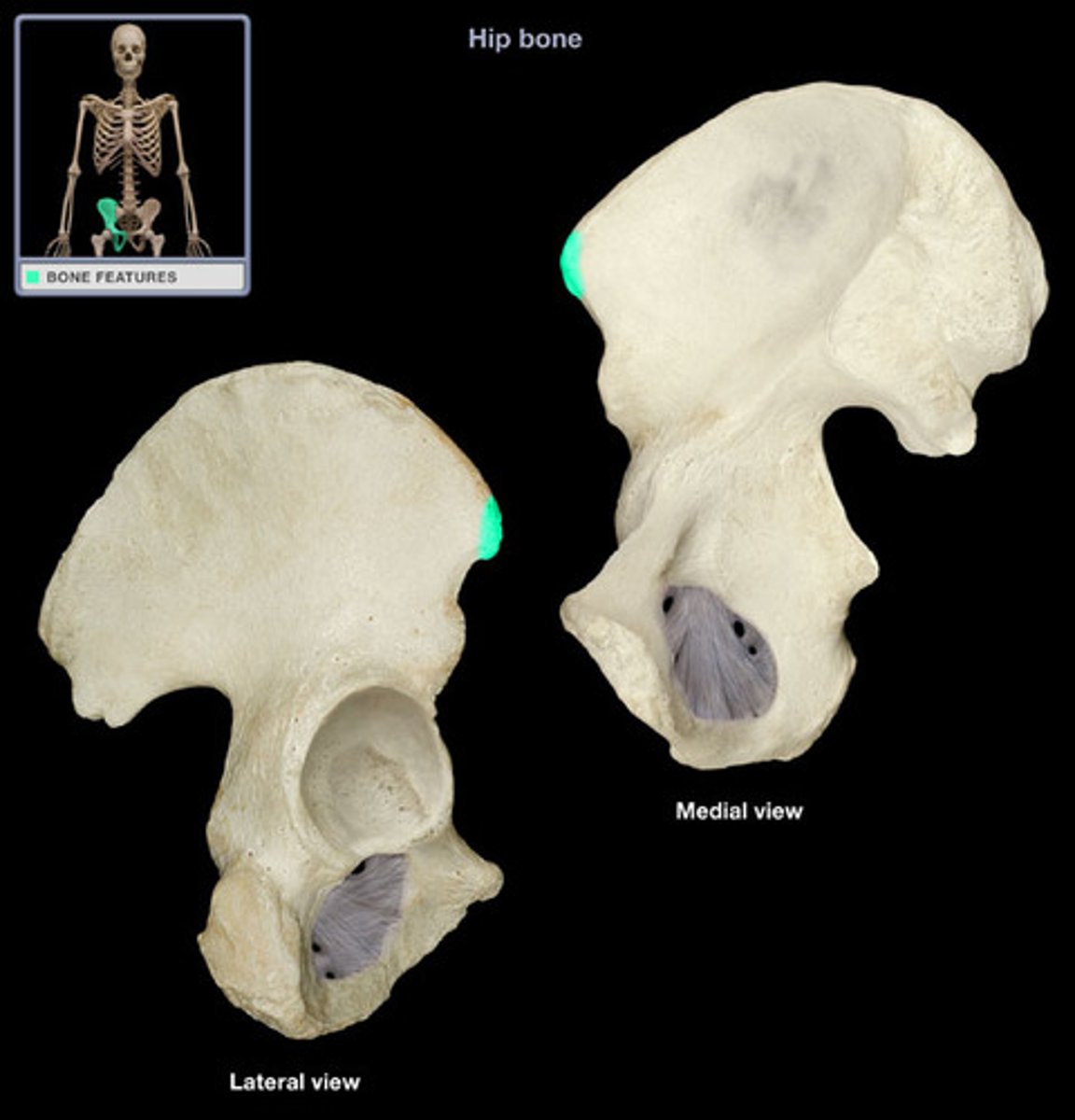
Anterior Superior Iliac Spine (Posterior Ilium)
bony projection at the anterior end of the iliac crest
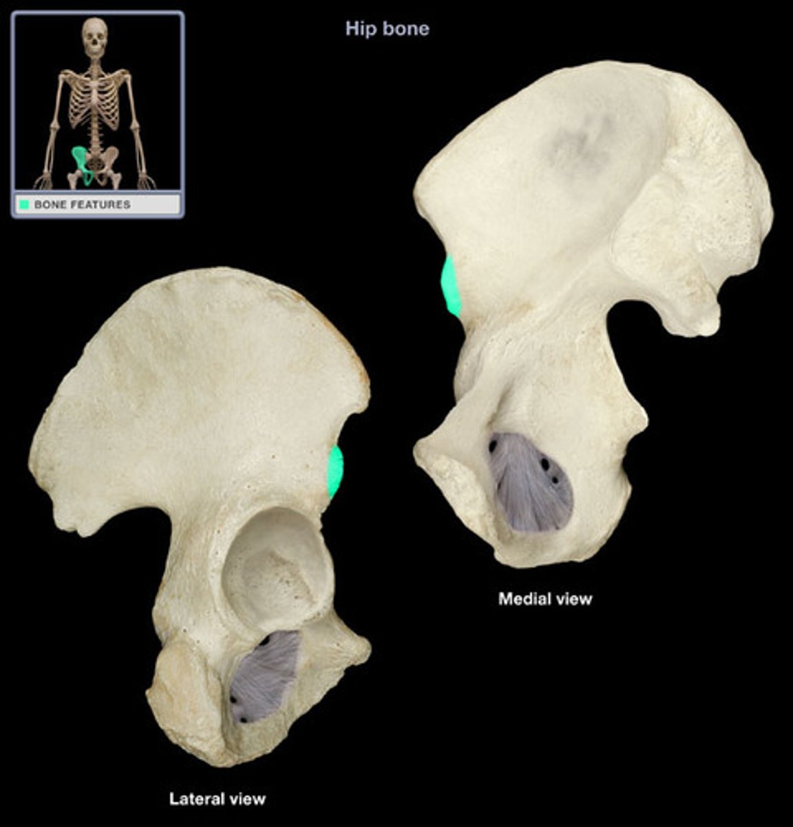
Anterior Inferior Iliac Spine (Posterior Ilium)
bony prominence of the ilium below the anterior superior iliac spine
Anterior Gluteal Line (Posterior Ilium)
longest of the three gluteal lines found on the external surface of the ilium
Posterior Gluteal Line (Posterior Ilium)
shortest curved ridge, located on the external surface of the ilium
Inferior Gluteal Line (Posterior Ilium)
faint, curved bony ridge located on the external surface of the ilium. starts behind the anterior inferior iliac spine and curves down to the greater sciatic notch
Posterior Superior Iliac Spine (Posterior Ilium)
the sharp posterior end of the iliac crest
Posterior Inferior Iliac Spine (Posterior Ilium)
small projection located just below the posterior superior iliac spine

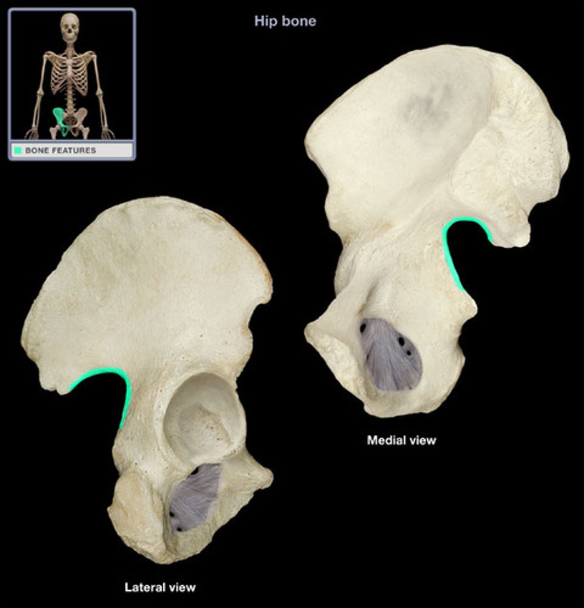
Greater Sciatic Notch (Posterior Ilium)
large, concave indentation on the posterior ilium of the hip bone
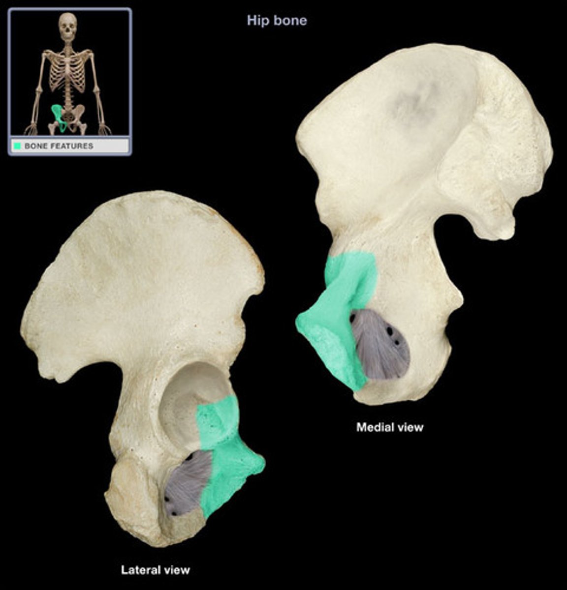
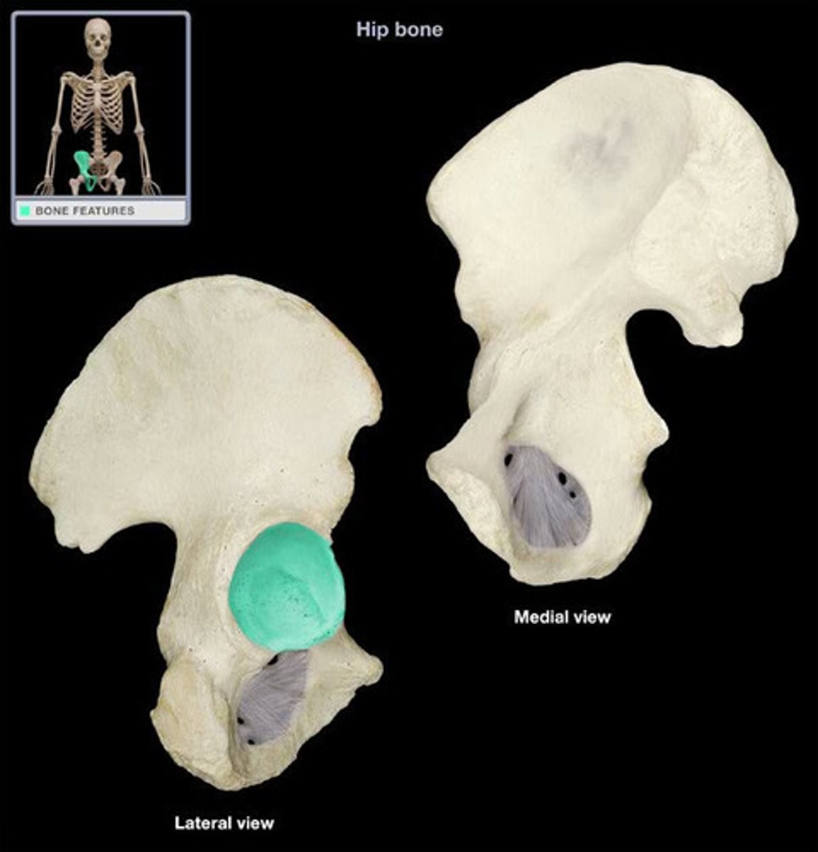
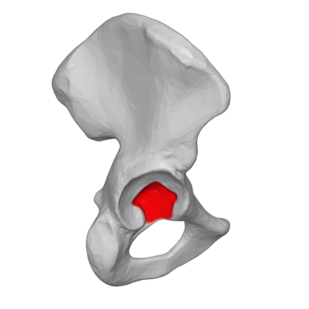
Acetabulum (Posterior Ischium, Ventral Pubis)
hip socket
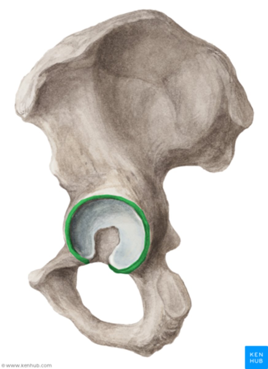
Acetabular Margin (Posterior Ischium, Ventral Pubis)
the lip (edge, rim) of the acetabulum
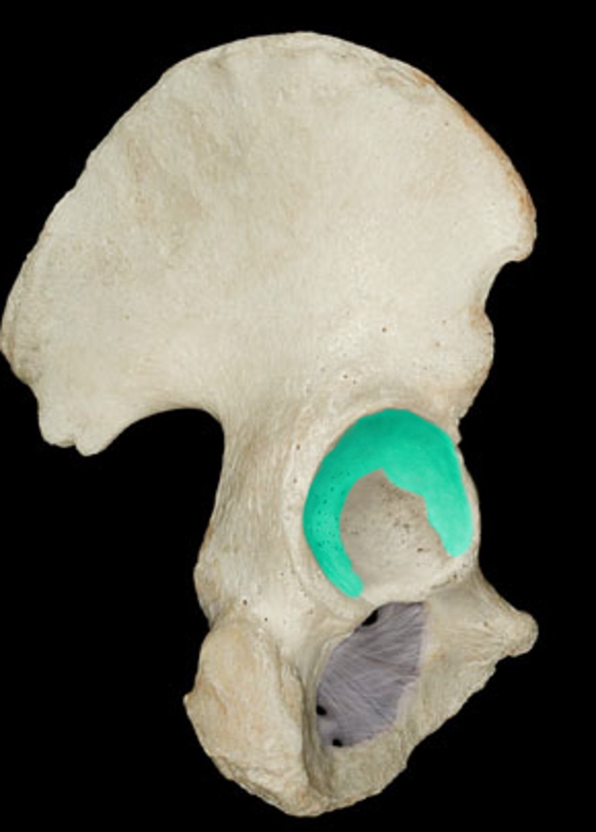
Lunate Surface (Posterior Ischium, Ventral Pubis)
smooth, cup shaped articular surface shaped like the moon
Acetabular Fossa (Posterior Ischium, Ventral Pubis)
circular depression located deep in the acetabulum
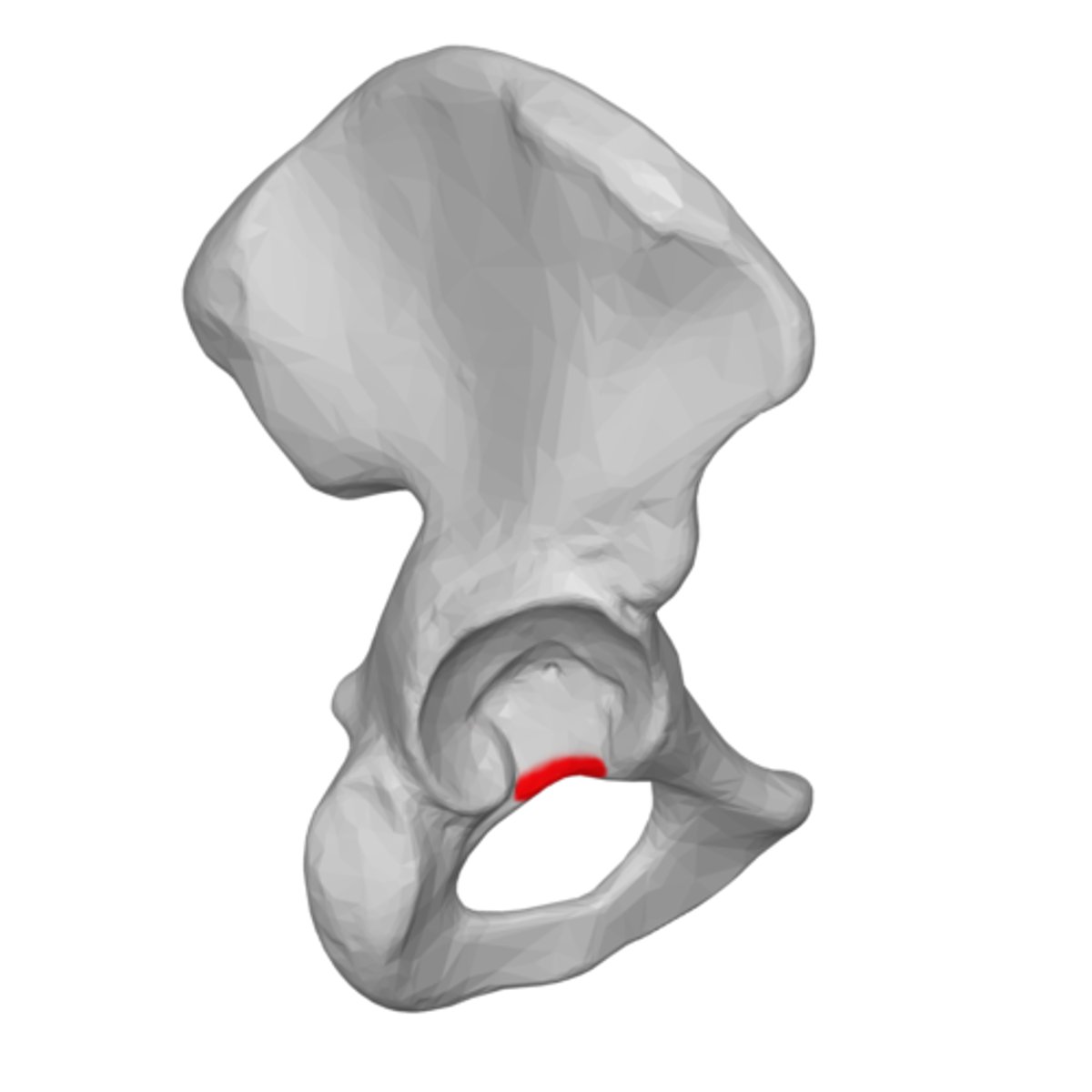
Acetabular Notch (Posterior Ischium, Ventral Pubis)
deep, distinct gap located in the inferior part of the rim of the acetabulum
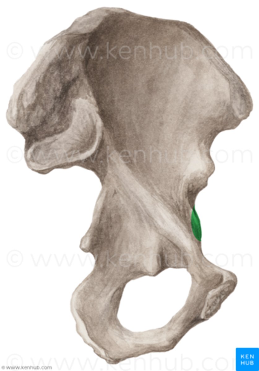
Iliopubic Eminence (Posterior Ischium, Ventral Pubis)
marks the point of union of the ilium and the pubis just lateral to the arcuate line
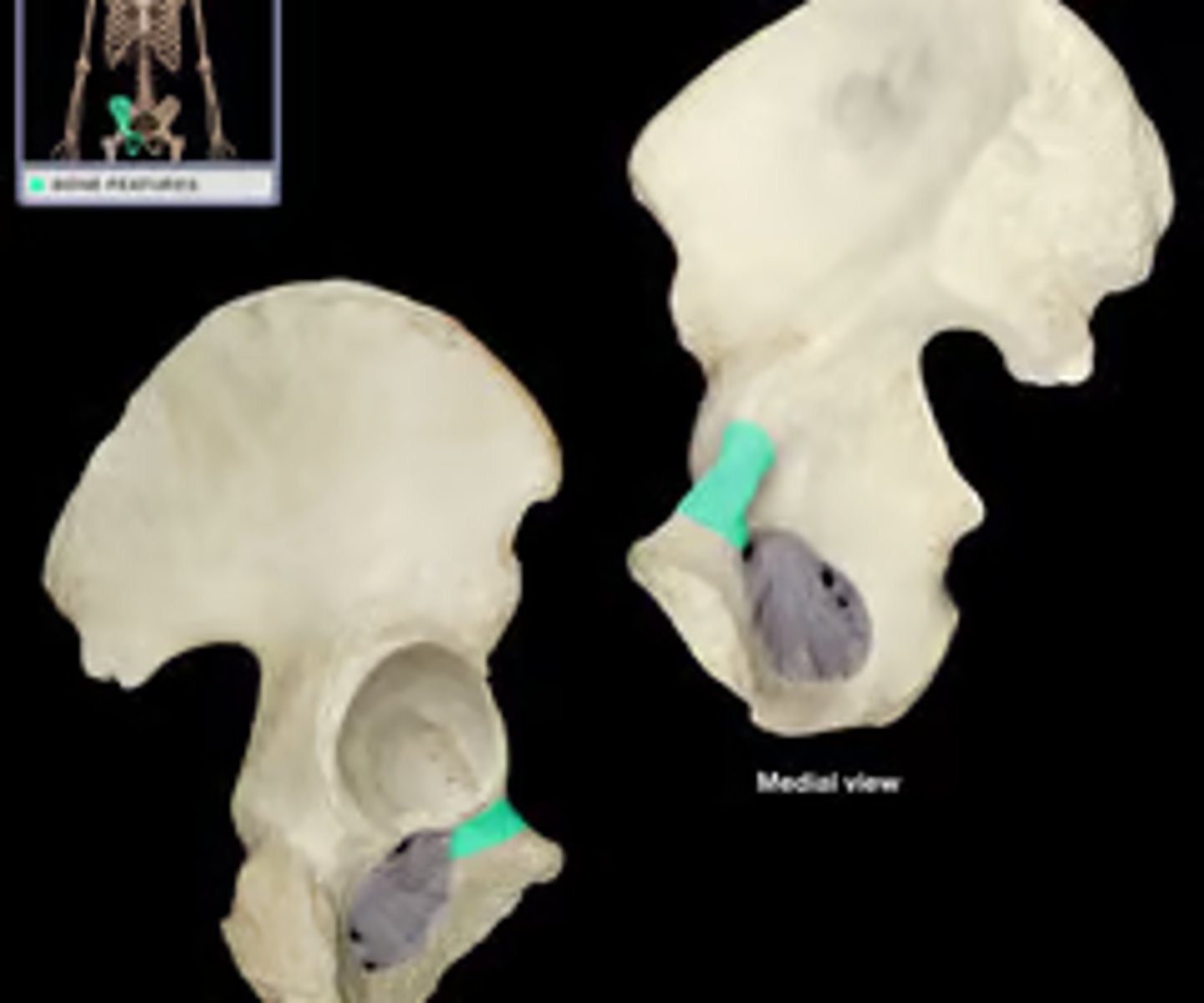
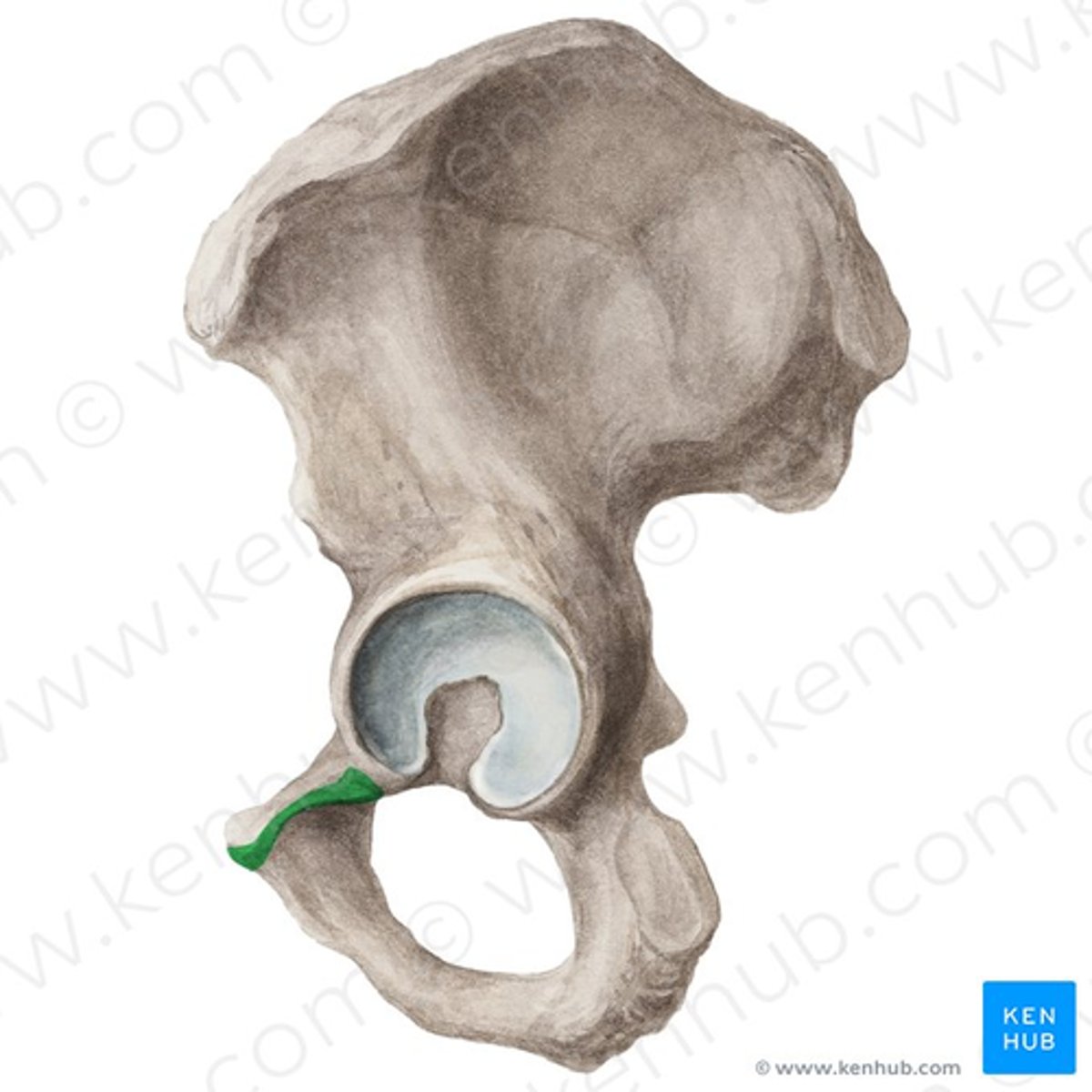
Iliopubic Ramus (Posterior Ischium, Ventral Pubis)
ridge of bone that bridges the ilium and pubis
Obturator Crest (Posterior Ischium, Ventral Pubis)
the sharp border between the pectineal and obturator surfaces of the pubis, at the anterior edge of the obturator groove
Pubic Body (Posterior Ischium, Ventral Pubis)
enlarged, medial portion of the pubis region of the hip bone
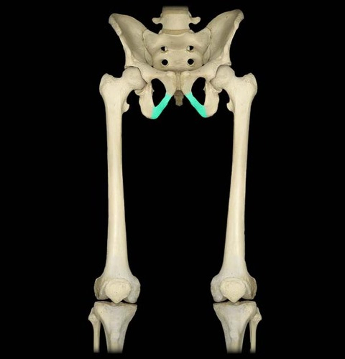
Ischiopubic Ramus (Posterior Ischium, Ventral Pubis)
narrow extension of bone that connects the ischial tuberosity to the pubic body; formed by the junction of the ischial ramus and inferior pubic ramus
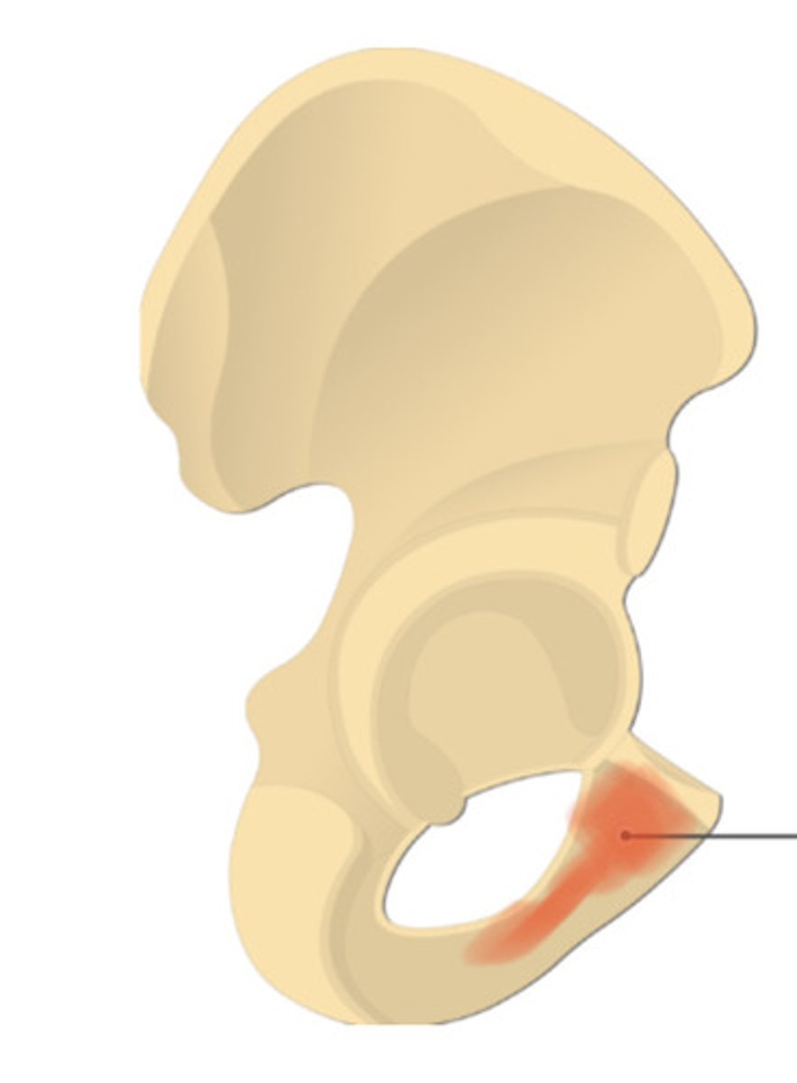
Ischial Tuberosity (Posterior Ischium, Ventral Pubis)
large bony prominence at the base of the pelvis that bears weight