Week 9- topic A- Anatomy of the urinary system
1/47
There's no tags or description
Looks like no tags are added yet.
Name | Mastery | Learn | Test | Matching | Spaced | Call with Kai |
|---|
No analytics yet
Send a link to your students to track their progress
48 Terms
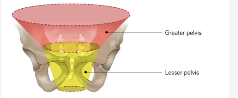
what 2 regions is the pelvis divided into
The pelvic region can be divided into a greater (false) and lesser (true) pelvis based on the bony pelvis. The lesser pelvis is what we know as the pelvic cavity.
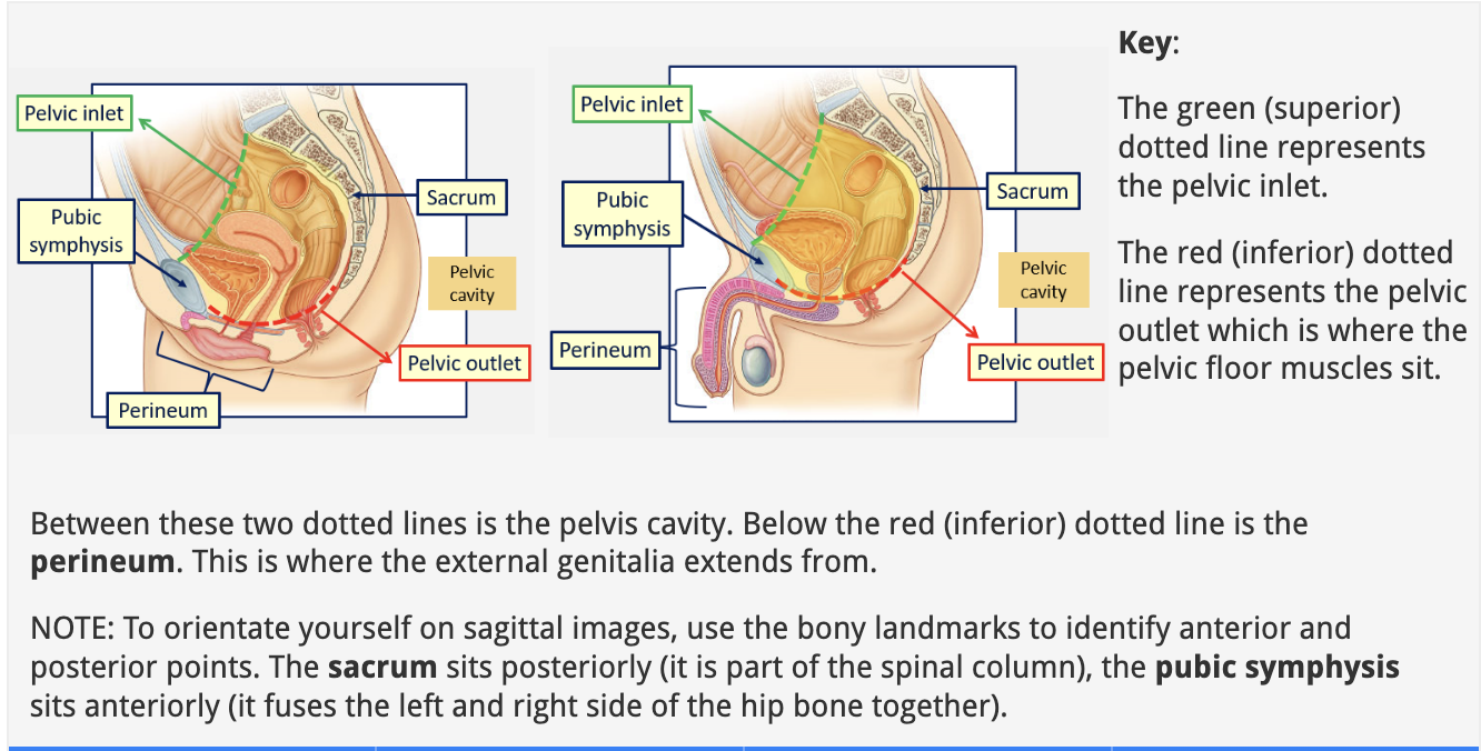
what sits between the pelvic inlet and pelvis outlet
the perineum- where the genitalia extend from
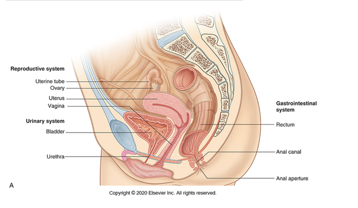
what does the female reproductive system consist of
The vagina
The uterus
The uterine (fallopian) tubes
The ovaries
The organs of reproduction sit in the lesser pelvis.
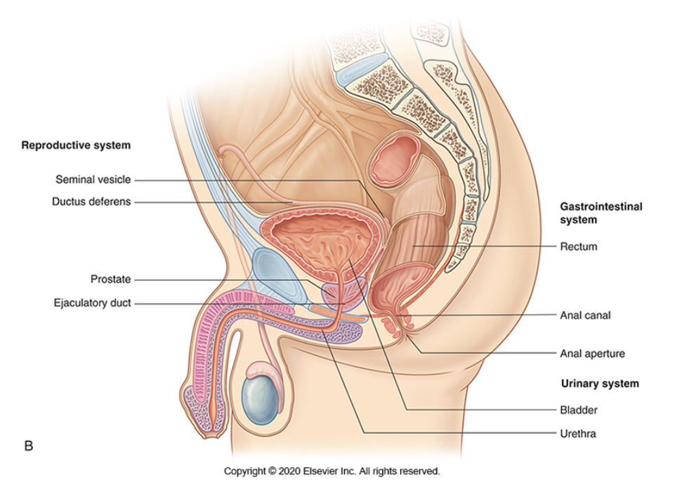
what are the internal organs of the male reproductive system
The testes (plural of testis)
The epididymides (plural of epididymis)
The ductus deferentes (pl. of ductus deferens)
The seminal vesicles
The ejaculatory ducts
The prostate
The bulbourethral glands
The testes sit outside the body in the scrotum. The other components sit within the lesser pelvis.
what does the urinary system consist of, what is its purpose
The urinary system consists of paired kidneys and ureters (right and left), a muscular urinary bladder and urethra. These structures sit in the abdomen, pelvic cavity and extend into the perineum/external genitalia.
The purpose of the urinary system is the excretion of urea and other toxins along with maintenance of blood volume and osmolarity.
what do the kidneys act to do (3)
Excrete most of the waste products of metabolism.
The kidneys filter the blood, removing waste products and producing urine.
The kidneys play a major role in controlling water volume and ion concentrations and in maintaining the acid/base balance of the blood.
describe the structure of the kidneys
The outer layer of tissue in the kidney is the cortex.
The inner layer of tissue in the kidney is the medulla.
Nephrons (see the next paragraph) in the cortex and medulla filter waste products from the blood, forming urine.
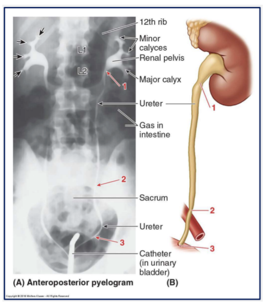
The pyramids of the cortex drain this into the minor calyces. Around 2-3 minor calyces drain into each of the 2-3 major calyces.
The major calyces drain into the renal pelvis (a funnel formed from the ureter leaving the kidney), which empties through the ureters towards the bladder.
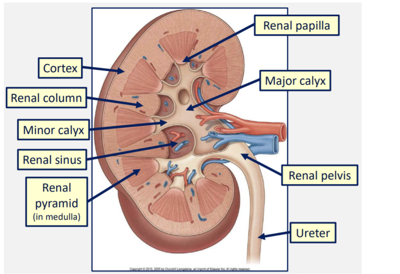
what is the hilum
The point where structures enter or exit the kidney is known as the hilum. This is circled in the image below.
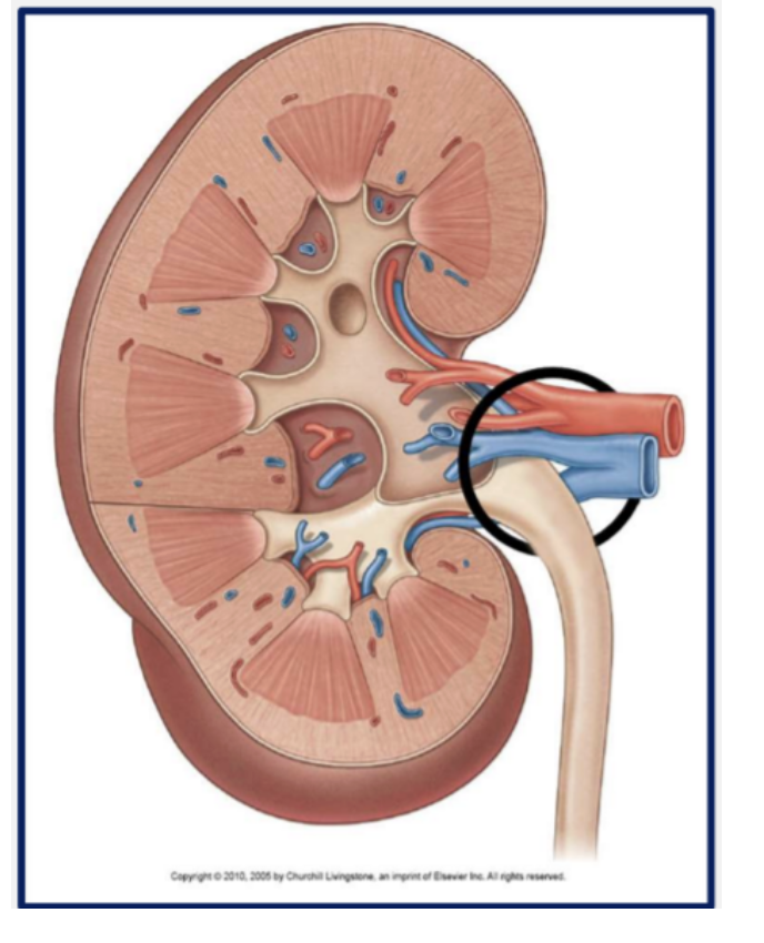
what structures enter/exit at the hilum
Renal vein
Renal artery
Ureter
Lymphatics & sympathetics

what is the nephron
The nephron is the functional unit of the kidney.
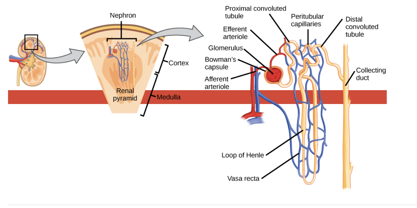
what happens in the glomeruli
Blood entering the kidneys for filtration passes through knots of capillaries called glomeruli.
The glomeruli are contained within the Bowman’s capsule.
Here, almost all of the constituents of plasma are filtered out of the blood, with the notable exception of large molecular weight proteins (>64KDa).
In particular, water and waste products are filtered out of the blood.
how are useful molecules like glucose and ions reabsorbed
In the proximal convoluted tubule, any substances that are useful to the body, such as glucose and ions, are reabsorbed.
In the Loop of Henle, water is reabsorbed.
In the distal convoluted tubule, any waste products that were not filtered out in the capsule are secreted into the lumen of the nephron.
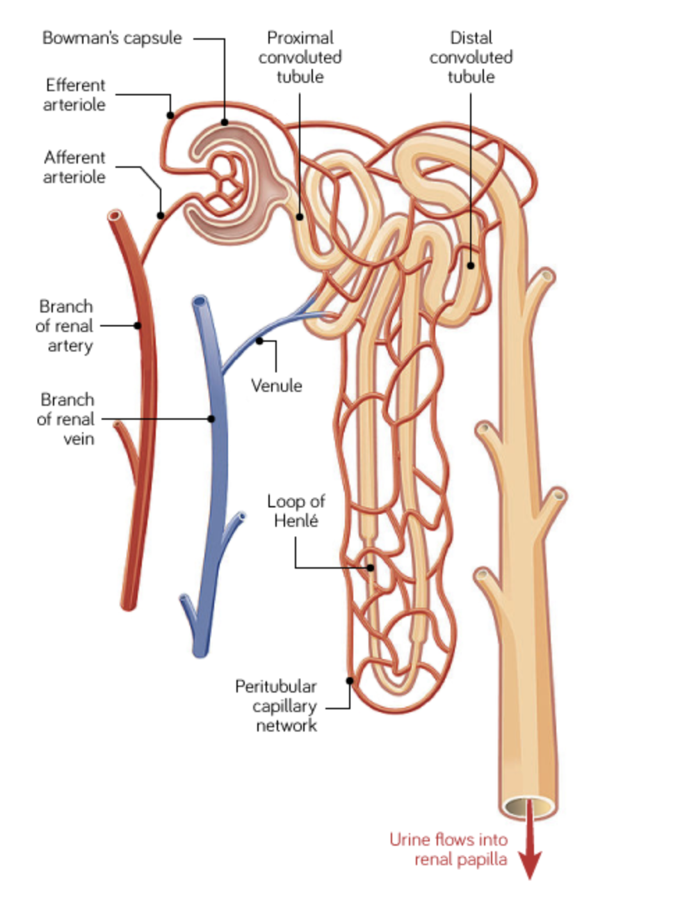
describe the microanatomy of the collecting duct
In the collecting duct, water and ions are reabsorbed into the blood.
It is reabsorption in the collecting duct that controls the volume of urine produced. The amount of water reabsorbed at the other sites remains constant.
The action of antidiuretic hormone, released from the posterior pituitary, on the collecting duct makes it more water permeable, allowing reabsorption of water.

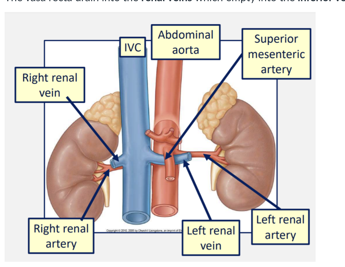
how is blood supplied to the kidney
The renal arteries are two large branches of the abdominal aorta that arise just below the level of L1.
These divide into afferent arterioles which feed into the glomeruli.
Efferent arterioles run from the glomeruli and wrap around the nephron.
Around the loop of Henle these are called the vasa recta.
The vasa recta drain into the renal veins which empty into the inferior vena cava.
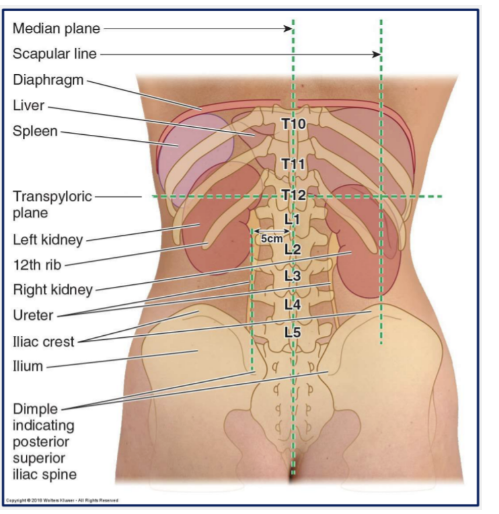
where are the kidneys located
The two kidneys lie retroperitoneally (behind the peritoneum) on the posterior body wall.
There is one on each side of the vertebral column
They sit at the level of T12 to L3 vertebrae
The kidneys are partially protected by ribs 11-12
The kidneys are related inferiorly to the diaphragm which also acts to separate them from the pleural cavities and 12th ribs

what is the difference between the 2 kidneys
The right kidney usually lies lower than the left due to the large right lobe of the liver

what is the shape and colour of the kidneys
In life, the kidneys red/brown in colour and are oval in shape (there’s a reason why kidney beans are called kidney beans!)
what relations do the kidneys have with other abdominal viscera (eg the diaphragm)
Both kidneys are related to the suprarenal, or adrenal, glands superiorly.
The right kidney is related to the liver, duodenum and ascending colon anteriorly.
The left kidney is related to the stomach, spleen, pancreas, jejunum and descending colon.
what are adrenal glands, where are they found
Located on the superior aspect of each kidney are the adrenal glands (also known as the suprarenal glands).
These are concerned with production of many of the body’s chemical messengers including adrenaline and steroid hormones.
what hormones are secreted from the adrenal cortex
Cortisol (glucocorticoid)
Aldosterone (mineralocorticoid)
Sex hormones (gonadocorticoids)
what hormones are secreted from the adrenal medula
Adrenaline (epinephrine)
Noradrenaline (norepinephrine)
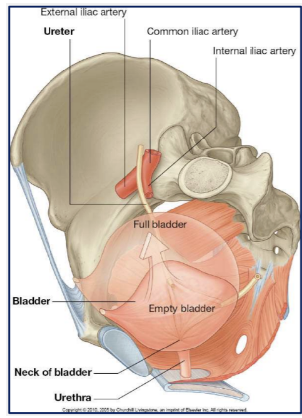
what are the left and right ureters
The left and right ureters are two muscular tubes that empty urine from their respective kidneys and carry it to the urinary bladder.

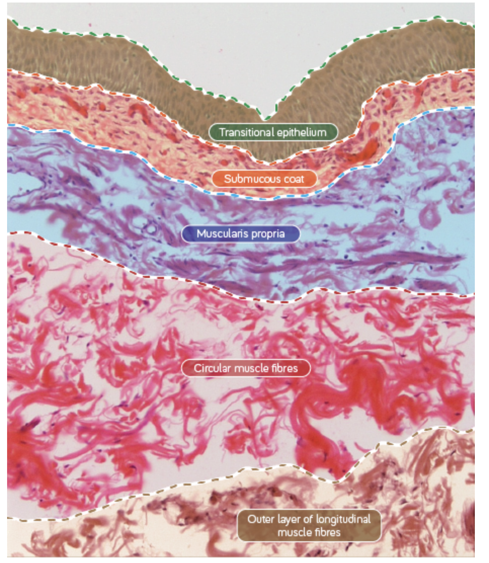
what makes up the right ad left ureter walls
Their walls consist of three layers of smooth muscle fibres that spiral around the tube and aid peristaltic contractions that force urine into the bladder.

where do the ureters descend from and enetr the bladder
They descend from the kidneys, behind the peritoneum, to enter the pelvis.
Their entrances to the bladder run obliquely through the muscle of the bladder wall.
When the bladder is full or contracting, these act as valves and prevent urinary reflux into the ureters.

whata re the 3 regions where ureters narrow
At the junction between the ureters and the renal pelvis
Where the ureters cross the brim of the pelvic bone
In the entrance of the ureters into the bladder
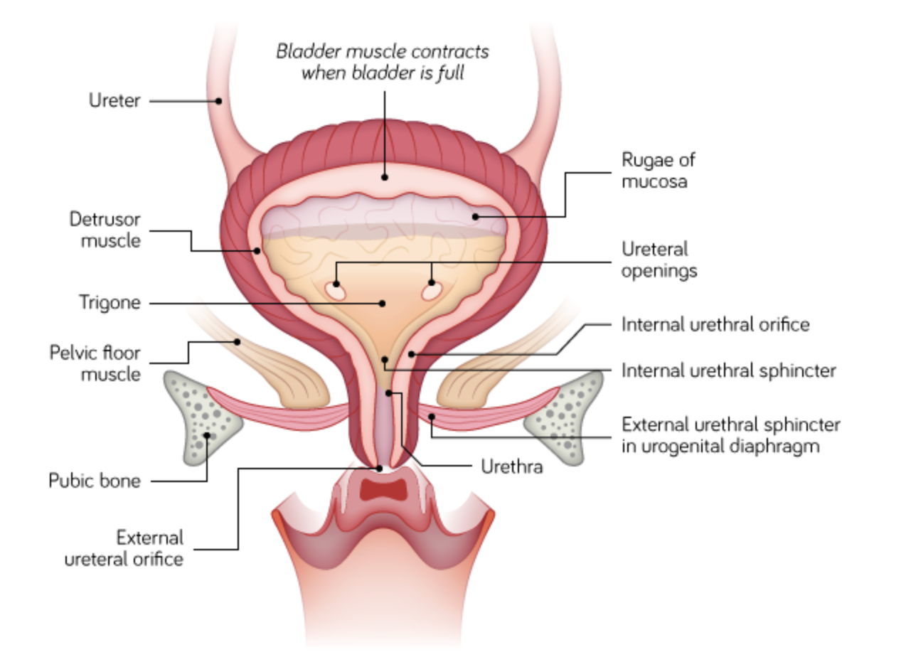
what are the basic features of the bladder
The bladder is a temporary reservoir for urine.
It can vary in size, shape, relations and position according to its content and the state of neighbouring viscera.
The bladder always contains some urine; the minimum amount is approximately 50 ml.
describe the microanatomy of the walls of the bladder
The wall of the bladder contains three layers of smooth muscle: an internal, middle and external layer. These form the detrusor muscle.
The fibres of the internal and external layers are in a similar longitudinal directional alignment. The middle layer is aligned in a roughly circular direction.
Bladder musculature is a distorted continuation of the three layers of spiral smooth muscle that surround the ureters.
what lines the urinary system from the renal pelvis to the uerthra
The urinary system from the renal pelvis to the urethra is lined with a specialised epithelium, transitional epithelium or urothelium.
what does the epithelium of the bladder have the ability to do
These cells have the ability to stretch, shift over one another and flatten.
The epithelium of a distended bladder may appear only two to three cells thick where as in an empty bladder it is usually five to six cells thick.
Non-distended urothelium has a cuboidal basal layer, polygonal celled middle layers and tall columnar cells in the surface layer.
where does the bladder sit in adults
In adults the empty bladder sits anteriorly in the lesser pelvis inferior to the peritoneum.
When full, it extends superiorly in the extraperitoneal fat of the anterior body wall.
where does the bladder sit in infants
In infants and children younger than six the bladder sits in the abdomen, even when empty.
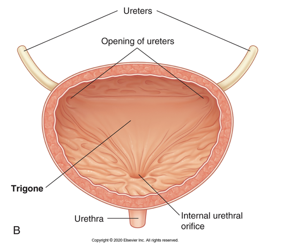
what is the trigone
Internally, on the posterior wall of the bladder between the ureteric orifices, there is a smooth triangular area, the trigone.
This is in contrast to the rugae (ridges) that line the majority of the empty bladder, flattening out as the bladder fills.
what is the internal urethral sphincter
At the junction between the bladder and the urethra is a smooth muscle sphincter, the internal urethral sphincter.
This is controlled by autonomic innervation.
The internal urethral sphincter is only found in males and it prevents ejaculatory reflux of semen into bladder.
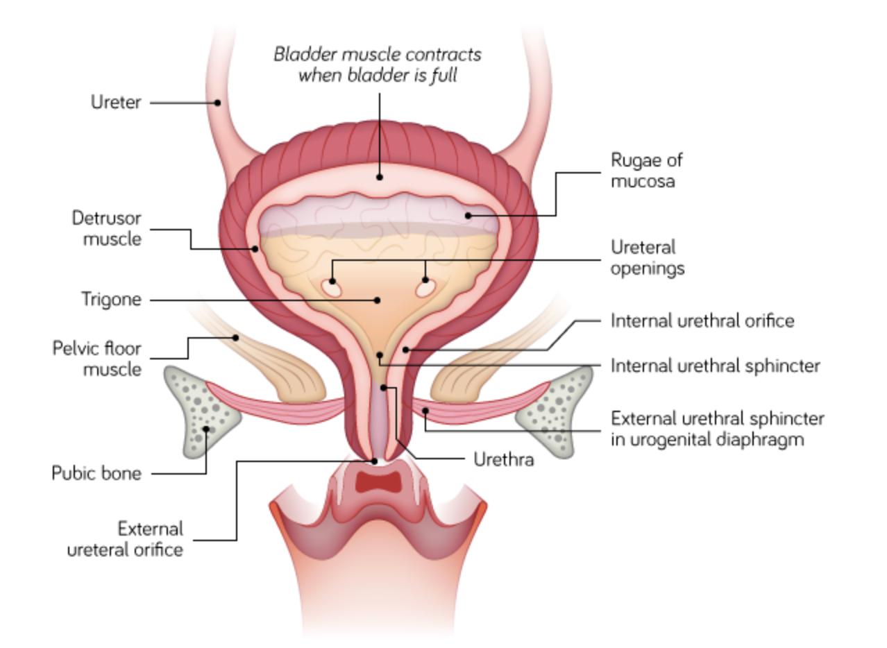
what is the urethra controlled by
The urethra is a continuation of the smooth muscle of the bladder but it also contains a skeletal muscle sphincter, the external urethral sphincter, which is under voluntary control.
what is the urethra
The urethra is a muscular-walled tube through which urine is expelled from the bladder during urination.
how is the urethra different in males and females
In males, the urethra also transports sperm and semen.
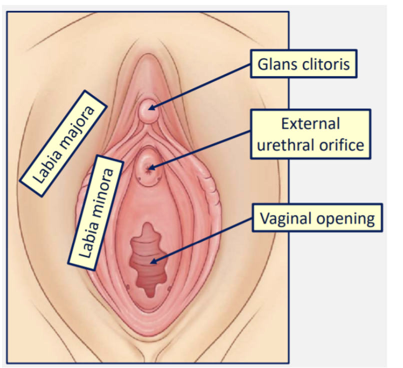
In females, there is a vaginal opening and a urethral opening (external urethral orifice)
In females, the urethra is short and straight. In males, it is longer.
The incidence of urinary tract infections is higher in females than males due to the shorter length of the female urethra and its proximity to the anus.
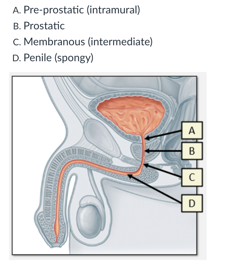
what are the 4 parts of the male urethra
Pre-prostatic (intramural)
Prostatic
Membranous (intermediate)
Penile (spongy)
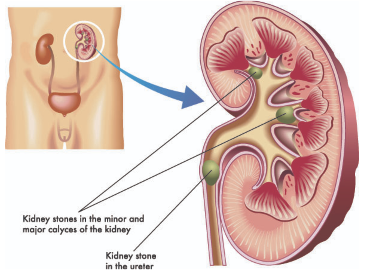
what are renal stones
Similar to gallstones blocking the outflow of the gallbladder, kidney stones (aka renal calculi) can form in the calyces of the kidneys and move into the ureters, blocking the outflow of the kidneys.
what is the difference in small and large kidney stones
Small kidney stones can pass through the urinary tract unnoticed.
Large kidney stones stretch the ureter and restrict the passage of urine, classically causing a severe flank pain that radiates to the ipsilateral groin
why might kidney stones damage the ureters
The stones may also cause some damage to the ureters as they pass through, leading to a microscopic amount of blood in the urine.
Certain sections of the ureters are usually narrower than others and it is in these sections that calculi can cause blockages.
what determines the treatment of kidney stones
The size, location, and composition of the stone determine the clinical treatment: smaller, more distal stones will be easier to pass naturally.
Options include shock wave lithotripsy to break down the stone into smaller fragments, surgery to stent the ureter, or pain relief and high fluid intake to encourage the stone to pass naturally.
what happens if kidney outflow is restricted by kidney stones
If the kidney outflow is restricted, waste products and pathogens can accumulate and cause infection of the kidney: this is pylenophritis.
Kidney infections can be severe and lead to sepsis, so the patient will need to commence treatment with antibiotics and be stabilised before the stone is managed.
what are urinary tract infections (UTIs)
Urinary Tract Infections (UTIs) are infections of the urinary tract which includes your bladder, urethra or kidneys.
what are causes of UTIs
Sexual intercourse
Being pregnant
Blockages within the urinary tract such as kidney stones
Not drinking enough fluids
Not keeping the genital region clean and dry
Antibiotics can be used as a treatment option.
what are the symptoms of lower urinary tract infections (LUTIs), who do they affect
Lower urinary tract symptoms (LUTS) are a common complaint for older men. The symptoms are generally classified as either:
Storage symptoms - difficulty storing urine in bladder. E.g. having to urinate frequently, urgently, and during the night.
Voiding symptoms - difficulty passing urine. E.g. weak stream that requires straining, starts and stops, and dribbling.
what are the most common causes of LUTIs
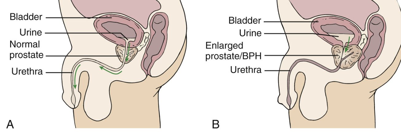
The most common cause of LUTS is a benign (non-cancerous) increase in the size of the prostate: benign prostatic hyperplasia (BPH).
If you recall that the urethra passes through the prostate, it makes sense that if the prostate is enlarged, the urethra is narrowed creating a higher pressure to urinate against.
why is a prostate exam performed
Importantly, some cases of prostate enlargement will be cancerous. Prostate cancer is very common, particularly with increasing age, but the majority of patients with prostate cancer will die from unrelated causes.
To help distinguish between BPH and prostate cancer a digital (meaning finger) rectal exam is performed.
what is the difference in the prostate when is growths are cancerous or benign
The posterior side of the prostate can be palpated through the anterior wall of the rectum.
Benign growths tend to feel smooth, symmetrical, and firm, whereas cancerous growths tend to be nodular, irregular and hard.