Dissection 2.1 - Superficial Thorax
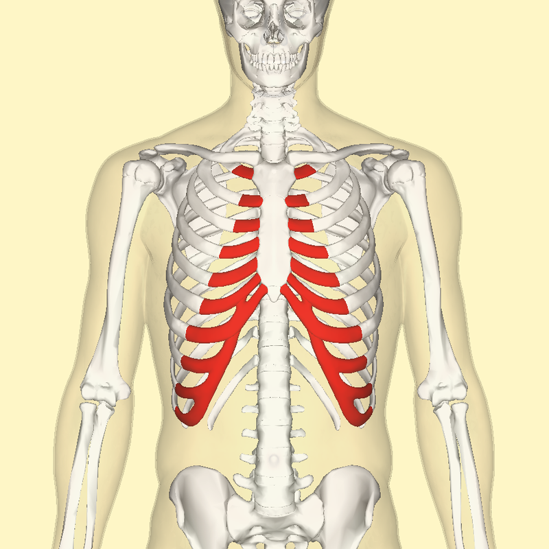
Body of the sternum
Part of sternum between manubrium and xiphoid process
Cephalic vein
Vein located between deltoid and pectoralis major
1/33
There's no tags or description
Looks like no tags are added yet.
Name | Mastery | Learn | Test | Matching | Spaced |
|---|
No study sessions yet.
34 Terms
Body of the sternum
Part of sternum between manubrium and xiphoid process
Cephalic vein
Vein located between deltoid and pectoralis major
Cephalic v. → Axillary v. → Subclavian v. → Brachiocephalic v. → Superior vena cava → Right atrium
Path of cephalic v. to R atrium
Clavicle
Bone located superficial to structures of the neck and thorax but deep to muscles of the thorax
SCM and Platysma
What muscles attach to the clavicle?
Corpus (body) of the mandible
Section of the mandible between the mental protuberance and ramus
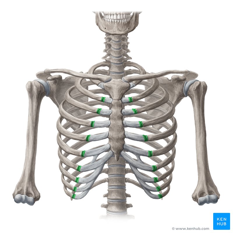
Costal cartilage
Hyaline cartilage that connects the ribs to the sternum
Costal groove
Groove located on the inferior border of ribs 2 to 12
By pulsations of the intercostal a.
How is the costal groove formed?
Intercostal VAN
What lays within the costal groove?
Inferior border of the ribcage
Costal margin
Costochondral joint
Joint between costal cartilage and rib
Dermatome
Strip of skin innervated by dorsal and ventral cutaneous nerves from a single spinal nerve
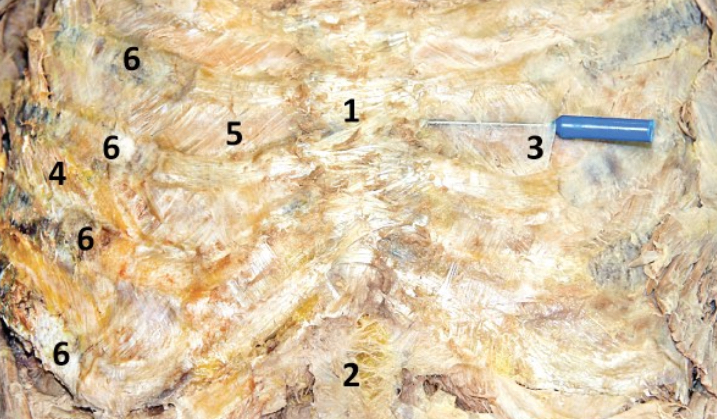
External intercostal membrane; 3
The most medial part of the external intercostal m. that is a semi-transparent membrane (directly superficial to the internal intercostal m.) - Include number in diagram

External intercostal muscle; 4
Muscles lateral to the costochondral joint - Include number in diagram

Elevate ribs and expand the chest cavity (inhilation)
Function of external intercostal m
Genu/symphysis of the mandible
Point of fusion where the 2 halves of the mandible join together during development (midline - near chin)
Gonial angle
Inferior angle of the mandible
Arch of aorta → L subclavian a. → Costocervical trunk → Intercostal a.
Path of intercostal a. from arch of aorta (L side)
Arch of aorta → R brachiocephalic a. → R subclavian a. → Costocervical trunk → Intercostal a.
Path of intercostal a. from arch of aorta (R side)
Ventral rami of 1st 11 thoracic n.
Where do intercostal nerves arise from?
Internal intercostal m.; 5
M. located deep to external intercostal membrane - Include number in diagram
Pull ribs down and in (reducing volume of thoracic cavity)
Function of internal intercostal muscle
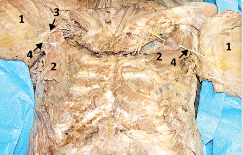
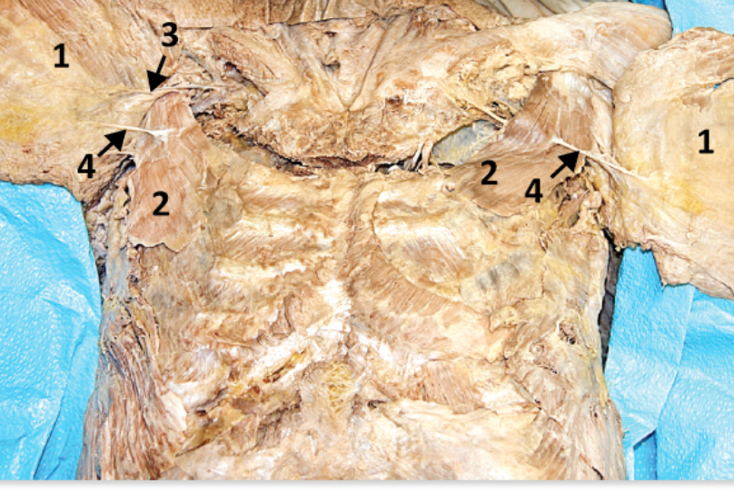
Lateral pectoral n.; 3
Supplies innervation to pectoralis major (located medially in pectoralis major) - Include number in diagram

Lateral cord of brachial plexus
What is the lateral pectoral nerve a branch of?
Medial pectoral n.; 4
Supplies innervation to pectoralis major (located laterally - pierces into to pectoralis minor) - Include number in diagram
Branch of medial cord of brachial plexus
What is the medial pectoral n. a branch of?
Pectoralis major; 4
Superficial muscle in thorax (innervated by medial and lateral pectoral nerves) - Include number in diagram
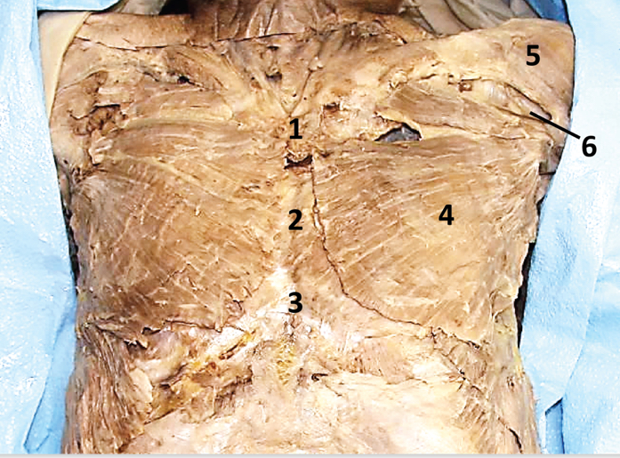
Pectoralis minor; 2
Deep to pectoralis major (innervated by medial pectoral n.) - Include number in diagram
Platysma
Thin muscle embedded in the superficial fascia deep to the skin of the anterior neck (innervated by CN VII)
Sternocostal joint
Joint between manubrium and costal cartilage which joins the anterior ends of the 1st rib
Subcostal n.
Ventral ramus of T12 that lies below the 12th rib
Suprajugular notch
Notch at the superior margin of the manubrium
SS, SS, ANS
Fiber types in ventral rami