Anatomy week 4: hearing, smell, and taste
1/37
There's no tags or description
Looks like no tags are added yet.
Name | Mastery | Learn | Test | Matching | Spaced | Call with Kai |
|---|
No analytics yet
Send a link to your students to track their progress
38 Terms
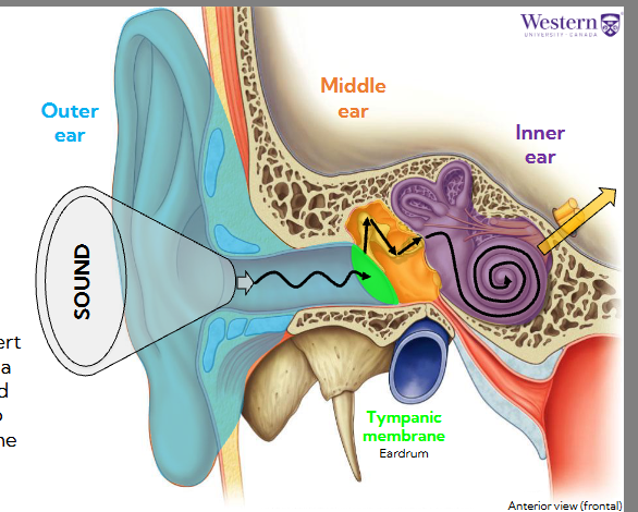
what are the 3 sections of the ear, and what is their general function
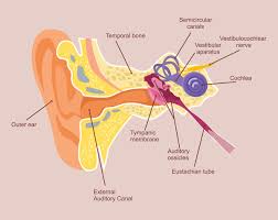
The ear is comprised of three sections, the outer, middle, and inner ear.
Their primary function is to funnel sound waves into the external auditory canal, convert those waves into vibrations via the tympanic membrane, and convert those vibrations into nervous impulses to send to the brain for hearing
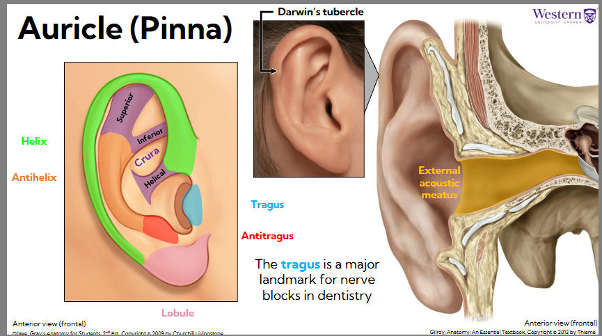
outer ear: auricle (pinna)
Auricle = actual ear on side of head
Some people have darwins tubercle, bump on top of helix, collects more sound
Auricle of auricle is to filter/funnel sound to external acoustic meatus
outer ear: arches of the auricle/pinna
Has main arches
Helix - outer most ridge
Has helical crura projection
Antihelix - smaller one inside
Has superior and inferior crura projections
The tragus is a major landmark for nerve blocks in dentistry: find TMJ and mandibular nerve
Antitragus: across the opening of the ear from the tragus
Lobule - skin dangling
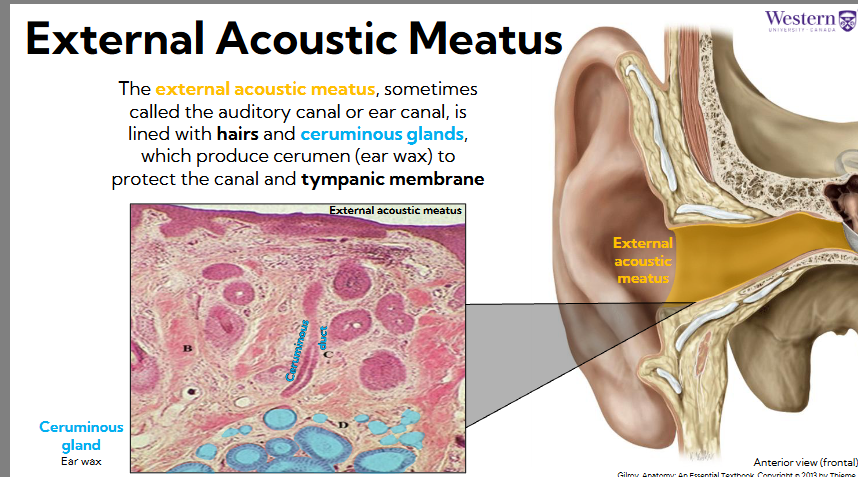
outer ear: external acoustic meatus and ceruminous glands
The external acoustic meatus, sometimes called the auditory canal or ear canal, is lined with hairs and ceruminous glands, which produce cerumen (ear wax) to protect the canal and tympanic membrane
Cerumen is funneled through ceruminous ducts to external acoustic meatus
Hairs also trap dust and debris, provides support structure for cerumen
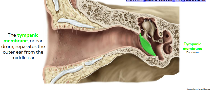
tympanic membrane
The tympanic membrane, or ear drum, separates the outer ear from the middle ear
It prevents debris from entering the middle ear, but its main job is to convert sound waves into vibrations
middle ear: tympanic membrane and ossicles
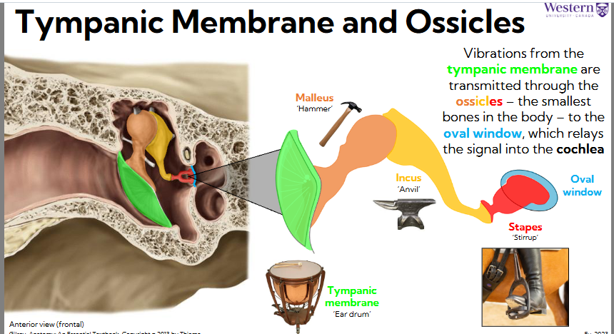
Vibrations from the tympanic membrane are transmitted through the ossicles – the smallest bones in the body – to the oval window, which relays the signal into the cochlea
Base of malleus connects to tympanic membrane, head of hammer is ball shaped head. It takes force from membrane and transmits it to incus (anvil)
Incus transmits force to stapes (stirrup), transmits force onto oval window membrane which goes to inner ear
middle ear muscles
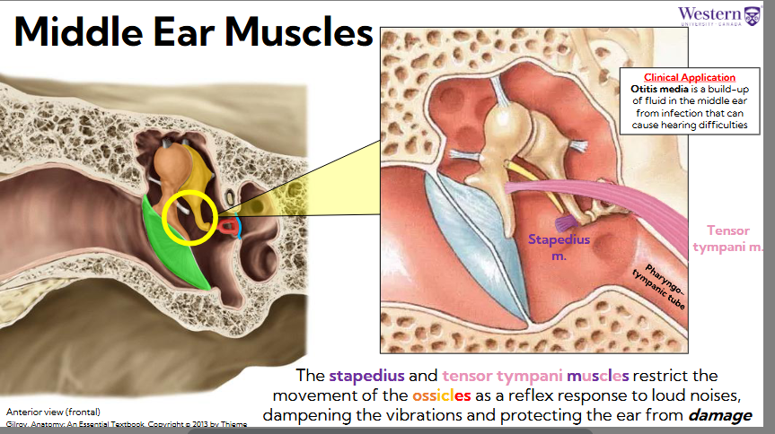
The stapedius and tensor tympani muscles restrict the movement of the ossicles as a reflex response to loud noises, dampening the vibrations and protecting the ear from damage
Tensor tympani pulls on base of the malleus to tighten tympanic membrane and prevents some of force from being transmitted onto incus
Malleus pivots away and force is reduced
These muscles respond to intense loud noises, prevents sound from damaging system
pharyngo tympanic tube (eustachian tube)
connects the middle ear to the nasopharynx
pathway for fluid, used to pop our ears
Diffuse (lower pressure) air creates a vacuum effect on ear drum and pulls it one way, air enters through pharynx to equilibrate outside air in pharynx and middle ear to match outside air, so tympanic membrane stays in neutral
otitis media
When sick, infected mucus can get into middle ear, causes ear infection
Otitis media, build up of fluid in the middle ear from infection that can impede these structures and cause hearing difficulties
inner ear: fluid filled spaces
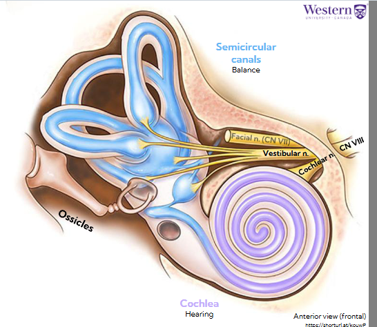
The inner ear is made up of two fluid-filled spaces – the semicircular canals, which are involved in balance, and the cochlea, which is the primary organ of hearing
Send signals back through vestibular cochlear nerve
Vestibular branch comes from semicircular canals
Cochlear branch comes from cochlea
They join to form CN VIII
inner ear: what are the semicircular canals
The system that controls balance is called the vestibular system which is how we determine where our head is in three-dimensional space
One in each plane
They sense:
Roll: coronal
Pitch: up or down, sagittal
Yaw: transverse movement
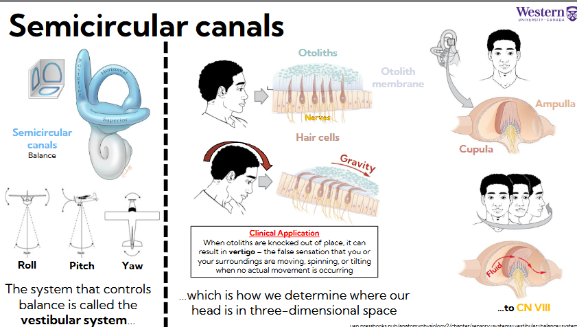
how do semicircular canals work (otolith membrane)
When head is neutral: hair cells project into otolith membrane (jellylike fluid), on top of jelly, have otolith crystals, add weight
When bend our head, causes top of membrane to sage one way or another, moving jelly and hair cells
Bending interpreted as nervous signal
Sense movement: ampulla (big open space) at end of semicircular canals, filled with fluid, they contain cupula projections,
As we move, fluid moves, pushes cupula, triggers nerve impulse

how does vertigo work
When otoliths are knocked out of place, it can result in vertigo – the false sensation that you or your surroundings are moving, spinning, or tilting when no actual movement is occurring
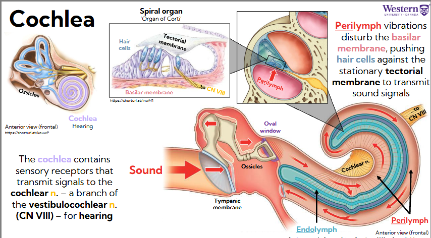
the cochlea
The cochlea contains sensory receptors that transmit signals to the cochlear n. – a branch of the vestibulocochlear n. (CN VIII) – for hearing
Signal goes through tympanic membrane, then ossicles, and then oval window (entrance to inner ear) which pusses on perilymph fluid
Moves through cochlea, all the way around and back out

how are frequencies detected in the cochlea (endolymph and perilymph)
Endolymph trapped within perilymph, moves
Different frequencies push and move endolymph on different areas
Perilymph pushes on spiral organ or organ of corti (within endolymph), force pushes onto basilar membrane, bends cilia (hair cells) against the tectorial membrane (rigid does not move), depending how they bend, sends signal through cochlear branch
Perilymph vibrations disturb the basilar membrane, pushing hair cells against the stationary tectorial membrane to transmit sound signals
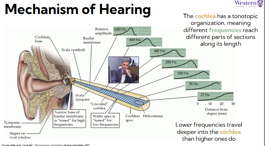
mechanism of hearing different frequencies
The cochlea has a tonotopic organization, meaning different frequencies reach different parts of sections along its length
Lower frequencies travel deeper into the cochlea than higher ones do
Higher frequencies hit endolymph earlier (have shorter sound waves)
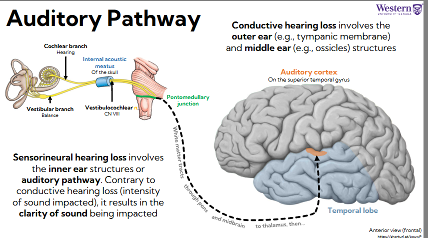
the auditory pathway
Vestibular and cochlear branch get bundled together and travel through internal acoustic meatus of the skull, becomes vestibulocochlear n and returns to pontomedullary junction. White matter tracts reach up to the thalamus, shoots those signals to the auditory cortex (on superior temporal gyrus)

sensorineural hearing loss vs. conductive hearing loss
Sensorineural hearing loss involves the inner ear structures or auditory pathway. Contrary to conductive hearing loss (intensity of sound impacted), it results in the clarity of sound being impacted
Conductive hearing loss involves the outer ear (e.g., tympanic membrane) and middle ear (e.g., ossicles) structures
Otitis media
Or permanent damage dampens amount of vibrations which effects amount of vibration
why are olfaction and gustation connected
Both use chemoreceptors
The olfactory (smell) and gustatory (taste) systems use chemoreceptors – specialized sensory cells that detect external chemical stimuli
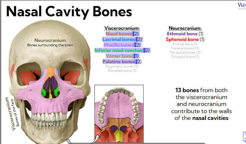
nasal cavity bones
13 bones from both the viscerocranium and neurocranium contribute to the walls of the nasal cavities
viscerocranium nasal cavity bones
Viscerocranium: face bones
Nasal bones (2) - bridge of nose
Lacrimal bones (2) - near tear ducts
Maxilla bones (2) - upper jaw, walls and floor of nasal cavity
Inferior nasal conchae (2) - project into nasal cavities from lateral walls
Vomer bone (1) - wall of nasal septum
Palatine bones (2 - floor of nasal cavity)
zygomatic and mandible not included in nasal cavity

neurocranium bones of nasal cavity
Neurocranium: bones surrounding brain
Ethmoid bone
Sphenoid bone- houses brain, posterior aspect of nasal cavity
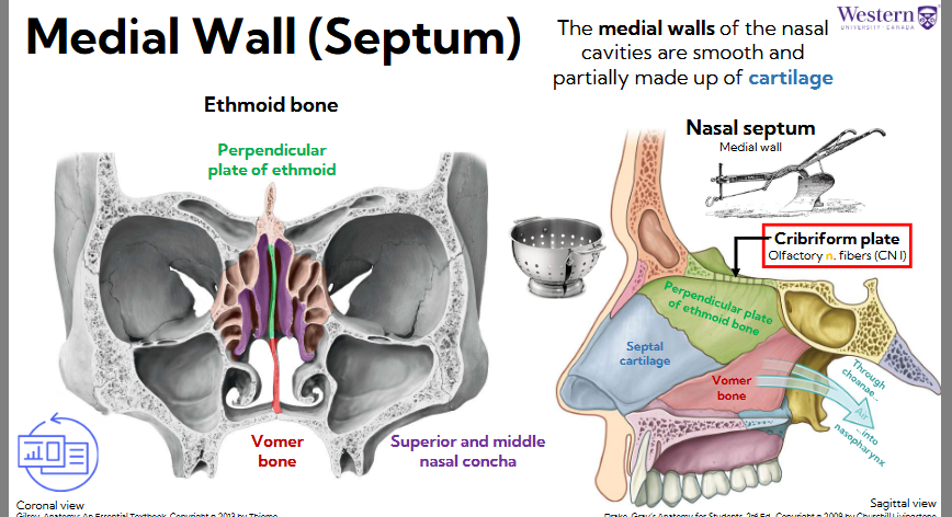
medial wall of nasal cavity (septum)
Huge portion made of ethmoid bone -medial wall (perpendicular plate of ethmoid)
Meets with vomer bone at bottom
Septal cartilage is huge chunk of medial wall
Air passes through choanae into nasopharynx
Moves past cribriform plate, top of ethmoid bone, with holes that run through it, where olfactory nerve fibers run, embedded in mucosa to sense
The medial walls of the nasal cavities are smooth and partially made up of cartilage
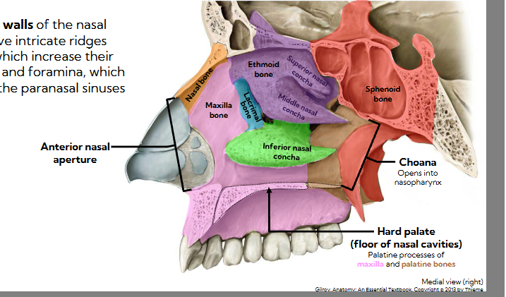
lateral walls of nasal cavity
The lateral walls of the nasal cavities have intricate ridges (conchae), which increase their surface areas, and foramina, which connect with the paranasal sinuses
Anterior nasal aperture -openings
Maxilla bone makes huge chunk of lateral bone
Nasal bone makes bridge
Lacrimal bone
Inferior nasal concha
Ethmoid bone: superior nasal concha and middle nasal concha
Hard palate: palatine processes of palatine bones and maxilla
Spehnoid bone has choana - opening into nasopharynx
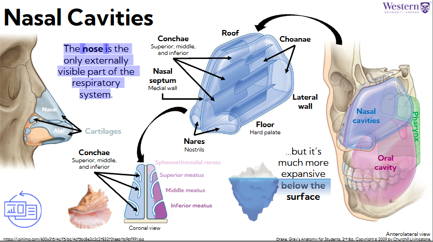
chonae projections in nasal cavity
The nose is the only externally visible part of the respiratory system - mostly cartilage
But its much more expansive below the face
Choanae create projections into nasal cavity, create pockets (inferior, middle, inferior meatus) creates turbulence in nasal cavity to help with olfaction
Olfaction happens in sphenoethmoid recess (roof of nasal cavity)
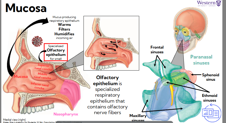
mucosa (smell)
Olfactory epithelium is specialized respiratory epithelium that contains olfactory nerve fibers
Mucosa producing respiratory epithelium:
Warms, filters, and humidifies incoming air
Paranasal sinuses (chambers within bones), lots of SA to perform these functions
Air passed by specialized olfactory epithelium for smell
Covid virus targets olfactory epithelium, can't smell
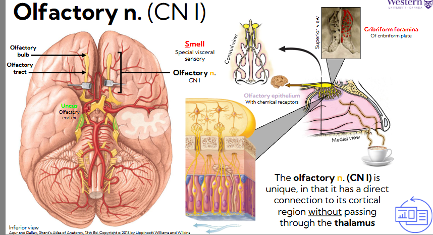
olfactory n (CN I)
The olfactory n. (CN I) is unique, in that it has a direct connection to its cortical region without passing through the thalamus
Has CNS neurons: olfactory n., bulb, and tract (located in uncus)
Olfactory bulb sits on inside of skull on the cribriform plate of ethmoid
Has little holes, cribriform foramina, where nerve fibers project through and embed in olfactory epithelium
Aromas pass odor molecules, mucosa dissolves those chemicals, and olfactory nerve transmits to nervous signals
Only sensory nerve that does not relay to thalamus, goes directly to cortex
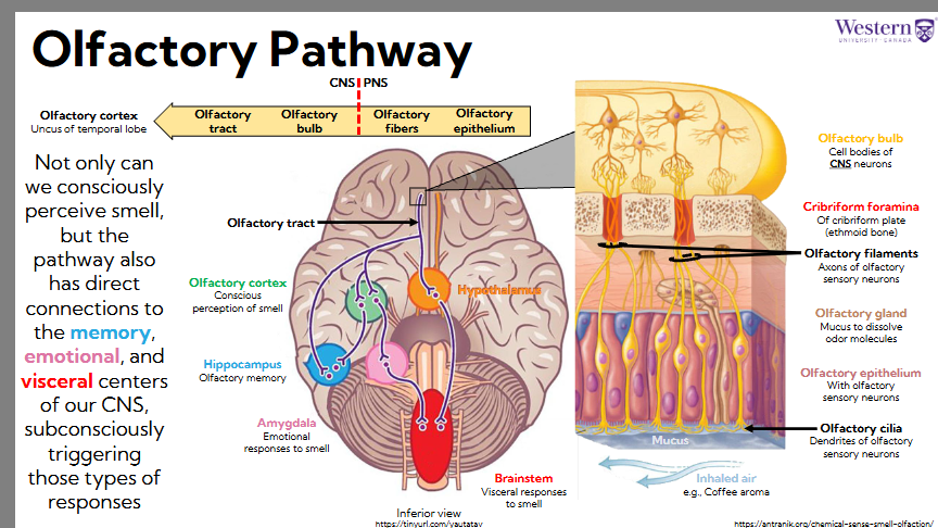
olfactory pathway
Not only can we consciously perceive smell, but the pathway also has direct connections to the memory, emotional, and visceral centers of our CNS, subconsciously triggering those types of responses
olfactory pathway in nasal cavities:
Nose: in order top down
Olfactory epithelium: with olfactory sensory neurons
Olfactory gland: mucus to dissolve odor molecules
Olfactory cilia: dendrites of olfactory sensory neurons
Olfactory filaments: axons of olfactory sensory neurones- toward brain (true olfactory nerves)
Cribriform foramina: of cribriform plate (ethmoid bone) - separates PNS from CNS
Olfactory bulb: synapse with cell bodies of CNS neurons

olfactory pathway in brain
Brain:
Olfactory cortex: conscious perception of smell- first place we send smell
Hippocampus: olfactory memory- directly connects to olfactory cortex, also connects to olfactory cortex (indirect connection)
Amygdala: emotional responses to smell
Brainstem: visceral responses to smell - reflexes like disgust
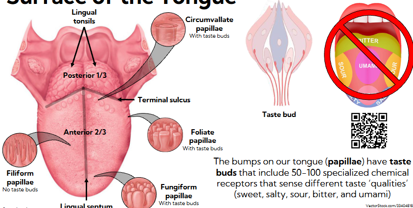
surface of the tongue
The bumps on our tongue (papillae) have taste buds that include 50-100 specialized chemical receptors that sense different taste ‘qualities’ (sweet, salty, sour, bitter, and umami)
Lingual septum divides tongue into 2 sides
Terminal sulcus, v shaped group, separates anterior ⅔ of tongue from posterior ⅓
Lingual tonsils - immune response
Filiform papillae - no taste buds, purpose to give us control over food, friction additive so we can move food

papillae with taste buds:
Fungiform: bulbous, little mushrooms
Foliate: squared off
circumvallate : circular suction cups, help bring food back as we swallow
taste bud
50-100 sensors that detect diff types of taste
Scattered throughout the tongue

innervation of the tongue
The tongue has three nerves with special sensory (taste) functions:
the chorda tympani (branch of facial n., CN VII), glossopharyngeal n. (CN IX), and vagus n. (CN X)
These nerves are also linked to parasympathetic functions related to digestion, hinting at the role of taste in preparing the body to receive and process food
Sensory includes general and special
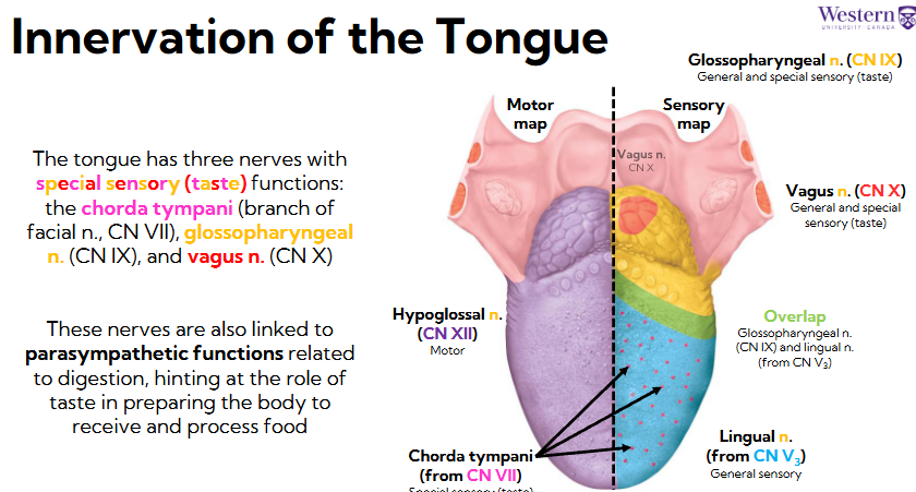
motor innervation of tongue
(CN XII) innervates

sensory innervation of tongue
Lingual n (from CN V#), innervates general sensory anterior ⅔
Chorda tympani from CN VII brings back special sensory for taste
Glossopharyngeal n (CN IX) does general and special sensory taste, in posterior ⅓
Vagus n (CN X) general and special sensory in posterior spot of tongue

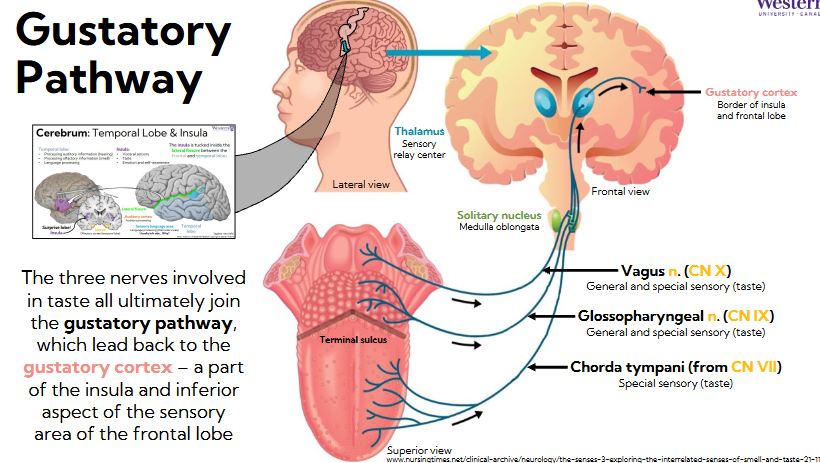
gustatory pathway
The three nerves involved in taste all ultimately join the gustatory pathway, which lead back to the gustatory cortex – a part of the insula and inferior aspect of the sensory area of the frontal lobe
Vagus n: general and special sensory (taste)
Glossopharyngeal n: general and special sensory (taste)
Chorda tympani (from CN VII): special sensory (taste)
Feed back to solitary nucleus in the medulla oblongata, those neurons feed back to thalamus (sensory relay center)
Thalamus sends signal to gustatory cortex, border of insula and inferior lateral aspects of frontal lobe
All of these nerves have parasympathetic functions:
Chorda tympani CN VII and glossopharyngeal CN IX create mucus
Vagus innervates digestive system
Means taste sensing nerves have direct line to parasympathetic response, explains when we taste we salivate
anosmia and ageusia
Ageusia = Loss of the sense of taste, inability to detect different flavours
These overlap and work together
Combined effect of taste and smell in perception
Anosmia and ageusia can be associated with the natural aging process, which is why our tastes evolve over time
Acquired tastes
Always evolving