Lower limb/lower limb bones
1/54
There's no tags or description
Looks like no tags are added yet.
Name | Mastery | Learn | Test | Matching | Spaced |
|---|
No study sessions yet.
55 Terms
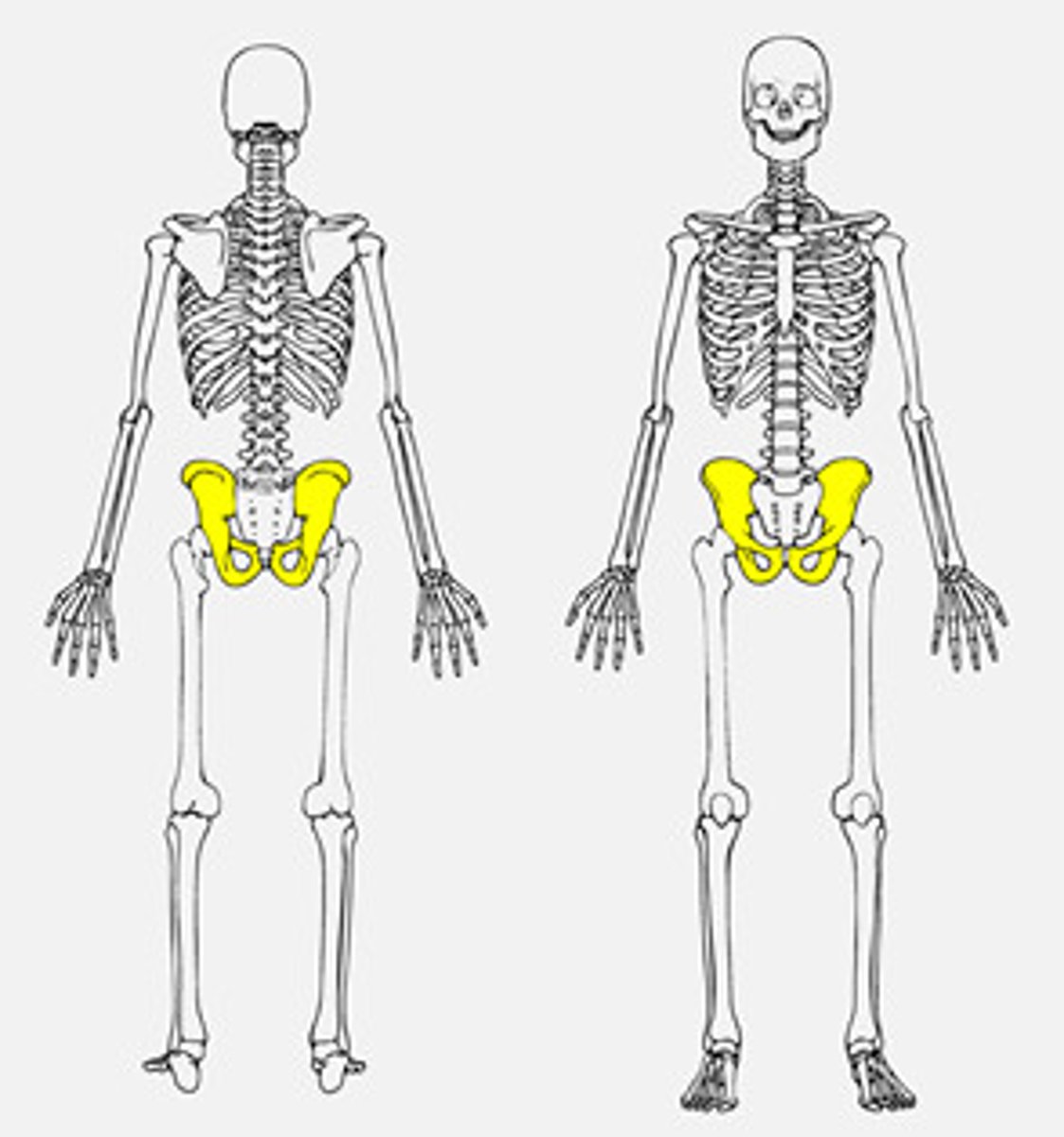
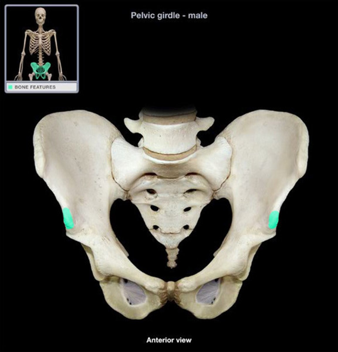
Os Coxae (hip bones)
Fusion of 3 bones:
o Ilium
o Ischium
o Pubis
o Fusion occurs at puberty; prior to this bones are held together by cartilage at
acetabulum
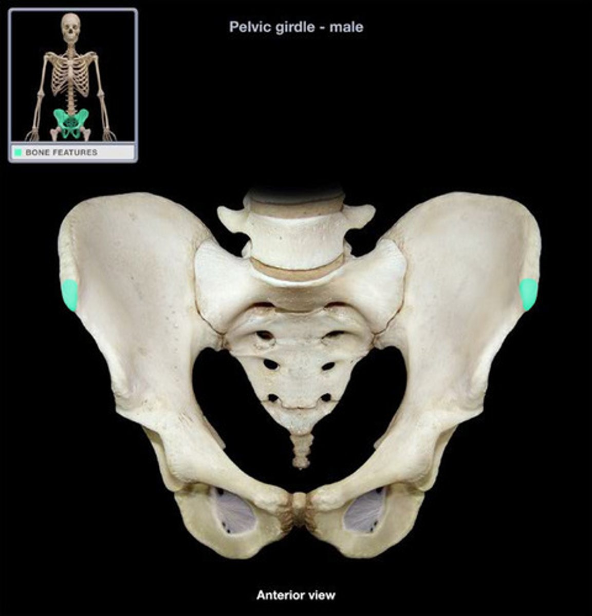
iliac blade
expansive spread part of fan-shaped region
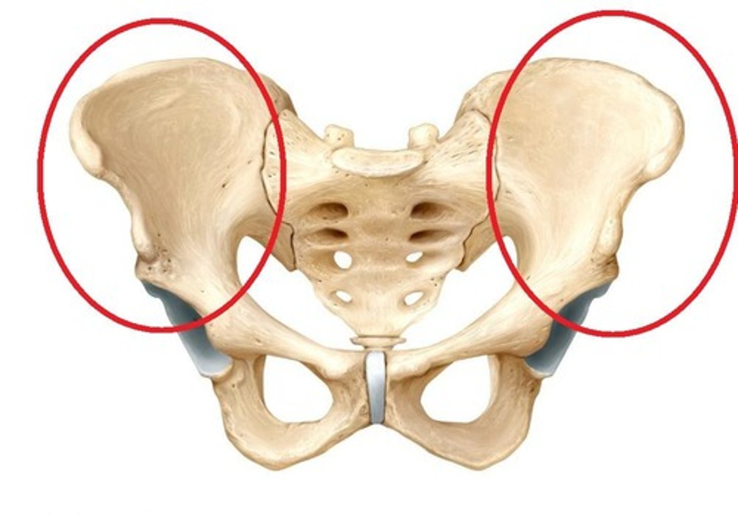
iliac crest of ilium
superior rim ilium
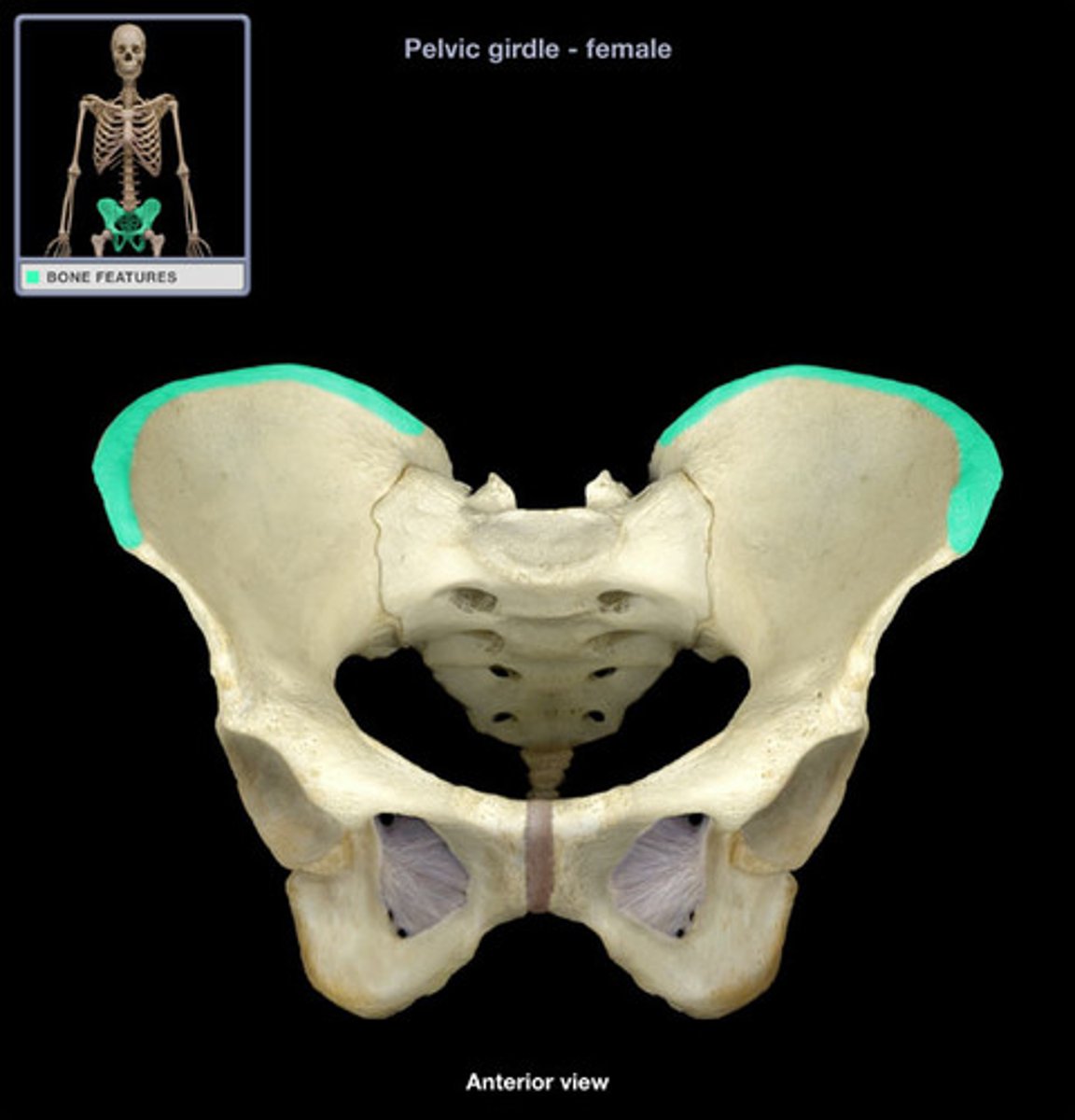
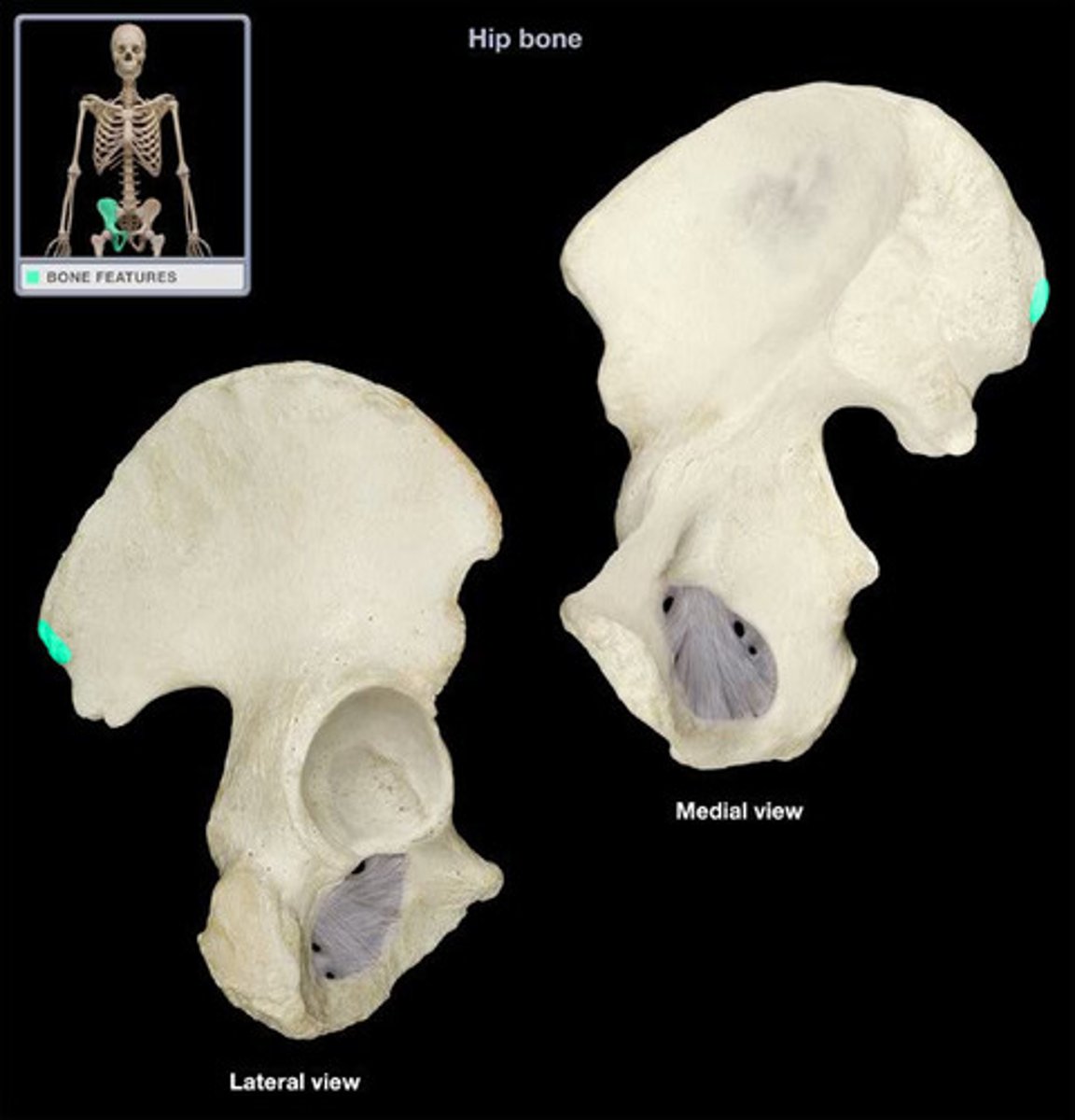
anterior superior iliac spine (ASIS)
projection on anterior end of iliac crest
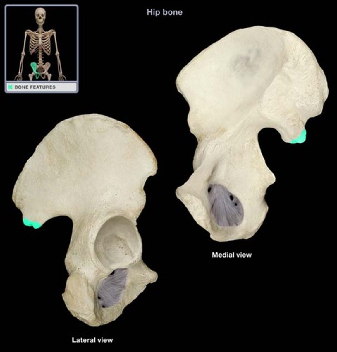
anterior inferior iliac spine (AIIS)
projection inferior to ASIS
Posterior superior iliac spine (PSIS)
projection on posterior end of iliac crest
Posterior inferior iliac spine (PIIS)
projection inferior to PSIS
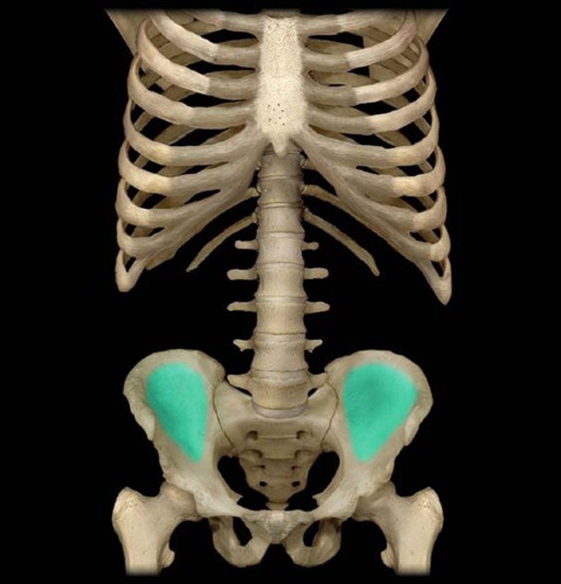
iliac fossa
medial concave part of ilium
body of ischium
contributes to acetabulum (behind the ischial spine shown in pics)-not likely to be tagged

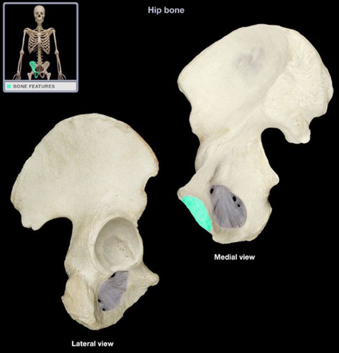
ischial ramus
joins inferior ramus of pubis to form ischiopubic ramus; forms portion of
obturator foramen
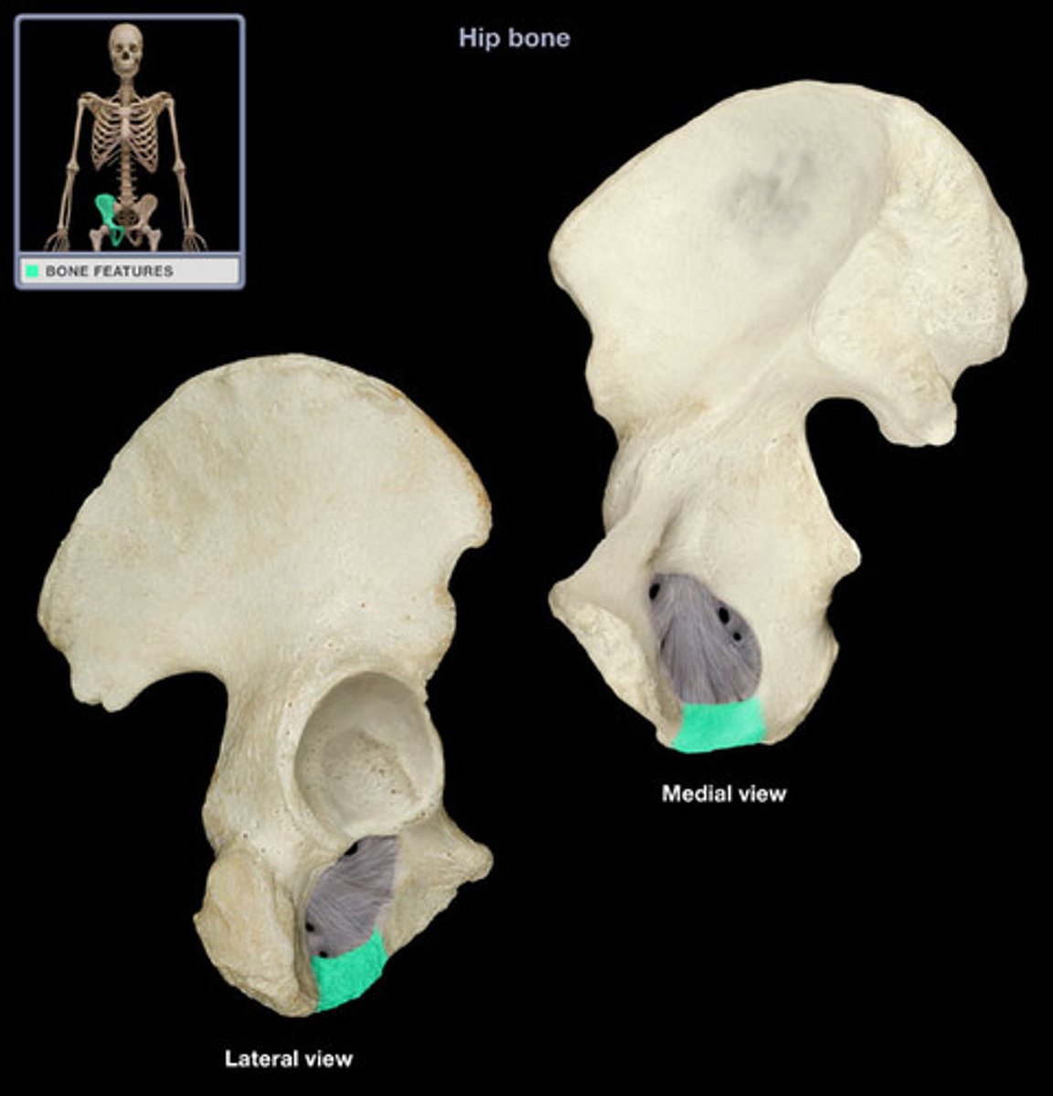
ischial tuberosity
large posteroinferior protuberance on ischial ramus, where hamstrings attach. Where we sit.
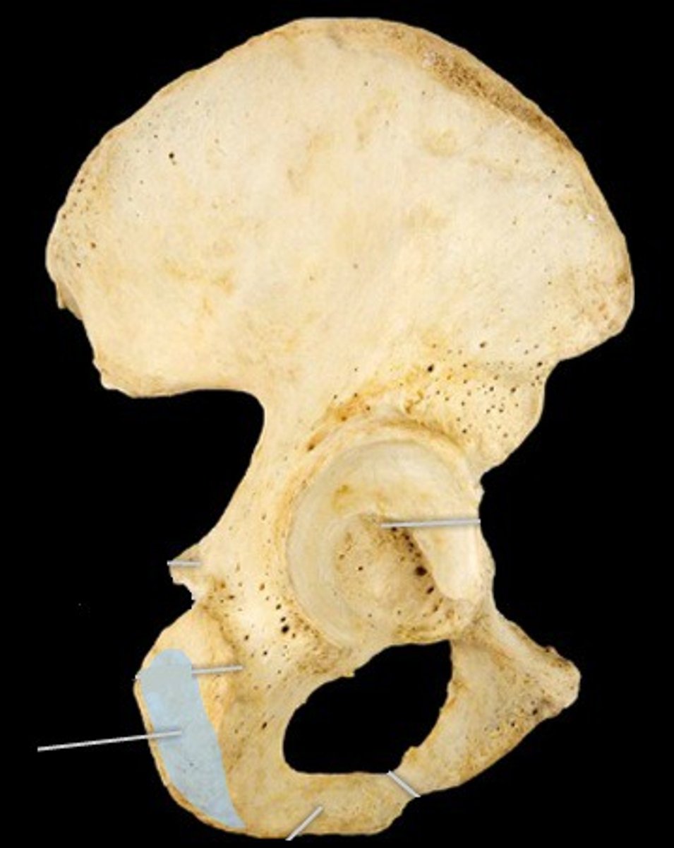
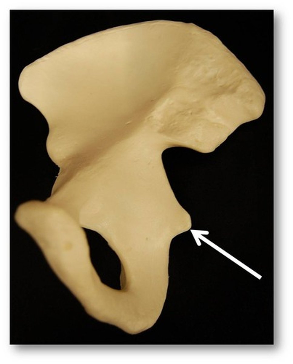
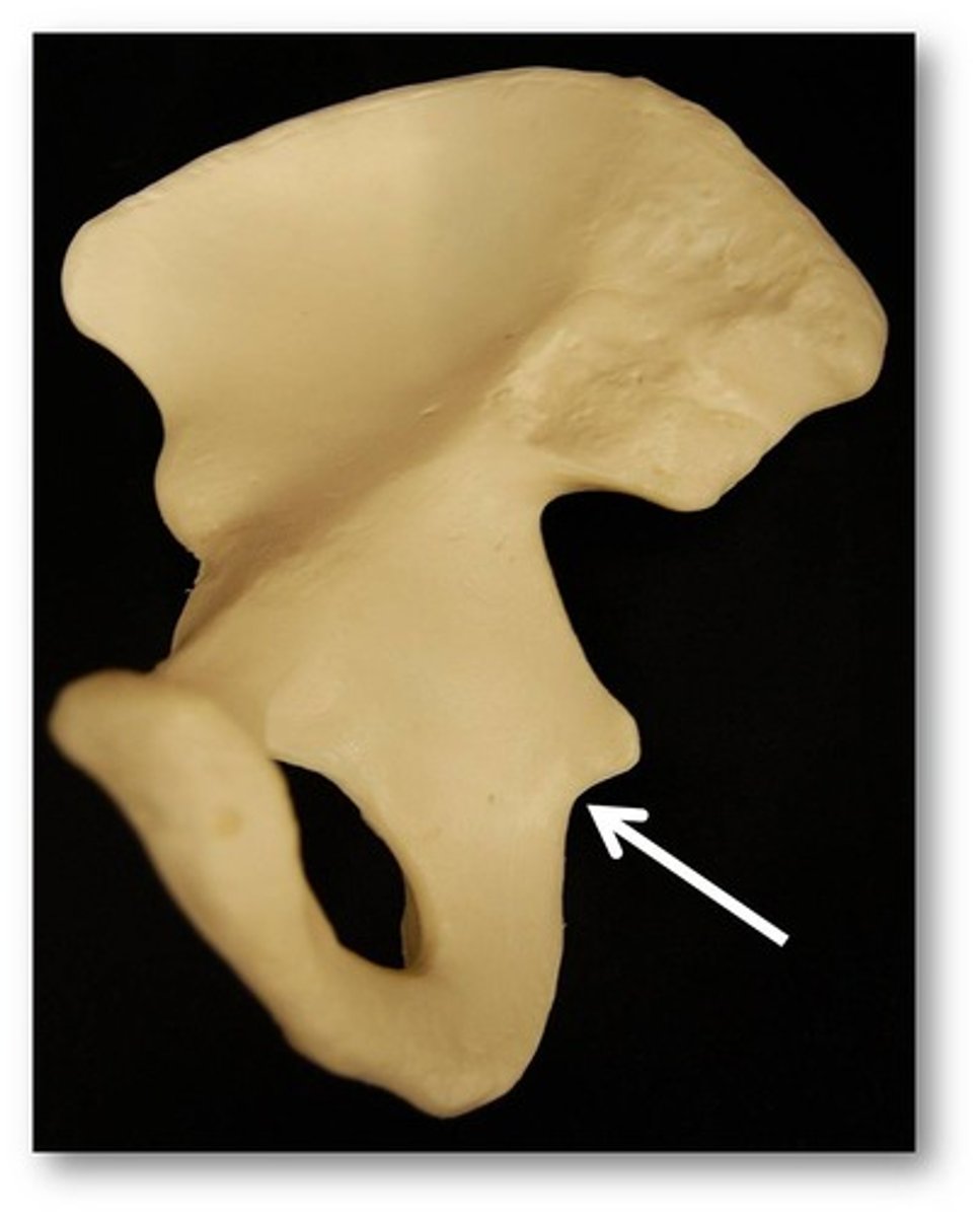
ischial spine
small, pointed projection near junction of body and ramus
lesser sciatic notch
concavity between ischial spine and ischial tuberosity
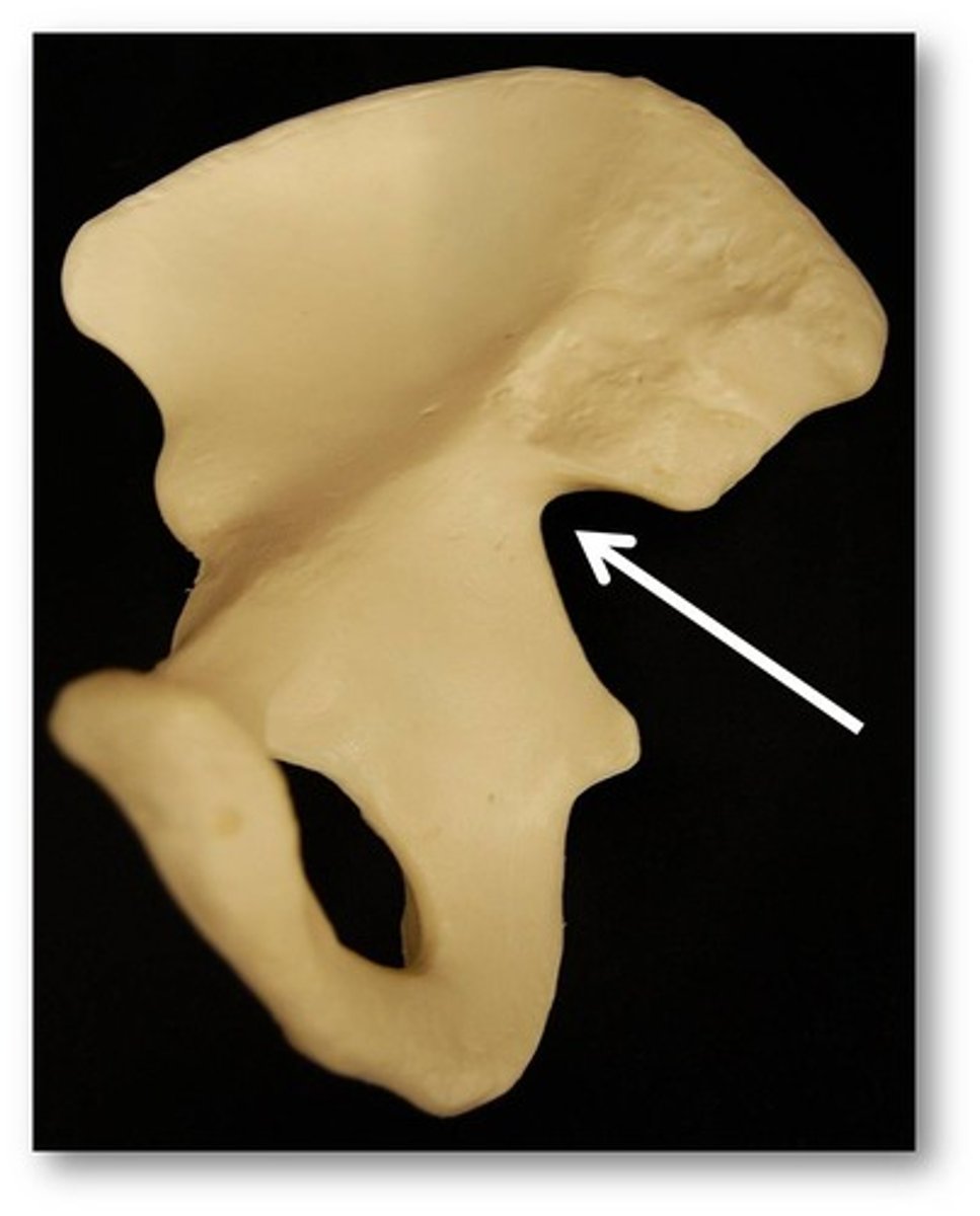
greater sciatic notch
concavity superior to ischial spine
body of pubis
flattened, medial part of bone, near the pubic symphysis where rami attach
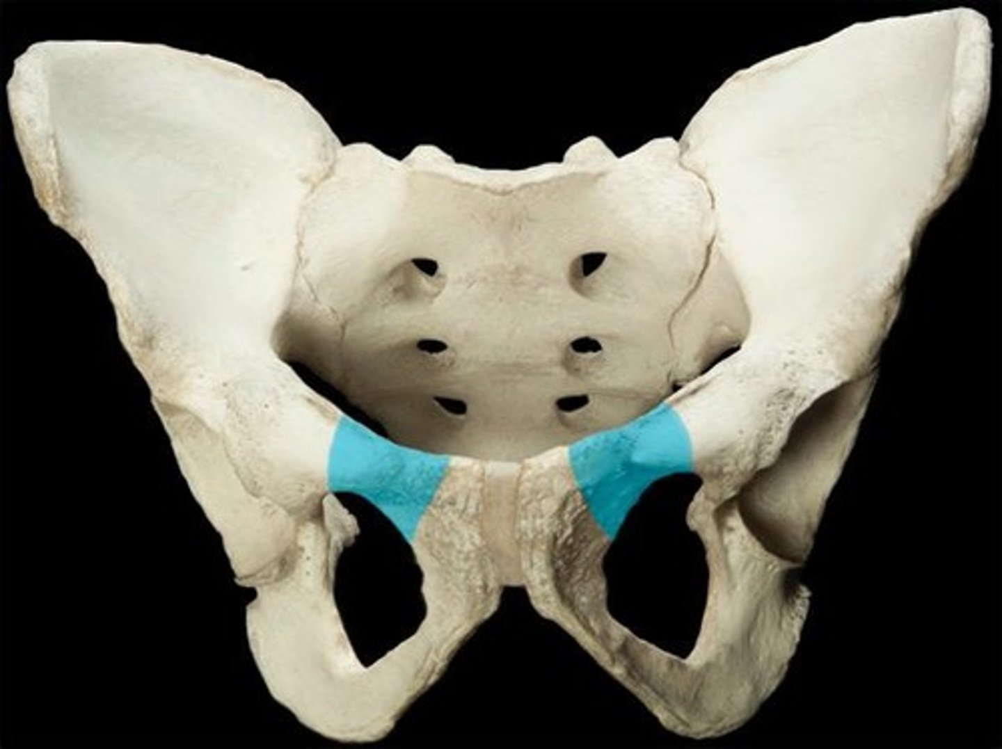
superior ramus of pubis
forms part of acetabulum
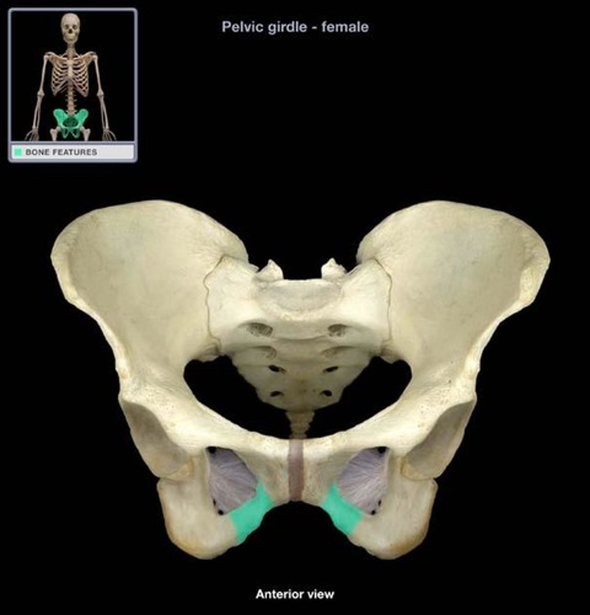
inferior ramus of pubis
- forms portion of obturator foramen
pubic symphysis
medial surface of body
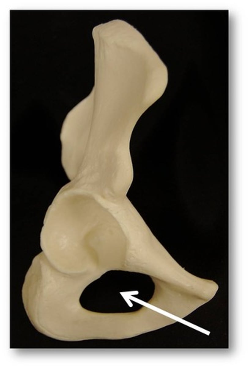
Obturator foramen
Large oval opening in hip bone
Surrounded by ischial and pubic rami
Closed by obturator membrane
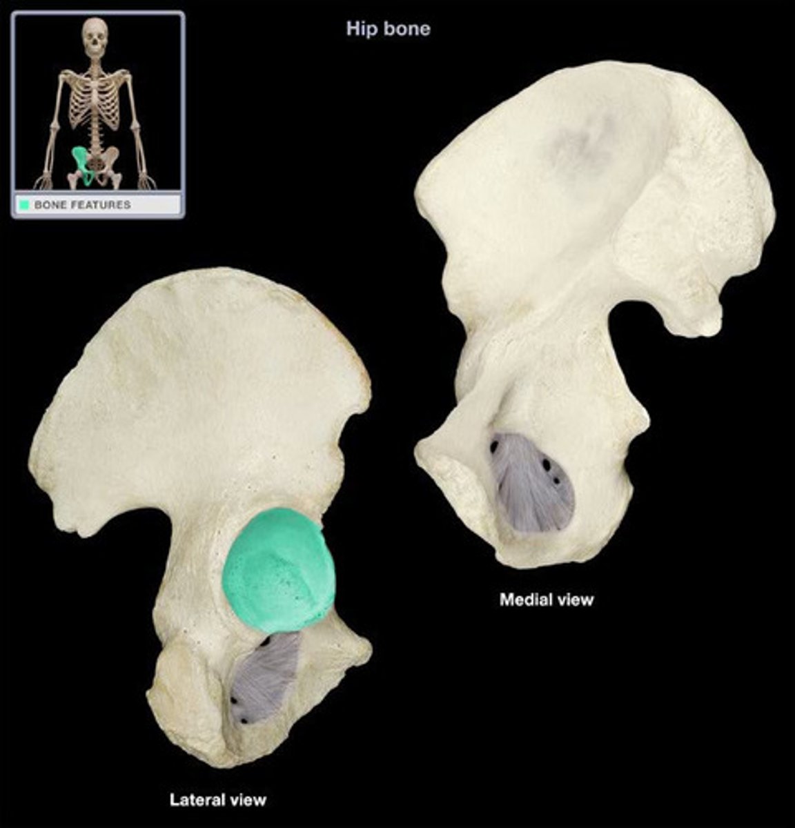
Acetabulum
Large, cup-shaped cavity (socket) on lateral aspect of hip bone
Articulates with femoral head
o Forms hip joint
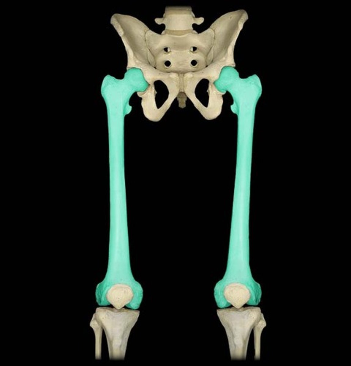
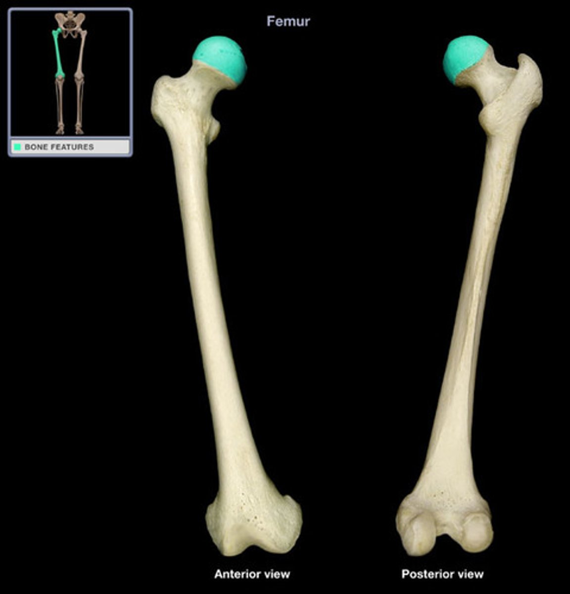
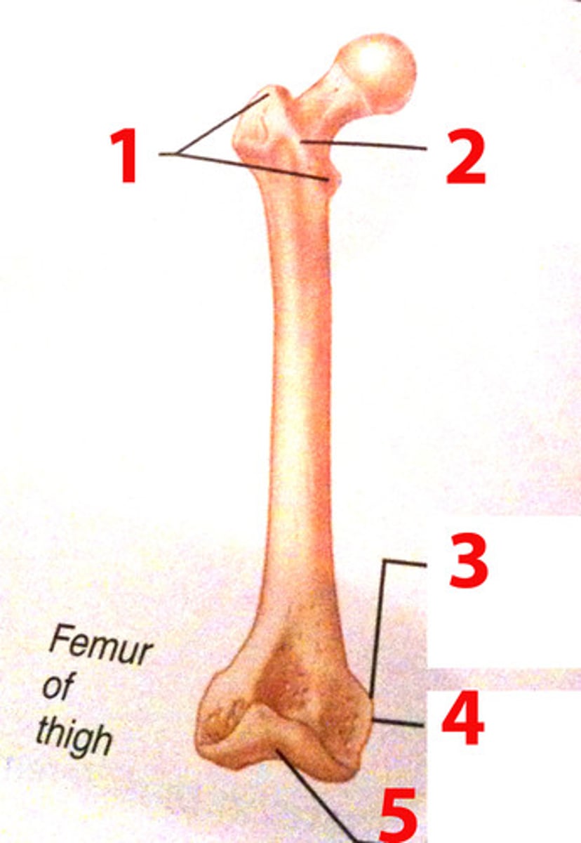
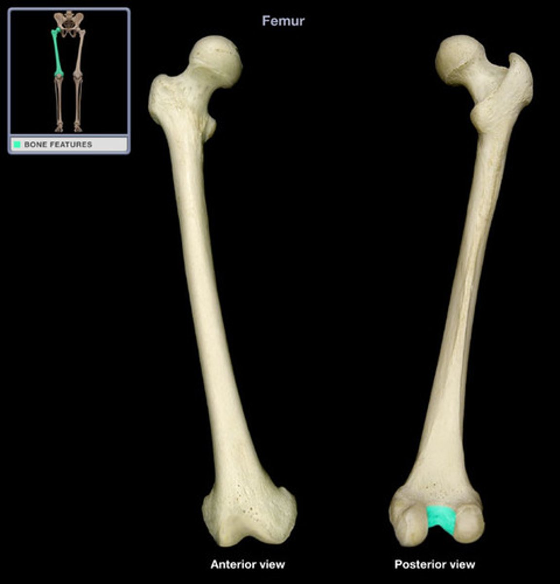
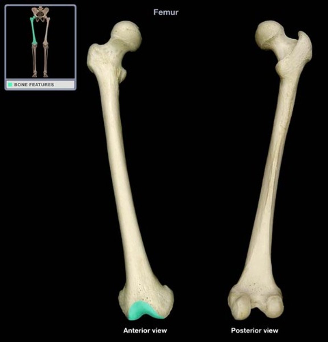
femur
Longest and heaviest bone in body
Transmits body weight from hip bone to tibia when standing
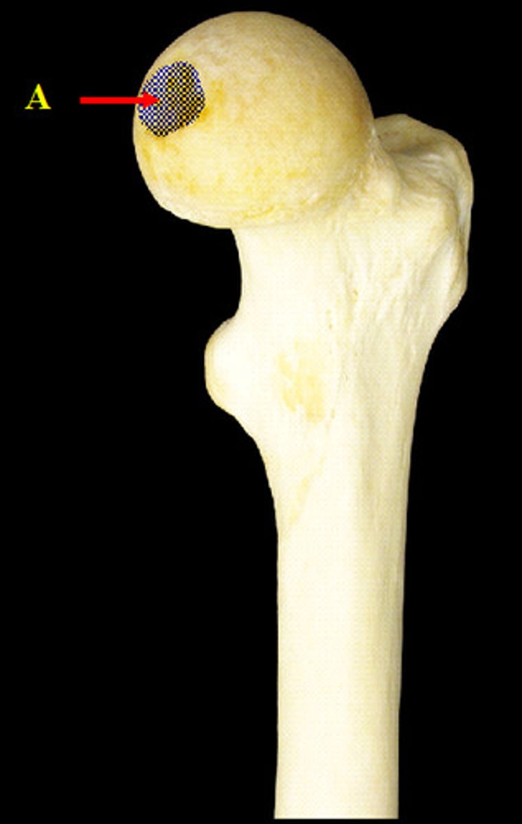
head of femur
proximal articular part
fovea of femur (fovea capitis)
depression in head for ligament to head
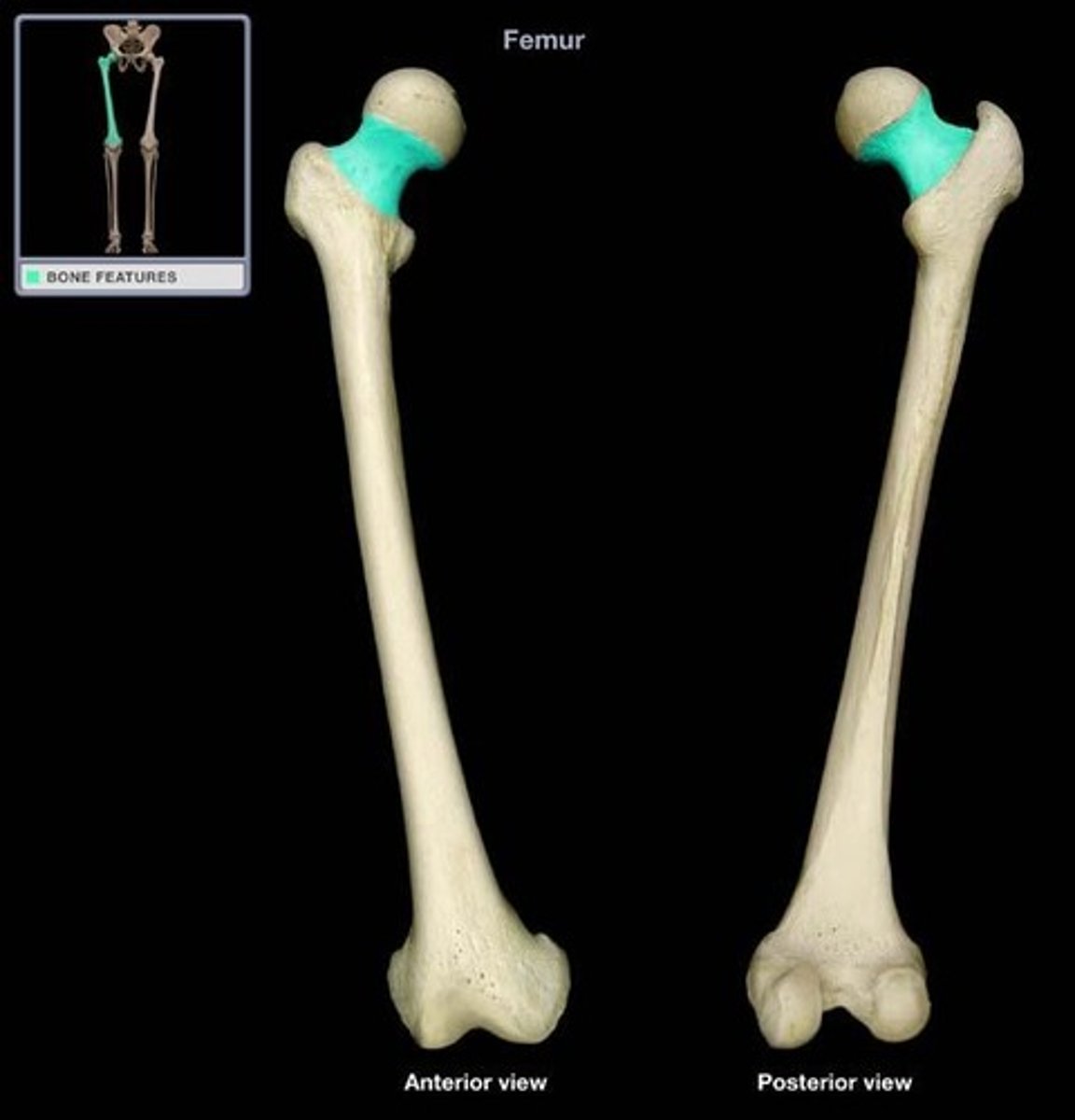
neck of femur
connects head to shaft
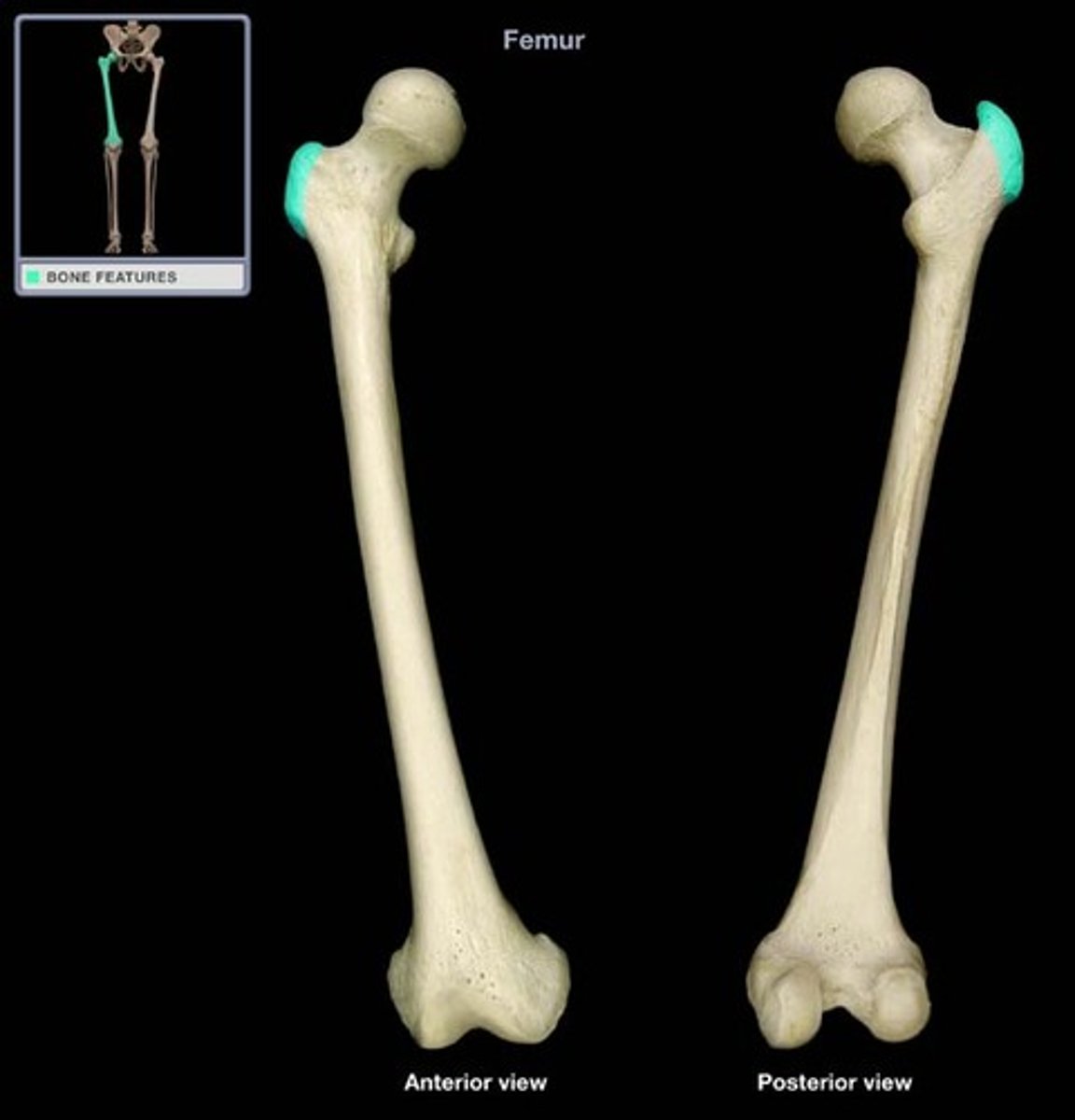
greater trochanter of femur
large, lateral mass that projects superior and posterior
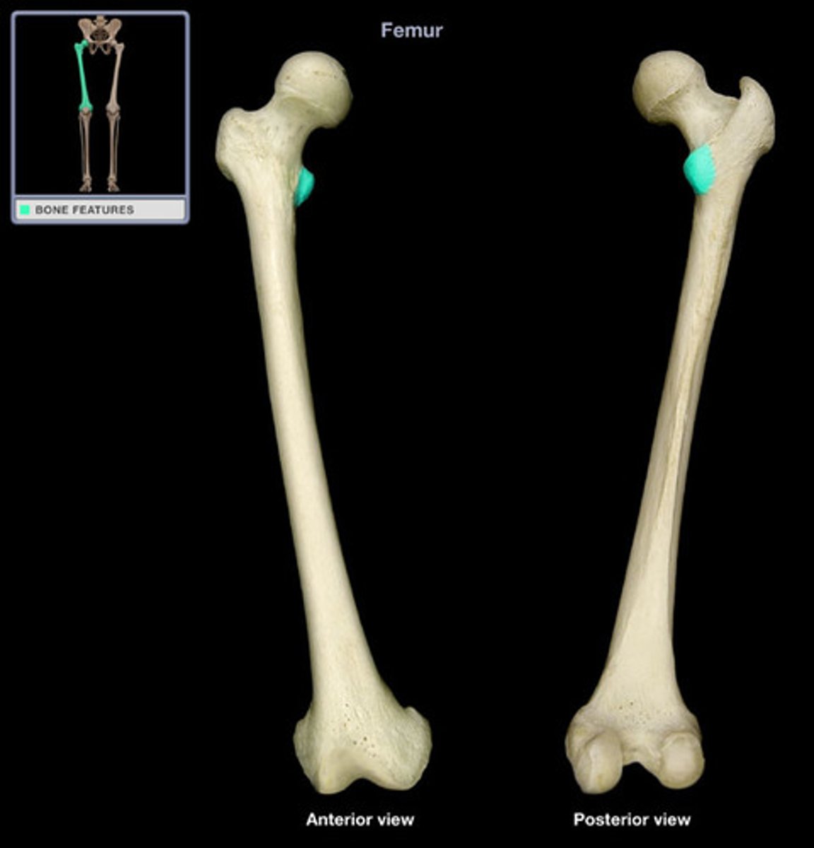
Lesser trochanter of femur
medial elevation at junction of neck and shaft
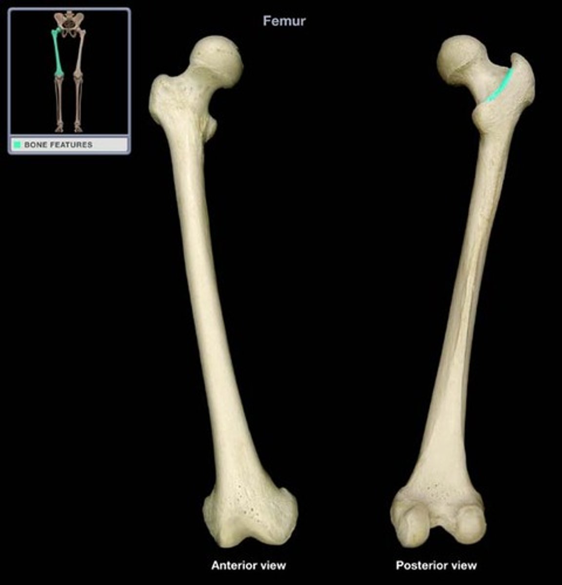
Intertrochanteric line of femur
anterior ridge between greater and lesser trochanters
o Continues posteriorly and inferior as spiral line (2)
intertrochanteric ridge of femur
- posterior ridge joining trochanters
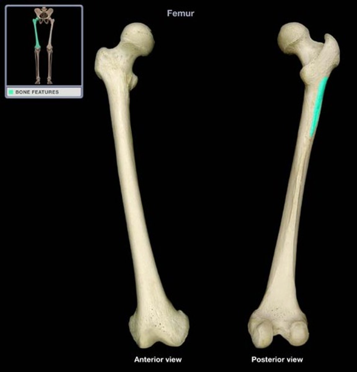
linea aspera
broad, rough line on posterior aspect of shaft

gluteal tuberosity of femur
top of the lateral lip of linea aspera
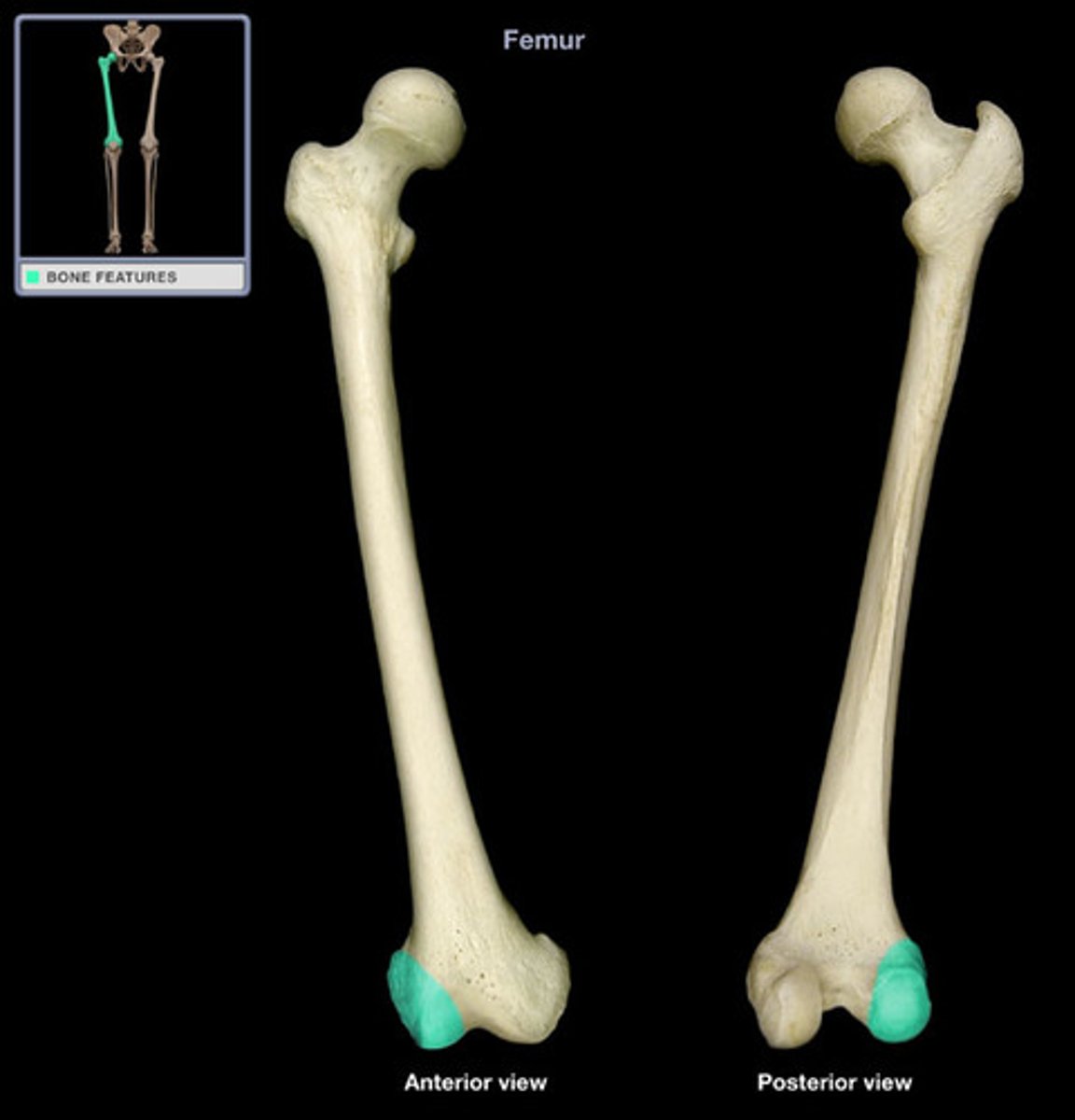
Medial and lateral femoral condyles of femur
large, inferior end of femur (pics show one)
Intercondylar fossa of femur
depression posteriorly between condyles
Patellar surface of femur
shallow depression anteriorly between condyles
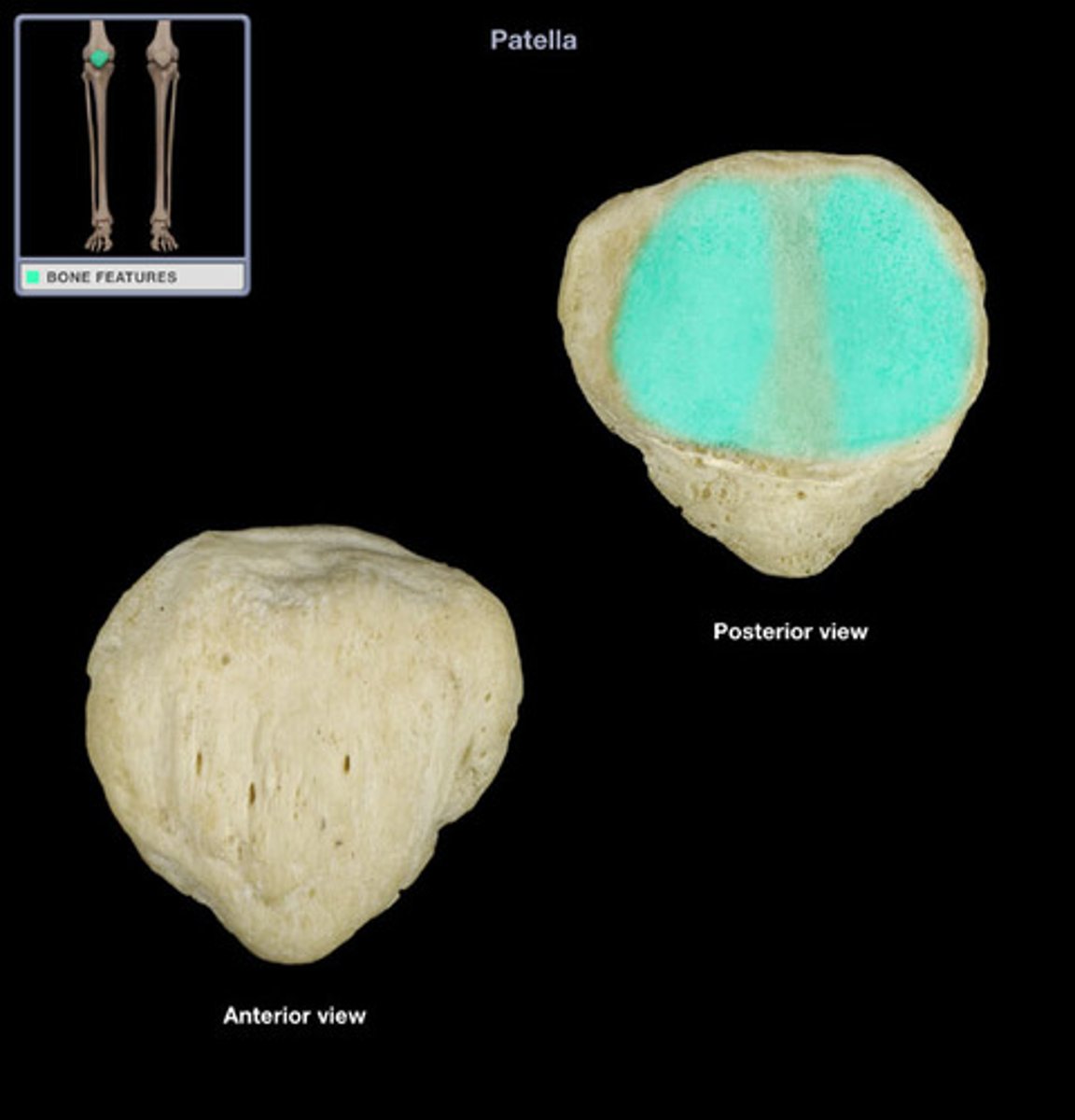
Patella
kneecap
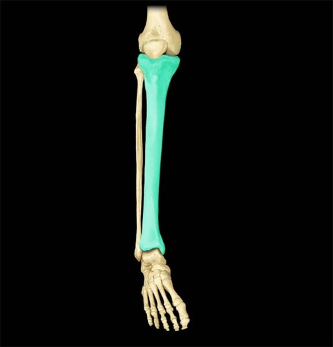
Tibia
Shin bone
On anteromedial side of leg
Articulates with femoral condyles superiorly
Articulates with talus inferiorly, fibula laterally
Transmits body weight to foot
Connected to fibula by interosseous membrane and syndesmoses
Medial and lateral tibial condyles
(only one shown in pic but on both sides)
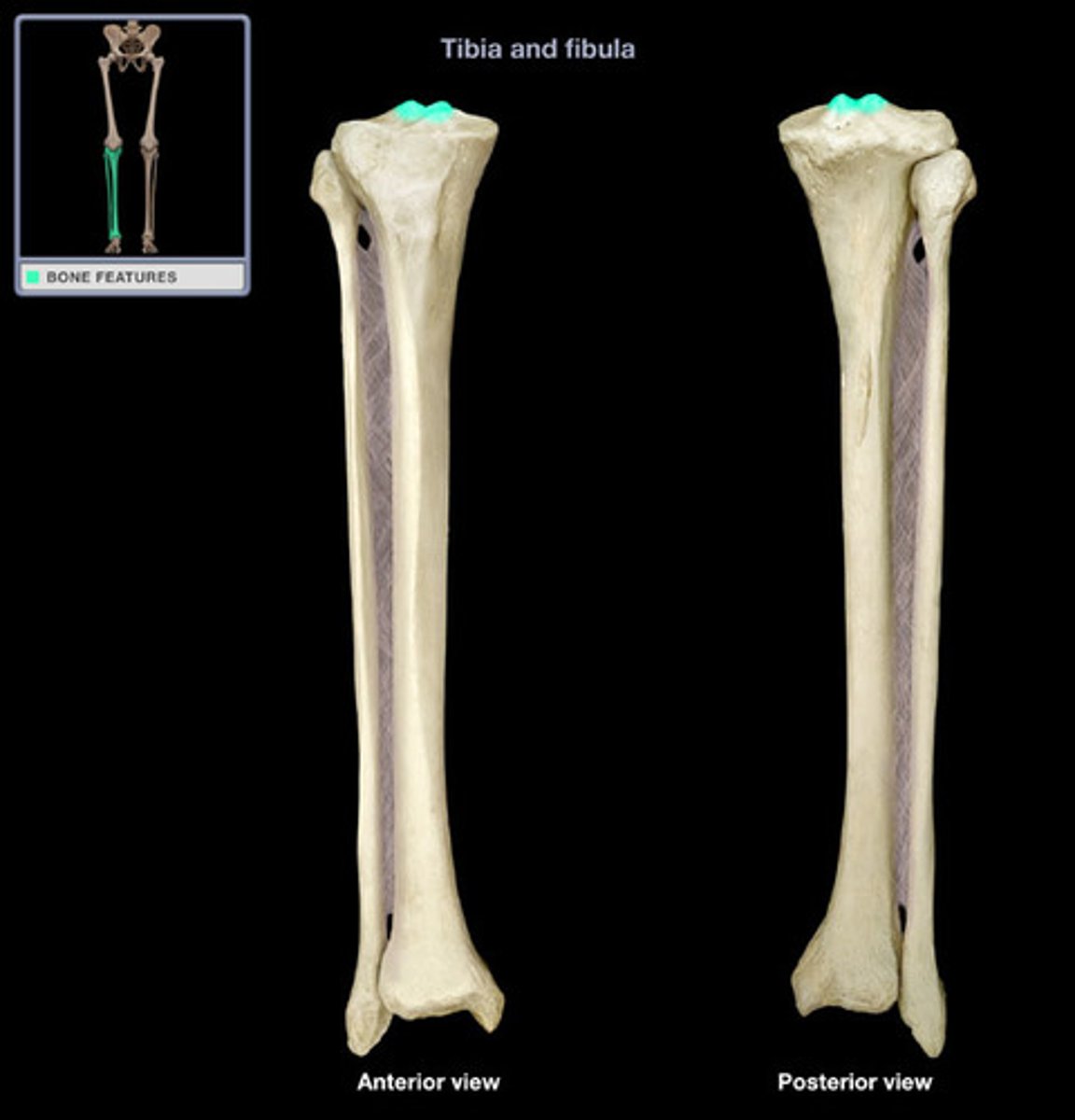
Intercondylar eminence of tibia
separates articular surfaces, little two bumps
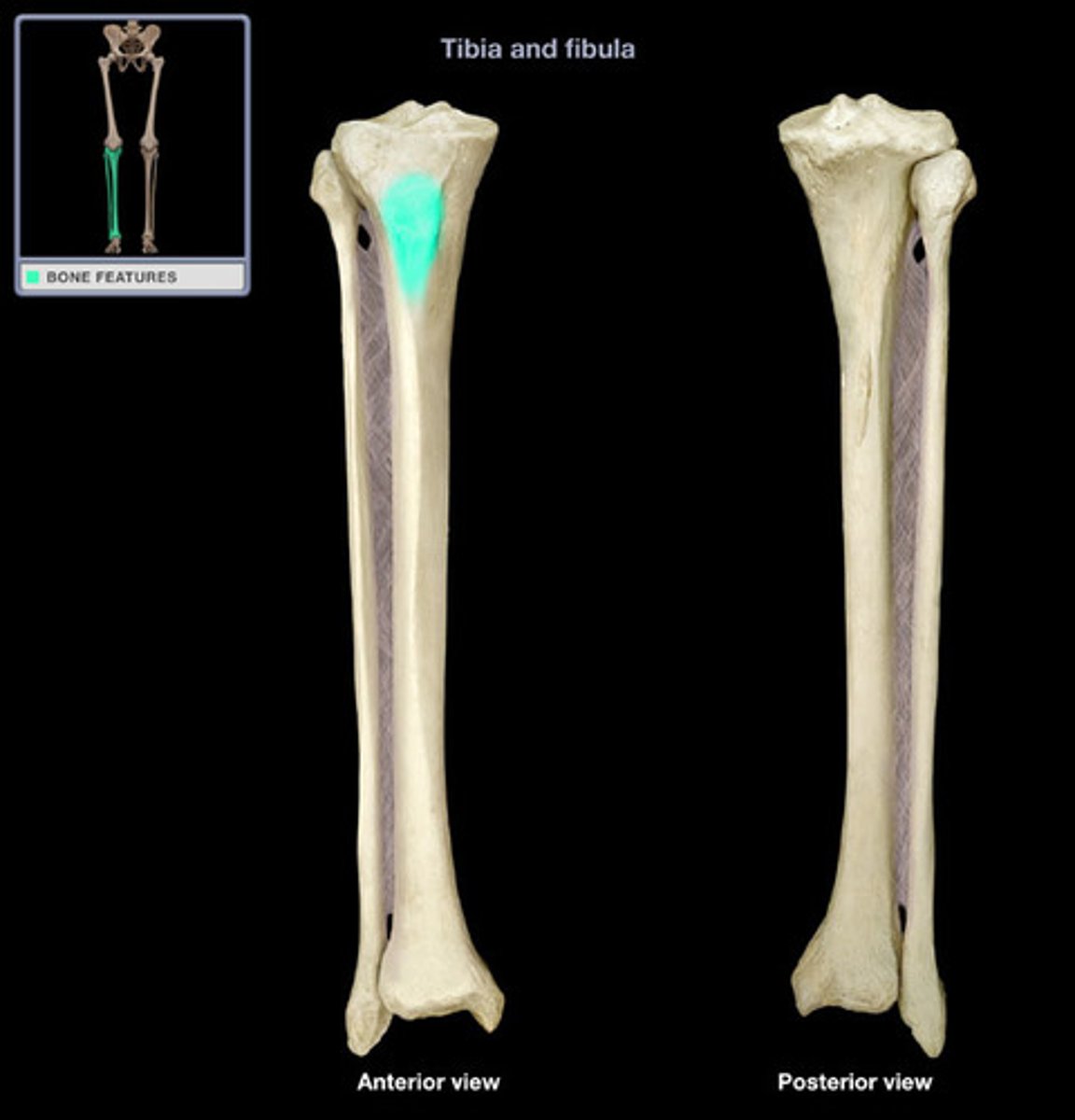
Tibial tuberosity
at superior part of anterior border, where quads attach
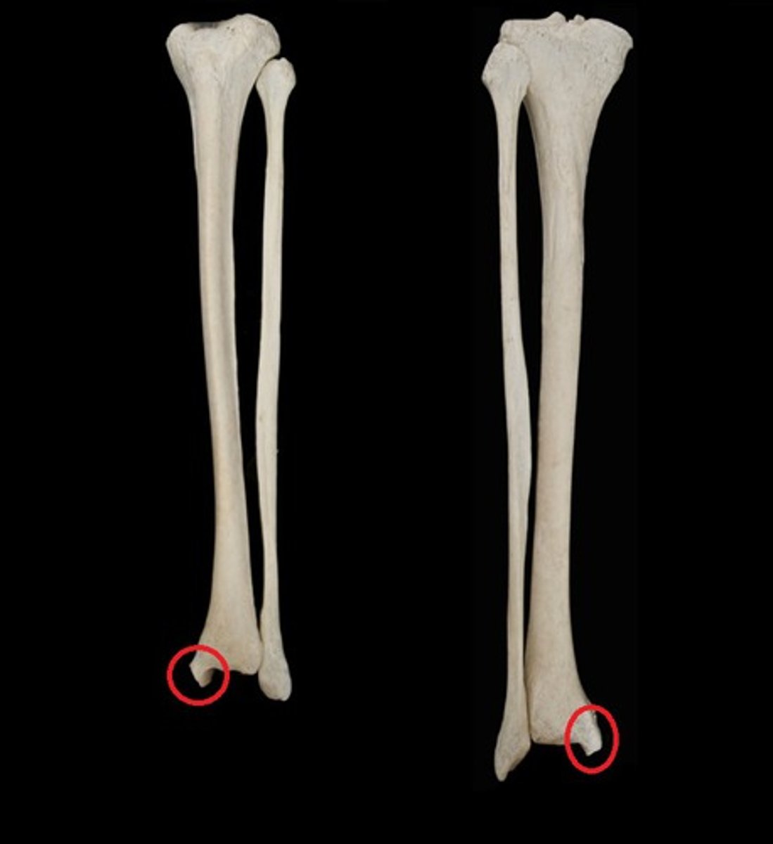
Medial malleolus of tibia
medial expansion inferior to shaft
fibula
Lies posterolateral to tibia
No function in weight-bearing
Articulates with tibia superiorly and inferiorly
Articulates with talus inferiorly
Attached to tibia via:
o Interosseous membrane
o Tibiofibular syndesmoses
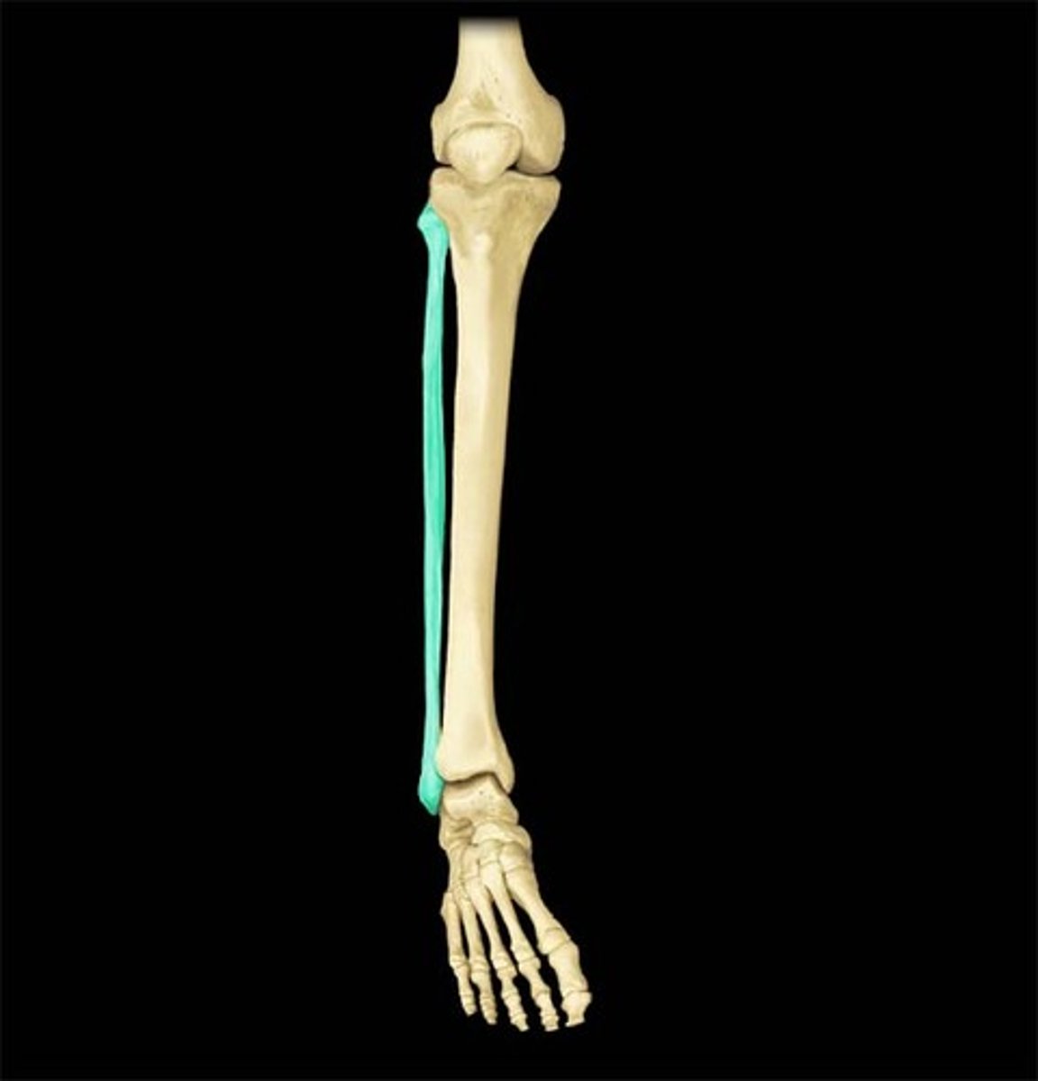
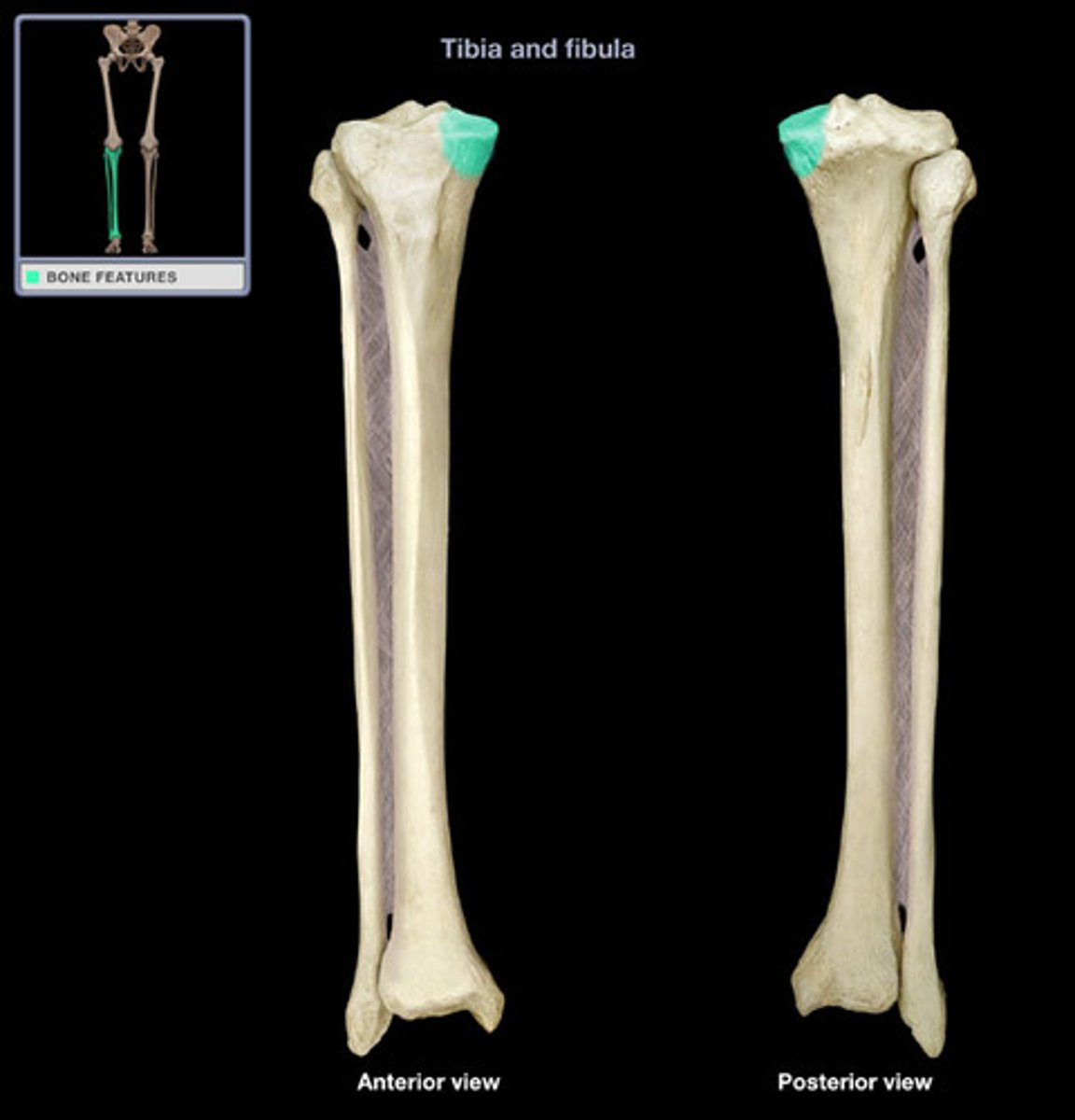
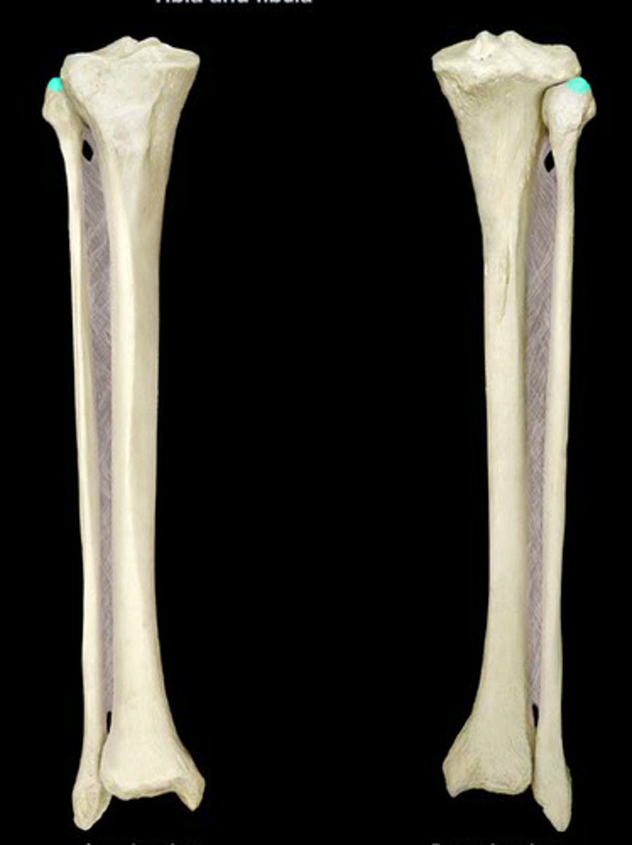
Head of fibula
most proximal portion of fibula, articulates with tibia
apex of fibula
the point on the top of the head
Lateral malleolus of fibula
enlarged distal end prolonged laterally and inferiorly

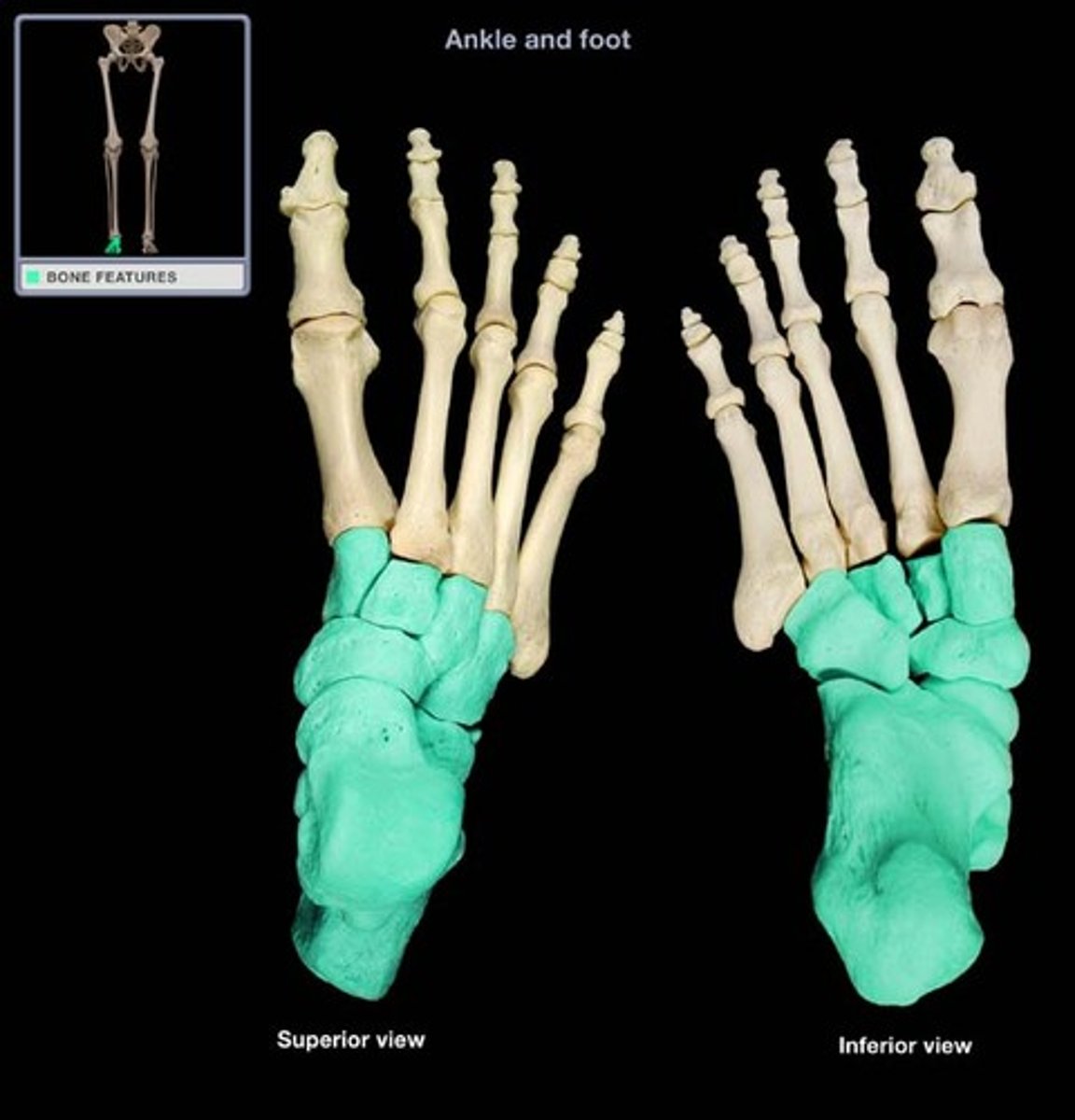
Tarsals
Compose hindfoot
7 bones total
talus
No muscular or tendinous attachment
Only bone to articulate with leg bones
Divides body weight between calcaneus and forefoot
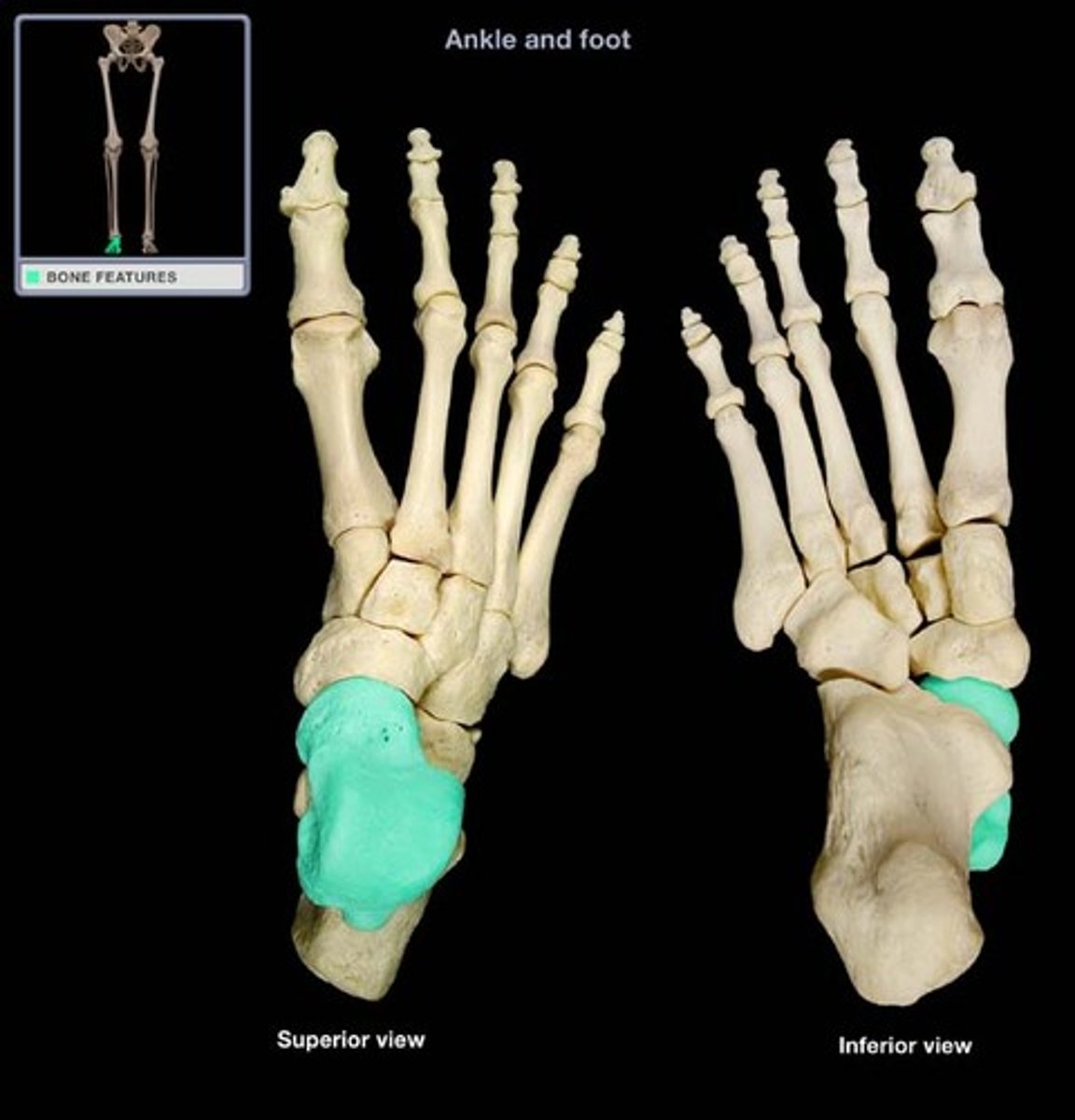
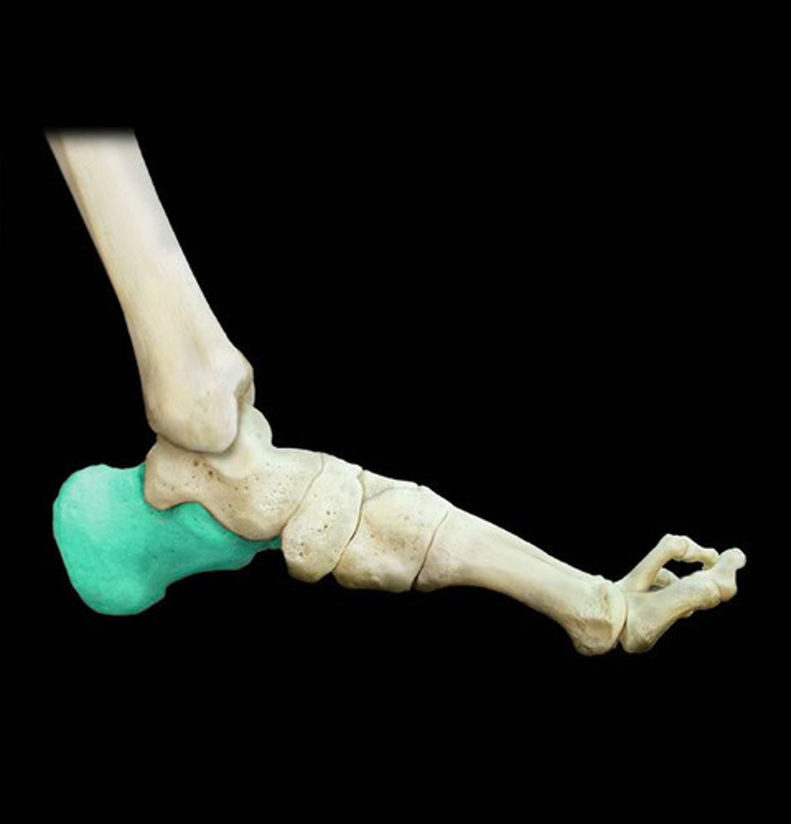
Calcaneus
Largest, strongest bone in foot
Transmits majority of body weight from talus to ground
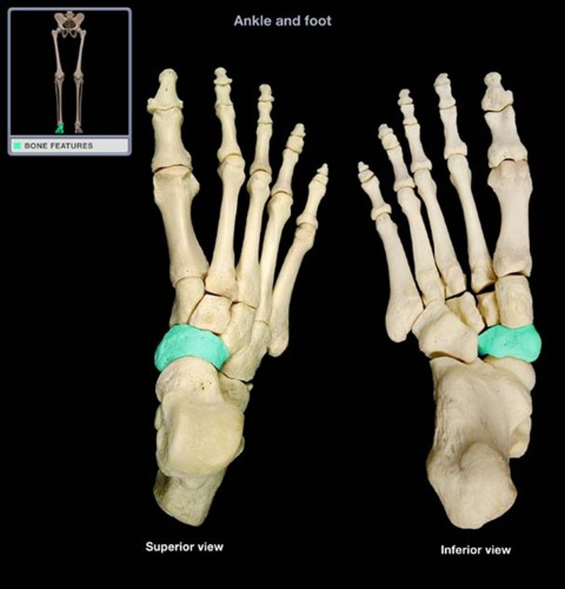
navicular bone
Flattened, boat-shaped bone
Between talus posteriorly and 3 cuneiforms anteriorly
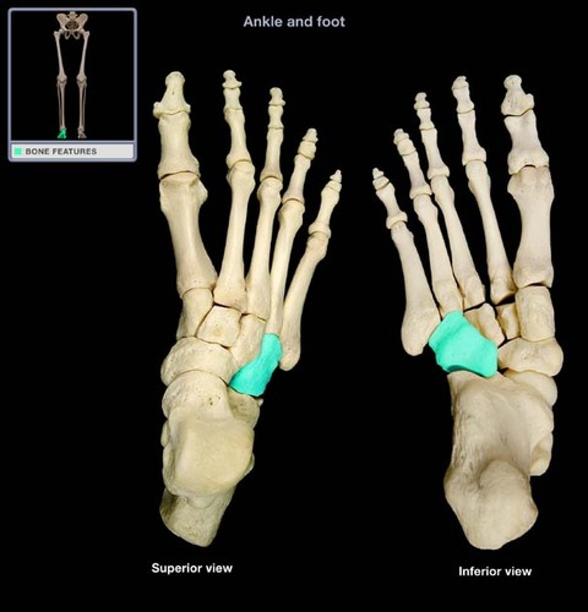
Cuboid
Most lateral bone in distal row of tarsals
Between calcaneus posteriorly and 4th-5th metatarsals anteriorly
Cuneiforms
Each articulates with navicular posteriorly and base of appropriate metatarsal anteriorly
Medial (1st) cuneform
largest of the 3 identically named bones in foot
Intermediate (2nd) cuneform
smallest of the 3 identically named tarsal bones
Lateral (3rd) cuneform
one of the three identically named tarsals that also articulates with cuboid
Metatarsals
Make up forefoot
5 numbered from medial to lateral
1st metatarsal is shorter and stouter
2nd metatarsal is longest (axis)
Phalanges
Make up forefoot
Each digit has 3 (proximal, middle, distal) except great toe which has 2 (proximal and distal)
Parts of phalanges
Base
Shaft
Head