Ch 3 - Nervous System Organization
 Call Kai
 Learn
Learn Practice Test
 Spaced Repetition
 Match
Match Flashcards
 Knowt Play
1/19

There's no tags or description
Looks like no tags are added yet.
Name | Mastery | Learn | Test | Matching | Spaced | Call with Kai | Chat |
|---|
No analytics yet
Send a link to your students to track their progress
20 Terms
Neuroanatomy
Nuclei = groups of NEURONS in CNS
Tracts = groups of AXONS in CNS
→ use specific terms to describe directions & positions in nervous system
CNS = brain & spinal cord
Neuroanatomy: Direction Terms
Rostral-caudal = nose → tail (front to back)
“anterior-posterior”
Dorsal-ventral = back to front axis of body (back to front)
“superior-inferior ONLY for brain”
Medial-lateral = twd middle or twd the side
top of brain = dorsal
bottom of brain = ventral
Neuroanatomy Structure terms
Ipsilateral = on same side of body
Contralateral = on opposite sides of body
ex. feeling on R arm is sent to the contralateral side of the brain (L brain)
Proximal = close together
Distal = far apart
Afferent = movement twd the CNS
ex. sensory info to brain
Efferent = movement away from CNS
ex. motor info to muscles
Anatomical organization

Nomenclature
many parts of the brain have a functional name + a structural name → either can be used
many names come from latin or greek or greek variants (don’t follow greek rules)
ex. the most posterior part of the frontal lobe, along the central sulcus: KNOW BOTH
Precentral gyrus (anatomical name)
Primary motor cortex (functional name)
NS Structure & Function
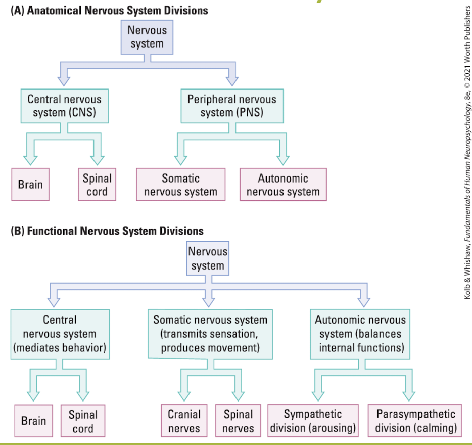
NS can be divided based on its structure or function:
→ CNS = spinal cord + brain
both protected by bone (skull & vertebrae)
→ PNS = projects out to the body + ISNT protected by bone
Somatic NS = provides interaction w external world (afferent&efferent)
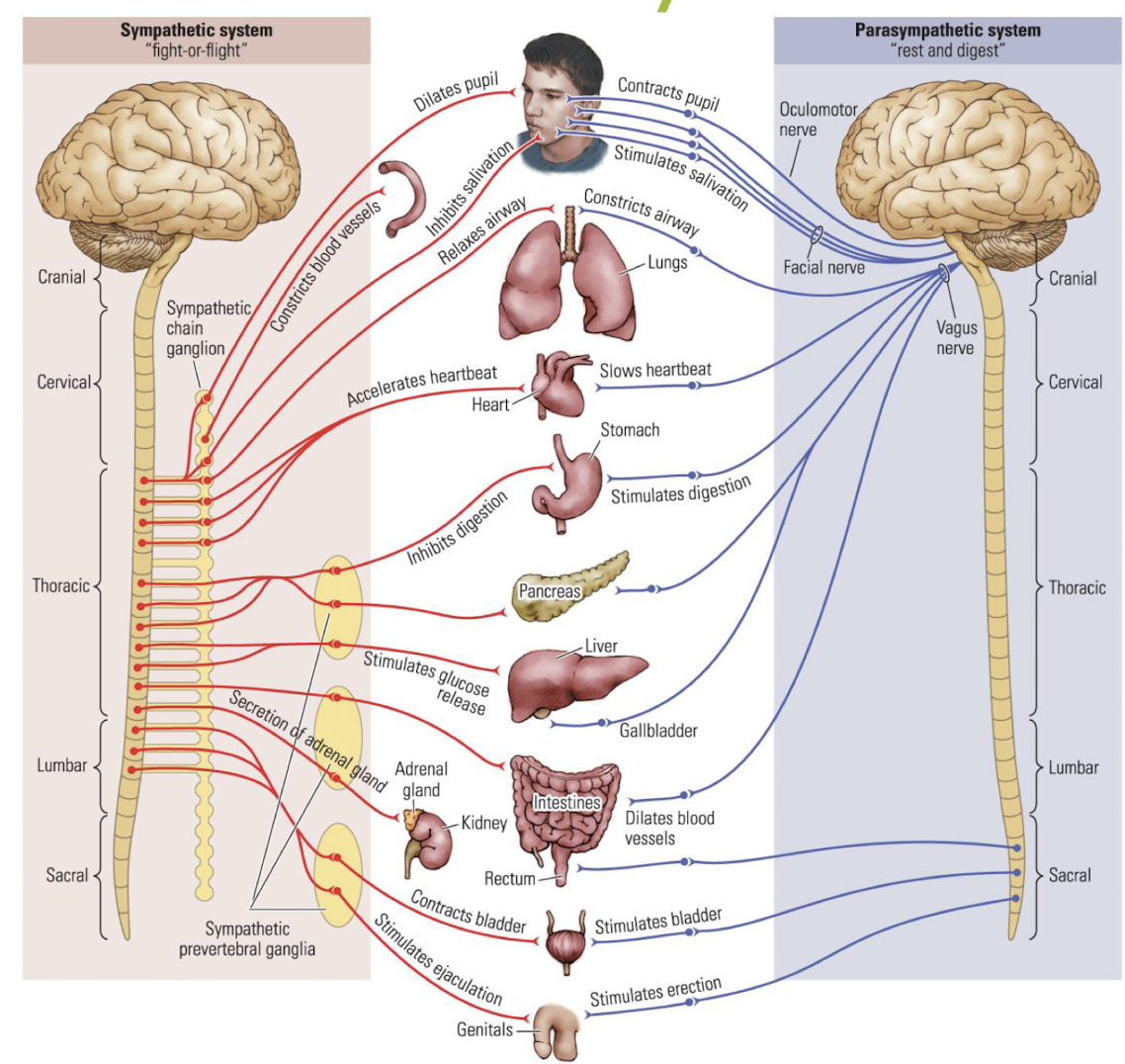
Autonomic system:
Sympathetic branch = flight-or-flight response
Parasympathetic branch = calming influence on body
Protection of the CNS
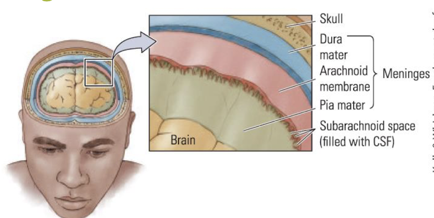
Bone (skull & vertebral column) surrounds the tissue
Meninges surround the CNS to hold it in place + to keep things out
Dura mater = tough OUTER covering
Arachnoid mater = weblike + connects layers + adds cushion
Pia mater = tough INNER layer + adheres to surface of brain (very thin)
note: see ridges when dura mater layer is removed
Cerebrospinal fluid around meninges absorbs shocks + carries waste away and nutrients in (salty)
Blood-brain barrier for chemical protection
formed by astroglia (astrocytes) that hold the cells of blood vessel tightly together
regulates what molecules can exit the blood supply to enter the brain
note: blood kills neurons
Blood Supply to Brain
→ multiple arteries supply blood to brain → ensures damage to one blood vessel doesn't interrupt blood flow to the entire brain
arteries branch to form smaller arteries → deliver to specific areas of brain
Stroke = interruption of blood supply to brain, bc artery is blocked or bursts (lack of oxygen & nutrients)
type of deficit observed after tells the clinician abt the location of brain damage (info abt what blood vessel is damaged)
Major Cerebral Arteries:
Anterior = along top of brain
Middle = middle of brain
Posterior = bottom of brain

Nuerons & Glia
brain starts as neural stem cells (undifferentiated)
stems cells divide → neuroblast cells → neurons & glioblasts → glia
overall:
stem cells
progenitor cells
blast cells (produce neuroblasts & glioblasts)
specialized cells (neurons & glia differentiate)
note: projecting neurons are sensory or motor neurons

3 Basic Types of Neurons
Sensory neurons = transduce info from the environ
PNS (afferent - twd CNS)
bipolar (one dendrite + one axon)
cell bodies sit outside CNS
note: somatosensory neuron has ONLY one projection from the soma (cell body)
Interneurons = connect sensory & motor neurons within the CNS
often have extensive branching of dendrites
MOST common
some in PNS
Motor neurons = project to muscle to carry out movement
PNS (efferent - away from CNS)
found in brainstem & spinal cord (cell bodies, but axons extend outward)
receive info @ dendrite

note: unipolar = 1 projection off of cell body
Types of Glia
Ependymal cell - secretes cerebrospinal fluid
small, ovoid
Astrocyte = nutritive & support functions + cognition + forms blood-brain barrier
Microglial cell = defensive function (CNS immune system)
used bc WBC cant provide defence since blood kills neurons
small, mesodermally derived
Oligodendroglial cell = forms insulating myelin around axons in CNS (brain&spinal cord)
can create myelin on 1+ axons at once
asymmetrical
Schwann cell = forms insulating myelin arounds nerves in PNS
creates myelin on ONLY 1 axon at once
asymmetrical
→ oligo & Schwann cells have same fxn but diff locations, thus they look different
Gray vs. White vs. Reticular Matter
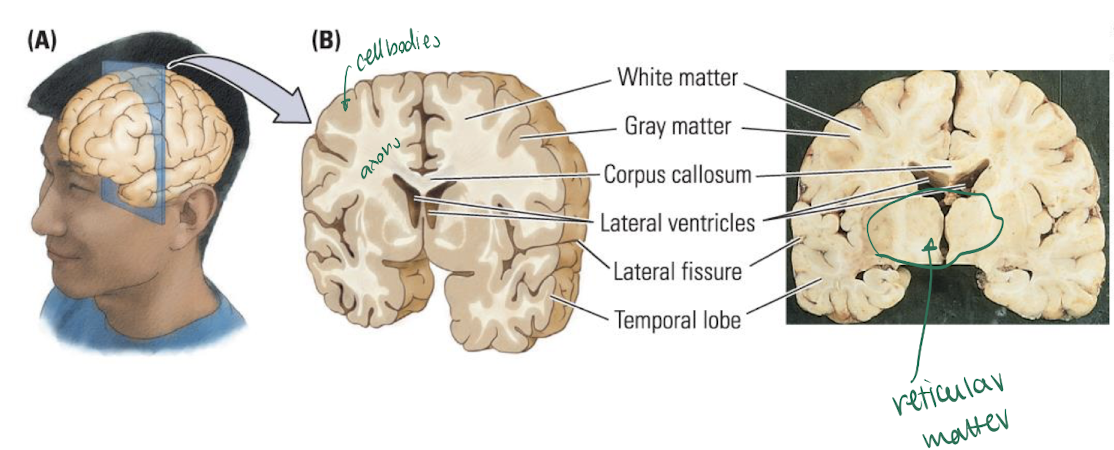
Gray matter = contains cell bodies & capillaries that supply them w blood
outer part of cortex (CLOSEST to blood)
note: cell bodies require blood the most
White matter = myelinated axons (fat)
underneath the cortex (inside of brain)
Reticular matter = netlike appearance & is a mix of gray/white matter
in brainstem (middle brain)
Development of the CNS
→ NS develops from 3 enlargements of the embryonic spinal cord
Prosencephalon = divides to form the telencephalon (cortex/brain) & diencephalon (thalamus + hypothalamus)
Mesencephalon = remains and becomes the midbrain
Rhombencephalon = divides to form the mesencephalon (pons + cerebellum) & myelencephalon (medulla oblongata)

The 4 Ventricles & their Locations
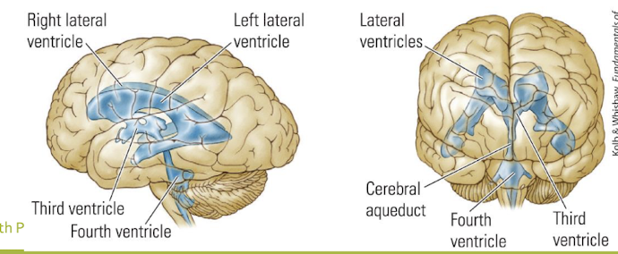
→ brain develops as a hollow tube → hollow parts become the ventricle filled with CSF
L Lateral ventricle (in telencephalon)
R lateral ventricle (in telencephalon)
3rd ventricle (midline of brain)
4th ventricle (b/w cerebellum & brainstem)
→ tube continues down the centre of the spinal cord
Spinal Cord
= gray matter surrounded by white matter
Anterior root = nerve roots branch from the cord to carry motor commands to the body
Posterior root = nerve roots branch from the cord to conduct sensory info into the CNS
→ 4 segments of the spinal cord are named based on the vertebrae the nerve roots pass to exit the CNS:
cervical
thoracic
lumbar
sacral
→ damage at any point effects any segment 2 and below it
→ sensory info enters @ back
→ motor info exits @ front
nerves = group of axons in PNS
Dermatomes = region of the body surface innervated by each nerve
Spinal Nerves & Spinal Reflexes
sensory neuron (cell body outside of spinal cord) brings sensory info from sensory receptors into posterior root (back) of spinal cord
sensory neuron in spinal cord talks to interferon or motor neuron
some info is sent to brain
motor neuron (cell body inside spinal cord) sends motor commands out the anterior root (front) to muscles

Reflxes = basic behaviours that occur w/o cognitive inout
spinal reflexes are generated in the spinal cord on posterior sensory input → direct connection onto the anterior motor output pathways
complex reflexes can use info across multiple spinal cord segments
Cranial Nerves
→ 12 pairs of nerves branch from the brain & brainstem to provide sensory & motor innervation to the head
PNS (outside of brain)
direct to brain (medulla) , NOT spinal cord
some are afferent (sensory), some are efferent (motor), some are mixed
→ access fxn of these cranial nerves to diagnose issues with NS
KNOW 2 CRANIAL NERVES:
olfactory = smell
oculomotor = movement of eyes

Autonomic NS
= interacts w internal organs & glands
→ Sympathetic branch = body ready for action
sympathetic ganglia are close to spinal cord → easier to activate than para
→ Parasympathetic branch = calms body
parasympathetic ganglia are near the target organs
Brainstem
= hindbrain + parts of midbrain
extends from where the spinal cords enters the skull to the forebrain
where the cranial nerves enter the brain
→ Hindbrain:
cerebellum = motor control & sensory integration (backup brain)
pons = connects cerebellum with rest of brain + sleep reg
medulla = regulations breathing & heart rate
reticular formation (spans pons/medulla) = sends projects to the cortex to maintain alertness & arousal
majority of brainstem (hindbrain & mid)
