PLAX and PSAX Anatomy + Direction
 Call Kai
 Learn
Learn Practice Test
 Spaced Repetition
 Match
Match Flashcards
 Knowt Play
1/30

There's no tags or description
Looks like no tags are added yet.
Name | Mastery | Learn | Test | Matching | Spaced | Call with Kai | Chat |
|---|
No analytics yet
Send a link to your students to track their progress
31 Terms
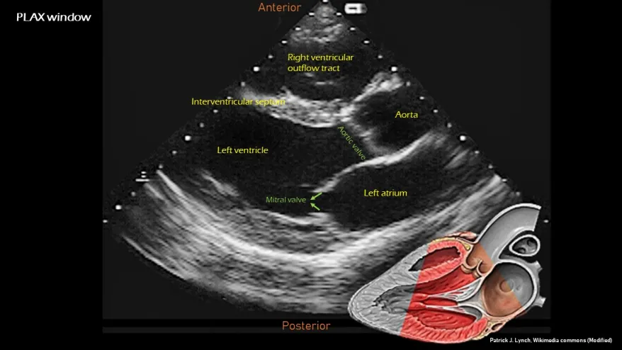
What image plane and acoustic window does this image represent ?
Parasternal Long Axis view
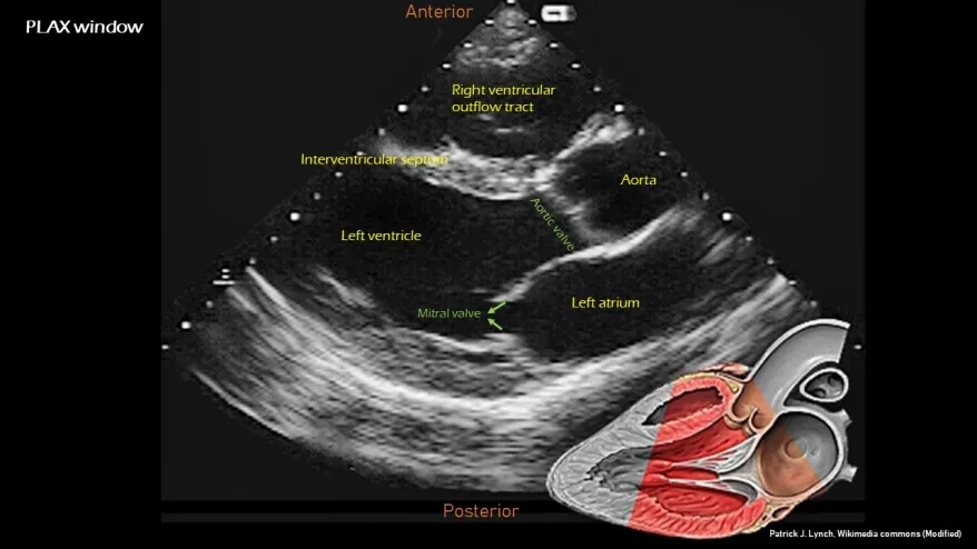
For this image define the probe location, index mark orientation, and how to obtain this image.
Probe Location:
Left sternal border
Index Mark:
➡ Toward the patient's right shoulder (10 o’clock)
How to Get There:
Place the probe at the left sternal border.
Orient the probe for the PLAX plane. ( 10’ oclock)
Adjust/angle until the characteristic PLAX anatomy is displayed.
What MAJOR structures should you typically see when in PLAX view?
Right Ventricle
Left Ventrical
Left Atrium
Aorta
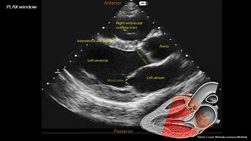
What MINOR structures do you typically see in PLAX view?
Interventricular septum
Aortic valve
Papillary muscle
Mitral valve
Right ventricular free wall
Aortic root
Descending aorta
Non coronary cusp and aortic sinus of valsava
Chordae tendineae
LV interolateral posterior wall
Pericardium

How would you know your in a PLAX imaging plane?
Look for the characteristic relationship:
Right ventricle→ Interventricular Septum → Left ventricle
and
Left Ventricle → Mitral Valve → Left Atrium
with the aortic root/AV also visualized.
⭐ Tip:
Don't memorize PLAX as a list. Learn the relationships between the structures. If you can point to the RV, IVS, LV, MV, LA and aorta on an unlabeled image, you actually know the view.

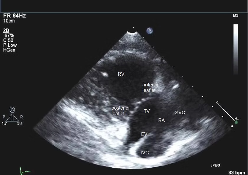
What image plane and acoustic window does this image represent ?
What is it?
A parasternal view that demonstrates the right ventricular inflow pathway.
Acoustic Window:
Parasternal
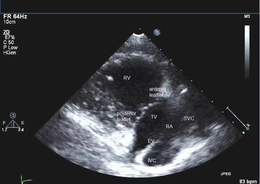
For this image define the probe location, index mark orientation, and how to obtain this image.
Probe Location:
Left parasternal region
Index Mark:
➡ Typically stays at 10 o’clock
How to Get There:
Start from the parasternal/PLAX position and angle the probe toward the patient's right hip to bring the RV inflow tract into view.
What MAJOR structures should you typically see when viewing the Right Ventricular Inflow Tract?
Right Ventricle
Right Atrium
Tricuspid valve
Inferior Vena Cava
Blood-flow pathway to remember: 🧠
Right Atrium → Tricuspid Valve → Right Ventricle

What MINOR structures should you typically see when viewing the Right Ventricular Inflow Tract?
Coronary Sinus orifice
Red and Green leaflets of the Tricuspid valve
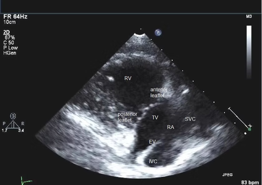
How would you know your in a RVIT imaging plane?
How to Know You're There: ⭐
You're looking for the right-sided inflow pathway, especially the relationship between the RA, tricuspid valve, and RV.
⭐ Tip:
Think:
RVIT = IN
Blood is coming into the RV through the tricuspid valve.
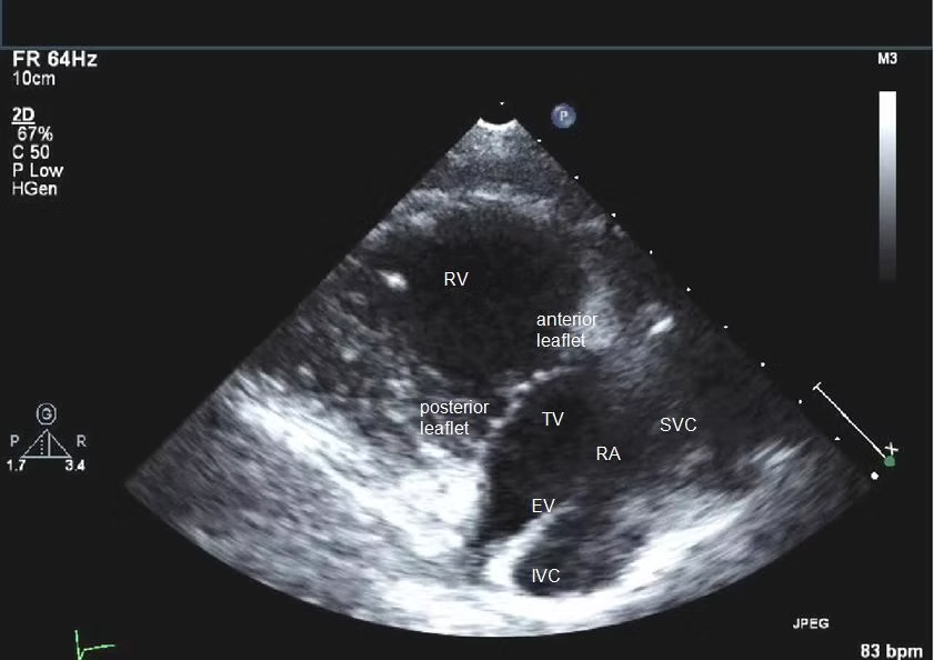
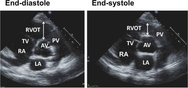
What image plane and acoustic window does this image represent ?
What is it?
A parasternal view demonstrating the pathway by which blood leaves the right ventricle. (RVOT)
Acoustic Window:
Parasternal

For this image define the probe location, index mark orientation, and how to obtain this image.
Probe Location:
Left parasternal region
Index Mark:
➡ 10 o’clock
How to Get There:
Tilt the face (head) of the probe downward and medially so the ultrasound beam points toward the patient's left shoulder.
What structures should you typically see when viewing the Right Ventricular Outflow Tract?
Right Ventricle
RVOT
Pulmonary valve
Main pulmonary artery branching into its left and right components (Bifurcation)
Blood-flow pathway: 🧠
Right Ventricle → Pulmonary Valve → Pulmonary Artery
How would you know your in a RVOT imaging plane?
How to Know You're There: ⭐
You should be able to identify the RVOT leading toward the pulmonary valve and pulmonary artery.
⭐ Tip:
RVIT = IN
RVOT = OUT


What image plane and acoustic window does this image represent ?
What is it?
A parasternal short-axis view obtained at the base/aortic valve level.
Acoustic Window:
Parasternal
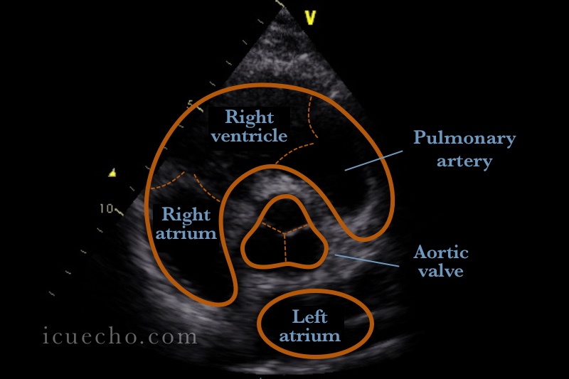
For this image define the probe location, index mark orientation, and how to obtain this image.
Probe Location:
Left parasternal region
Index Mark:
➡ Rotate clockwise to 1 o’clock
How to Get There:
Start from PLAX and rotate the probe to obtain the short-axis plane, then adjust/angle to the aortic valve/base level.
What MAJOR structures should you typically see when in PSAX AVL/BASE view?
Right Atrium
Left Atrium
RVOT/RV
Aortic Valve

What MINOR structures should you typically see when in PSAX AVL/BASE view?
Tricuspid valve
Pulmonary artery
Right, Left, and Non-Coronary Cusps
Main Pulmonary Artery Trunk
Left Main Coronary Artery
Interatrial Septum
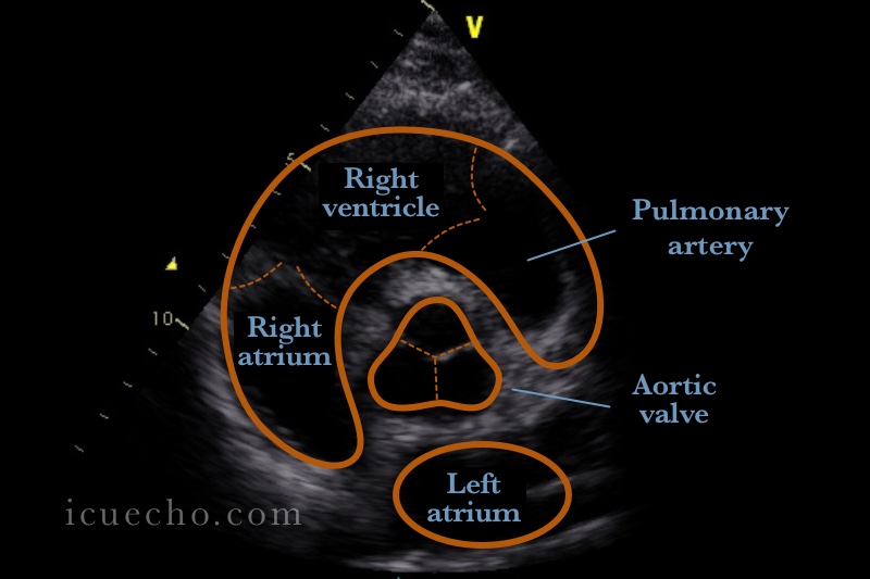
How would you know your in a PSAX AVL/BASE imaging plane?
How to Know You're There: ⭐
The aortic valve should have its characteristic short-axis appearance.
⭐ Tip:
PSAX isn't one single view.
Think:
PSAX = slicing the heart at different levels.
This card is specifically the base/aortic valve level.


What image plane and acoustic window does this image represent ?
What is it?
A parasternal short-axis view at the mitral valve level.
Acoustic Window:
Parasternal

For this image define the probe location, index mark orientation, and how to obtain this image.
Probe Location:
Left parasternal region
Index Mark:
➡ We are going from 1 o’clock TO 3 o’clock
How to Get There:
From the PSAX position, adjust/angle the probe to move from the base toward the mitral valve level.
What MAJOR structures should you typically see when in PSAX MVL view?
Structures You Should See:
Mitral valve (leaflets)
LV
RV
IVS
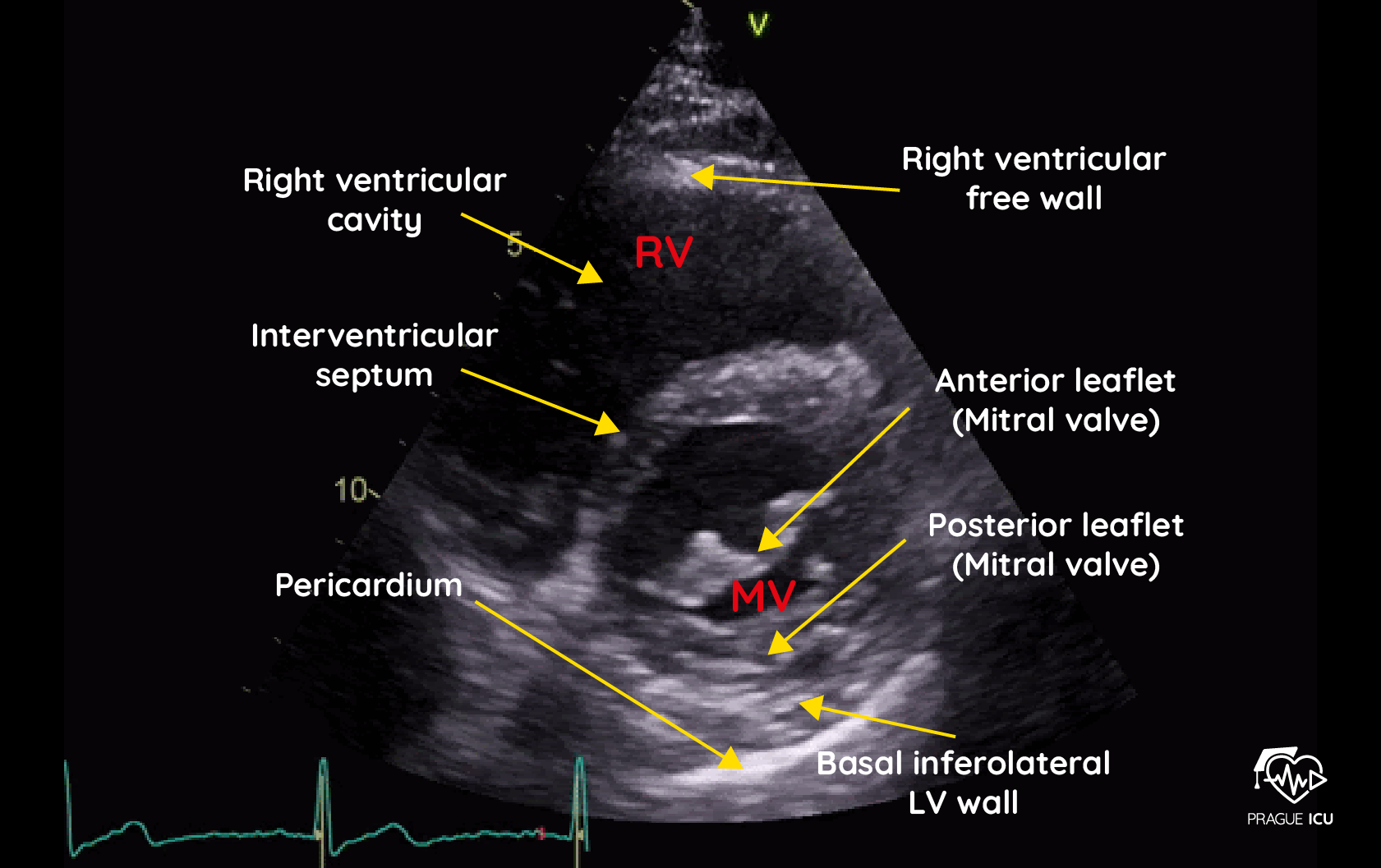
How would you know your in a PSAX MVL imaging plane?
You are in the PSAX mitral valve level when you see the left ventricle as a circular "doughnut" containing a "fish-mouth" opening formed by the mitral valve leaflets


What image plane and acoustic window does this image represent ?
What is it?
A parasternal short-axis view through the mid-LV/papillary muscle level.
Acoustic Window:
Parasternal

For this image define the probe location, index mark orientation, and how to obtain this image.
Probe Location:
Left parasternal region
Index Mark:
➡ Use your course's PSAX orientation
How to Get There:
Start at the Mitral Valve Level (3 o'clock fish-mouth view).
Keep the probe rotation locked.
Tilt the probe tail toward the patient's head.
This angles the ultrasound beam inferiorly toward the apex.
Stop when the fish-mouth disappears and two muscular lumps appear inside the left ventricle ring. [1]
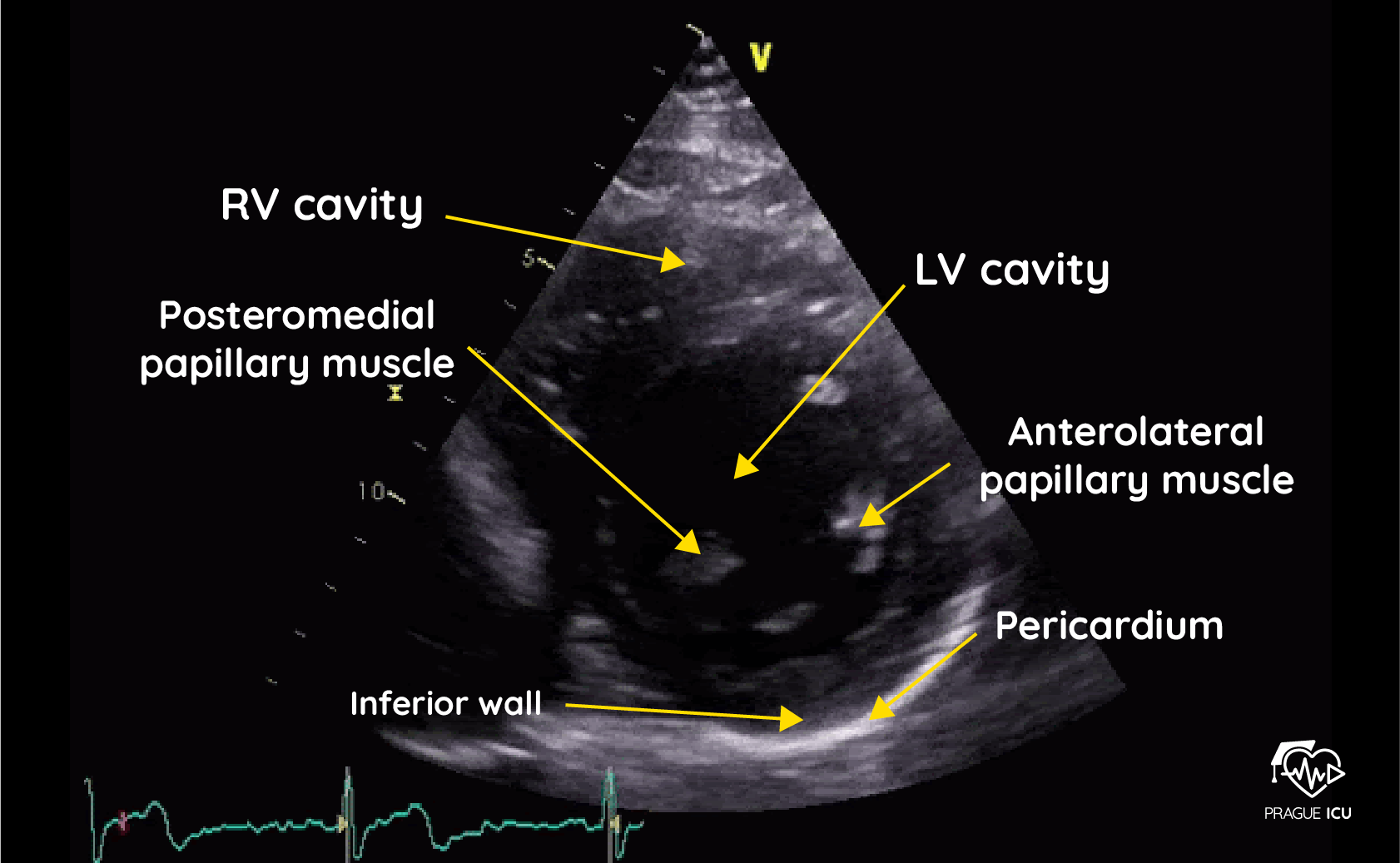
What MAJOR structures should you typically see when in PSAX PML view?
Structures You Should See:
LV
Anterolateral Papillary Muscle
Posteromedial Papillary Muscle
RV

How would you know your in a PSAX PML imaging plane?
How to Know You're There: ⭐
You know you are in the PSAX Papillary Muscle Level imaging plane when you see a perfectly round, donut-shaped Left Ventricle with two distinct muscle pillars protruding into the center.
⭐ Tip:
This is where thinking about the LV as a circle becomes really helpful.

What image plane and acoustic window does this image represent ?
What is it?
A parasternal short-axis view obtained toward the apical level of the LV.
Acoustic Window:
Parasternal
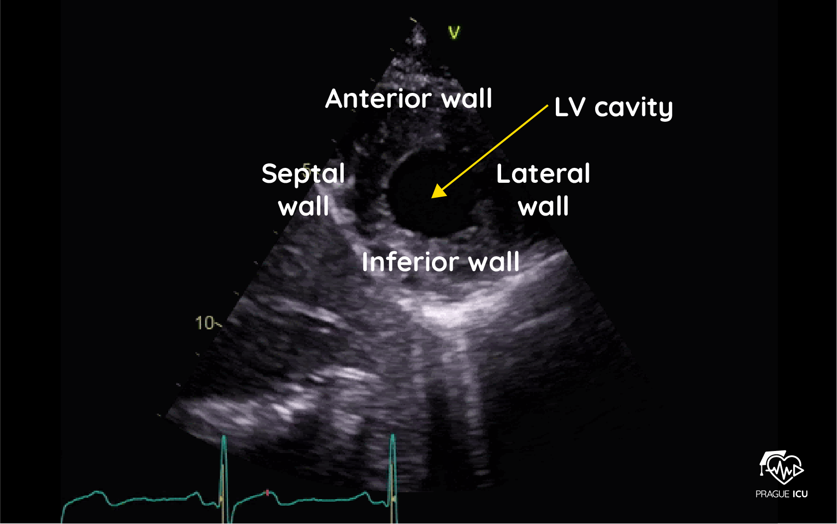
For this image define the probe location, index mark orientation, and how to obtain this image.
Probe Location:
Left parasternal region
Index Mark:
➡ 1-3 o’clock
How to Get There:
Start at the Papillary Muscle Level view.
Keep the probe rotation locked.
Tilt the probe tail even further toward the patient's head.
This angles the ultrasound beam further downward into the very tip of the heart.
Stop when the papillary muscles disappear and the central cavity shrinks to a small, solid circular button. [1]
What MAJOR structures should you typically see when in PSAX LV APICAL view?
Structures You Should See:
Left Ventricle cavity
LV myocardium
The anterior, inferior, septal, and lateral apical walls.

How would you know your in a PSAX LV Apical imaging plane?
Tiny central cavity: The open space inside the left ventricle ring is at its smallest diameter.
No papillary muscles: The two muscular pillars from the mid-level have completely vanished.
No mitral valve: The "fish-mouth" leaflets are completely gone.
Disappearing right ventricle: The RV crescent at the top-left becomes extremely small or disappears.
Symmetrical round shape: The apex looks like a perfect circle, not an oval.
