Introduction to Histology (Epithelial Tissue)
 Call Kai
 Learn
Learn Practice Test
 Spaced Repetition
 Match
Match Flashcards
 Knowt Play
1/91

Earn XP
Description and Tags
Basic Science Core - Histology Exam 1
Name | Mastery | Learn | Test | Matching | Spaced | Call with Kai | Chat |
|---|
No analytics yet
Send a link to your students to track their progress
92 Terms
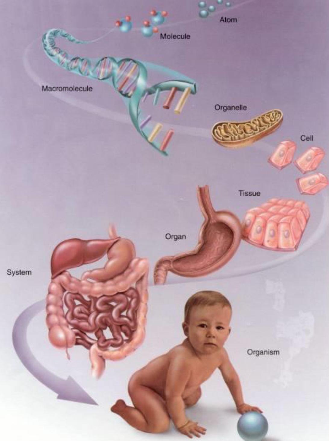
Structural Hierarchy of the Human Body
1) Atoms
2) Molecules
3) Macromolecules
4) Organelles
5) Cells
6) Tissues
7) Organs
8) Organ systems
9) Human body
Four Basic Tissue Types
1) Epithelial tissue
2) Connective tissue
3) Muscle tissue
4) Nervous tissue
Epithelial tissue
Refered to as Epithelium
Likes to form layers (Like a sheet)
Covers body surfaces
Lines body cavities
Prominent in glandular tissue
Epithelial tissue function
Protection, secretion, absorption, excretion
Lacks blood vessels (avascular)
Rests on a basement membrane
Part of the MUCOSA of many organ wall
Types of Epithelium (7)
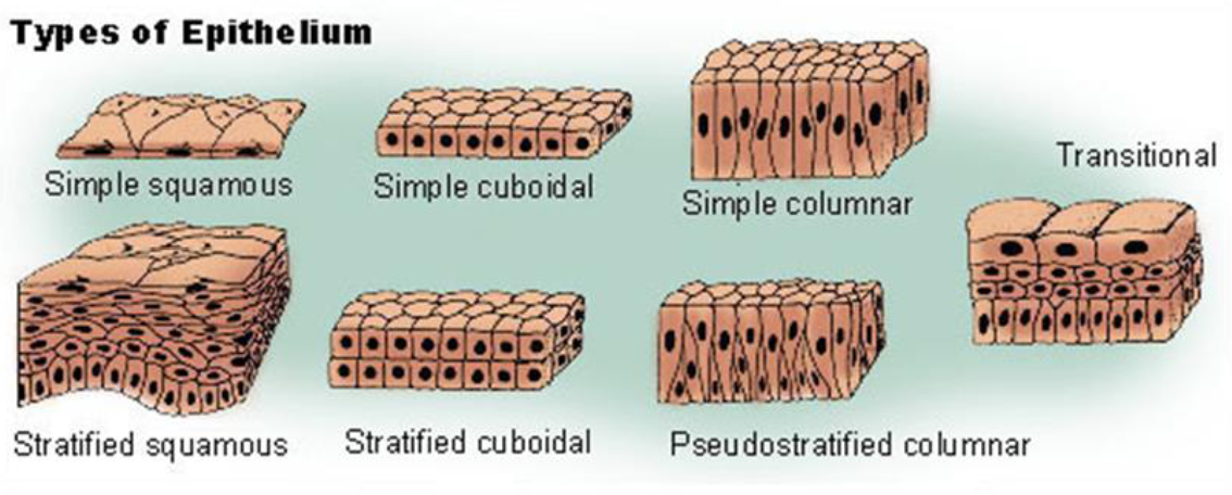
Simple vs Stratified
Simple is 1 layer
Stratified is multiple layers
Pseudostratified vs transitional
Pseudostratified → Looks like any cells but it is not
Transitional →
Connective Tissue (Function)
Supports other tissues
.
Mechanical reinforcement (tendons, ligaments), protection & support (bone, cartilage), immune surveillance (blood), transport of nutrients/wastes (blood), energy storage (adipose)
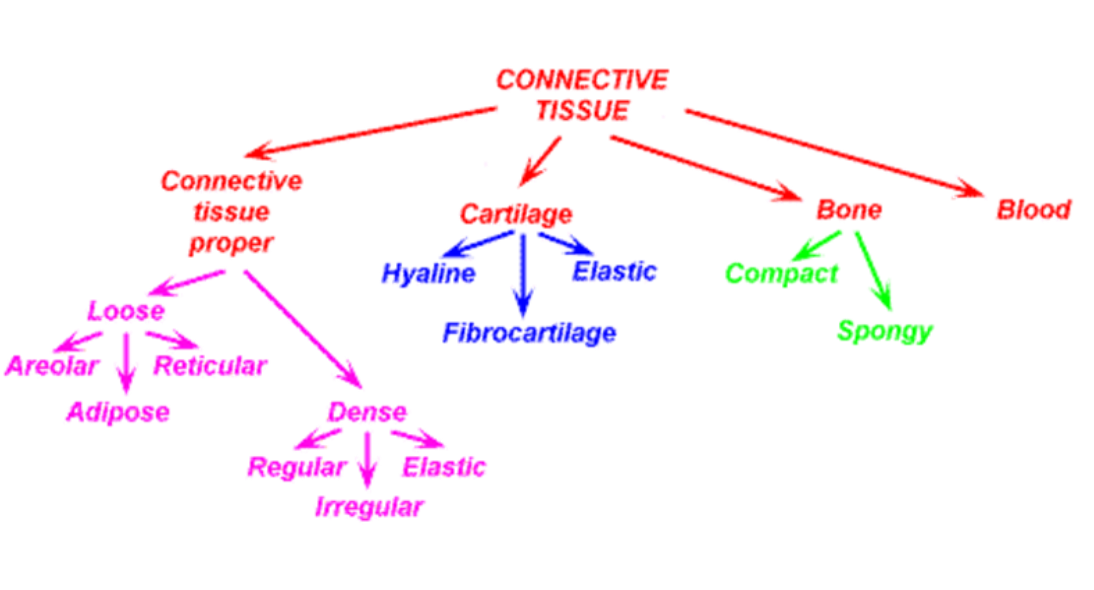
Types of Connective Tissue (4 Big Types
1) Blood
2) Bone
3) Cartlidge
4) Connective Tissue Proper
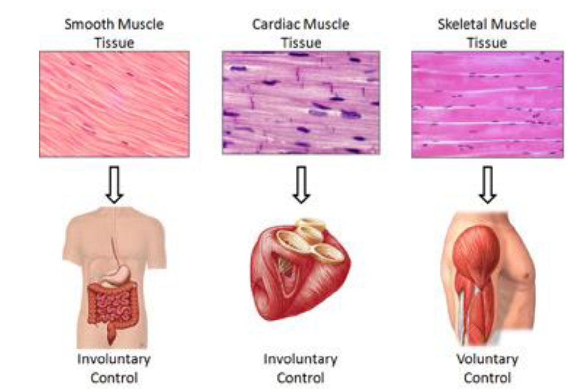
Muscle Tissue (And Function)
Consists of excitable, contractile cells
Movement, motility
Heat production
Skeletal, cardiac and smooth
Types of Muscle (3)
Nervous Tissue
Consists of excitable cells capable of
electrical transmission (conduction)
as a communication tool
Brain, spinal cord, nerves, sensory organs
Neurons, neuroglia
Epithelial Tissue Functions (5)
Secretion
Mechanical protection
Absorption / Reabsorption
Receptor transduction
Transportation
Secretion
hormones, gastric juice, pancreatic juice, saliva, bile
Mechanical protection
skin (epidermis), linings of GI tract
Absorption / Reabsorption
nutrients, electrolytes, vitamins, water
Receptor transduction
taste buds, hair cells, photoreceptors (rods & cones)
Transportation
cilia (oviducts, certain airways)
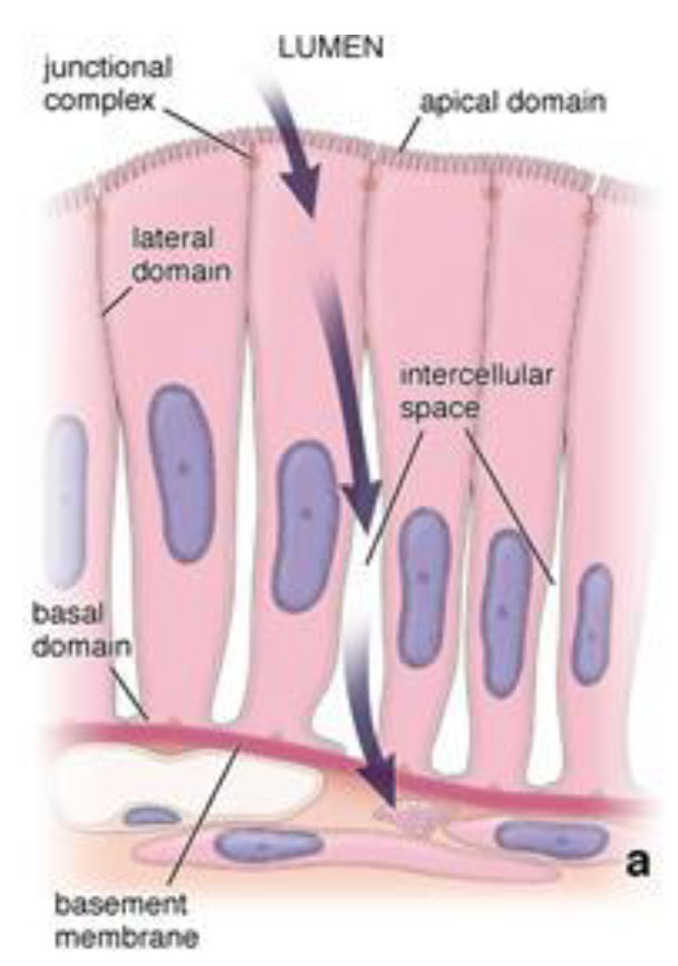
Surface Domains of Epithelial Cell (3)
Apical surface
Lateral surface
Basolateral (Basal) surface
Apical surface
faces lumen,
may have specialized features
(cilia, microvilli, stereocilia)
Lateral surface
may form
“tight junctions”
Basolateral (Basal) surface
attaches to the BASEMENT
MEMBRANE
Epithelial Cell Turnover / Renewal (4)
Rate of turnover is specific to the type
of epithelium
Replenished by a “stem cell” population
\
1) Surface epithelial cells in the stomach every 1-2 days
2) Enterocytes in the small intestine every 4-6 days
3) Oral epithelial cells every 14-21 days
4) Liver hepatocytes every 200-300 days
Apical Surface Variations
MICROVILLI
CILIA
STEREOCILIA
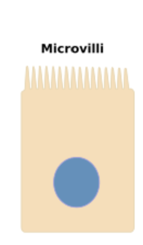
MICROVILLI
finger-like projections
that increase surface area and facilitate
absorption; found in the small intestine
and kidneys
CILIA
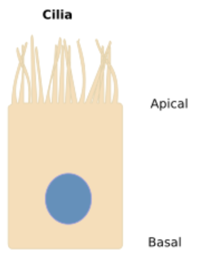
hair-like structures that can
move in a coordinated manner to propel
substances along a surface; only found in
the oviducts and certain airways
(trachea, bronchi, non-respiratory
bronchioles)
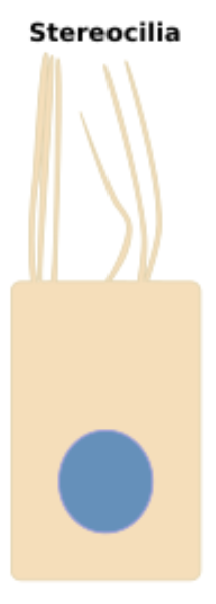
STEREOCILIA
long, non-motile
structures that are thicker than cilia and
involved in sensory transduction
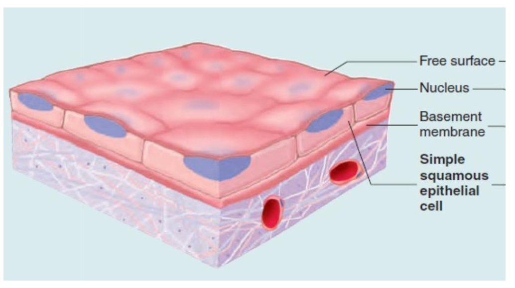
Simple Squamous Epithelium
Single layer of flat, thin cells
Allows for rapid, efficient
movement of substances by
diffusion
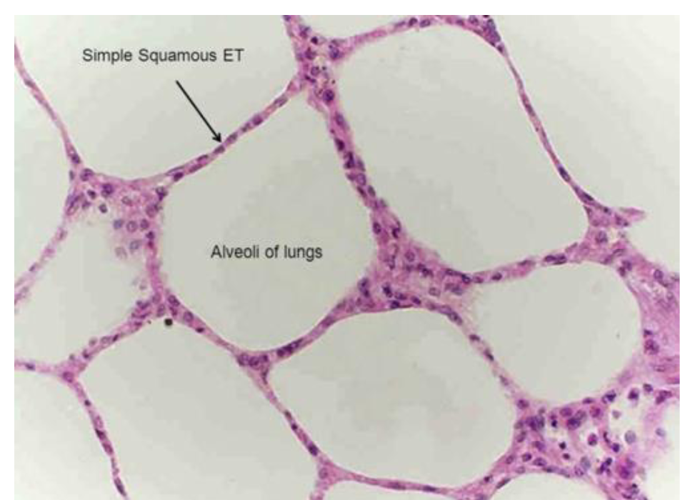
Simple Squamous Epithelium (Where in the body)
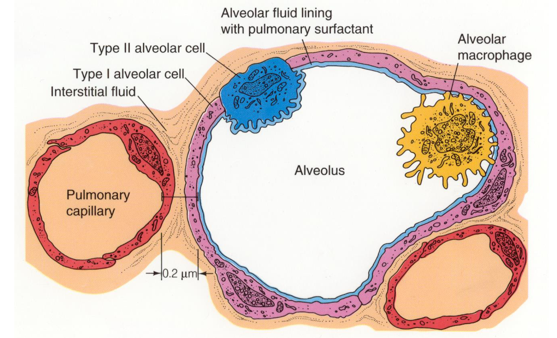
Lung alveoli
Endothelium
Lining of lymphatic vessels
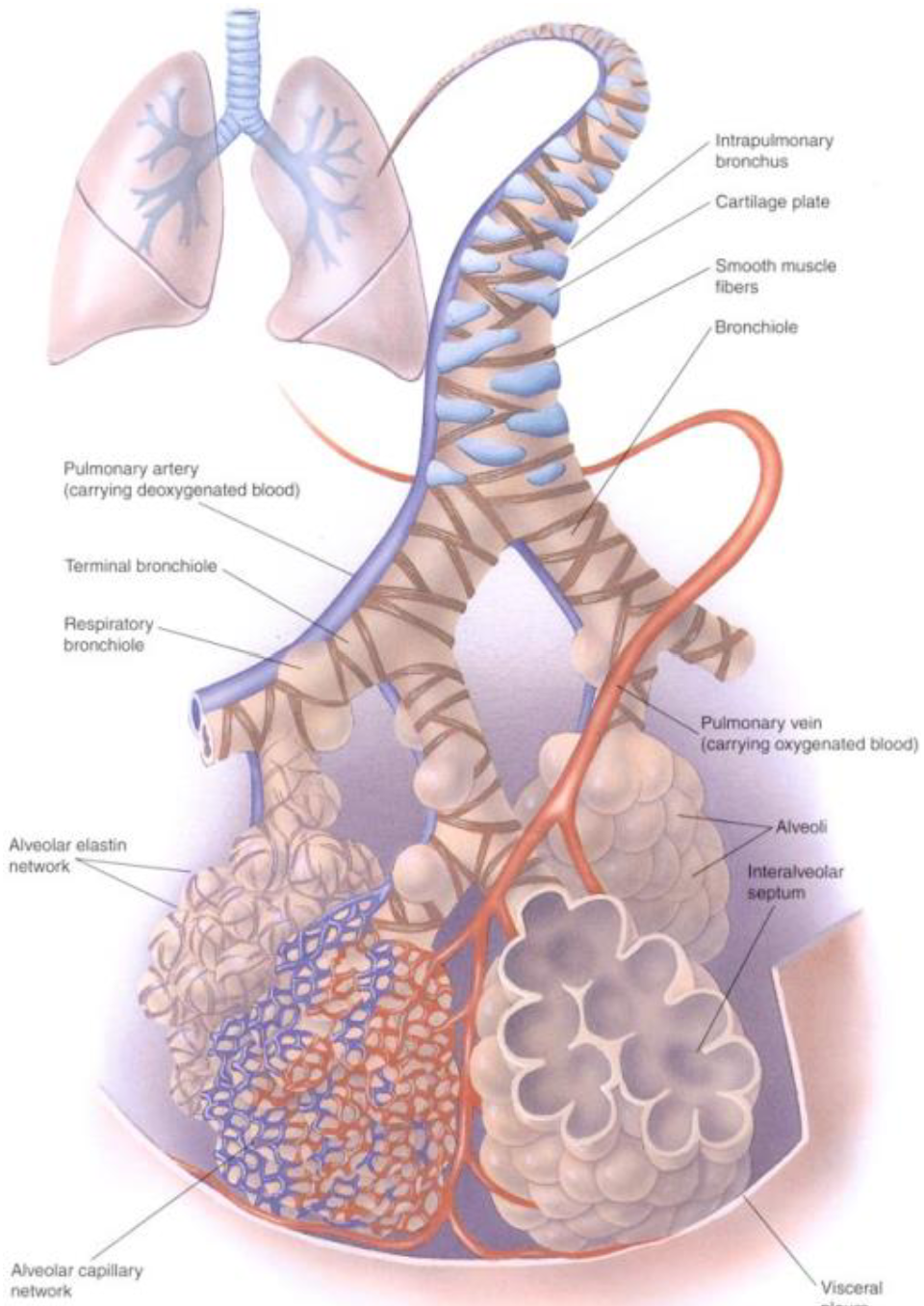
Alveoli
Overwhelming major of gas exchange occurs within the alveoli -- smaller amount occurs with respiratory bronchioles
.
Alveoli are richly surrounded by pulmonary capillaries
.
Each lung has a total of approx. 300 million alveoli
Alveolar-Pulmonary Capillary Interface
Physiological significance lies with the interface’s THINNESS & small amount of interstitial fluid
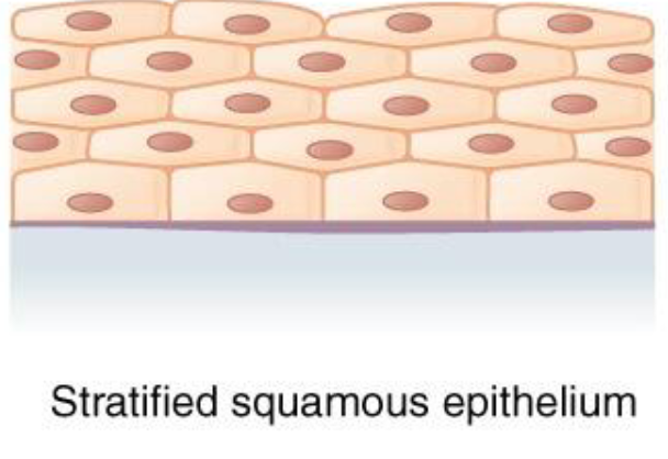
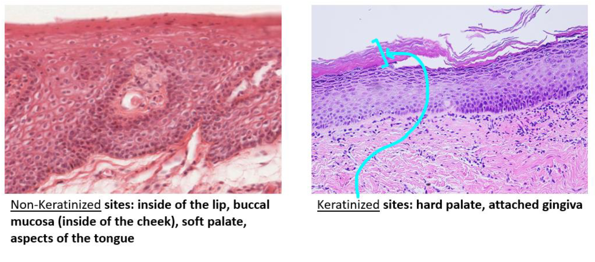
Non-Keratinized Stratified Squamous Epithelium
Multiple layers of flat, thin cells
Provides a barrier against mechanical stress and abrasion
Lubrication (due to mucus production) – helps to
maintain moisture, preventing drying and cracking
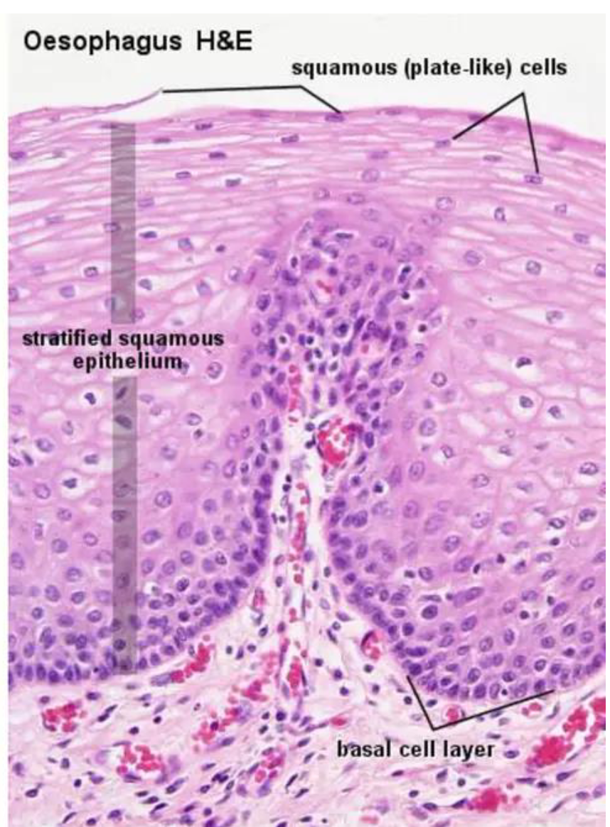
Non-Keratinized Stratified Squamous Epithelium Examples
Examples:
• Esophagus
• Oral cavity (buccal lining, inside of the lips, soft
palate, floor of the mouth, some parts of the tongue)
• Cornea
• Vagina
• Anus
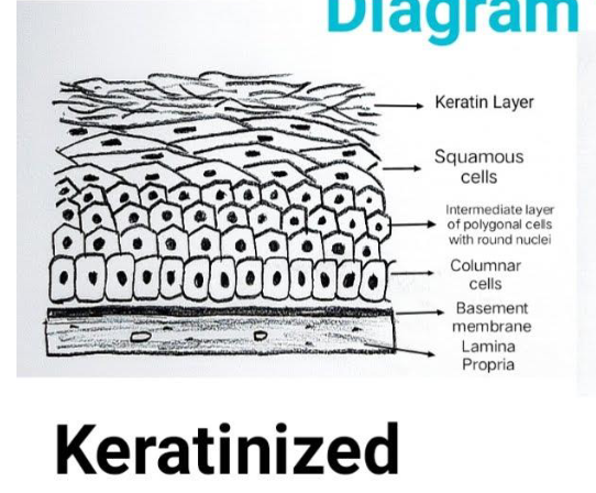
Keratinized Stratified Squamous Epithelium
Multiple layers of flat, thin cells BUT with a layer of KERATIN on top
.
Provides a barrier against mechanical stress and abrasion
Keratinized Stratified Squamous Epithelium in Oral Health Care
The gingiva, hard palate and parts
of the tongue (especially the dorsal
surface) have this type of
epithelium, which helps them
withstand the forces of chewing
and the tongue’s movements
Keratinized Stratified Squamous Epithelium Examples
Epidermis of the skin
• Oral cavity (hard palate,
gingiva)
• Some parts of the tongue
Two Types of Stratified Squamous Epithelium
Keratinized Stratified Squamous Epithelium
Non-Keratinized Stratified Squamous Epithelium
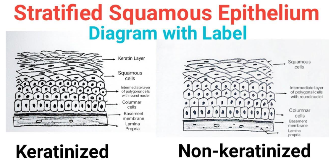
Oral Cavity Epithelium
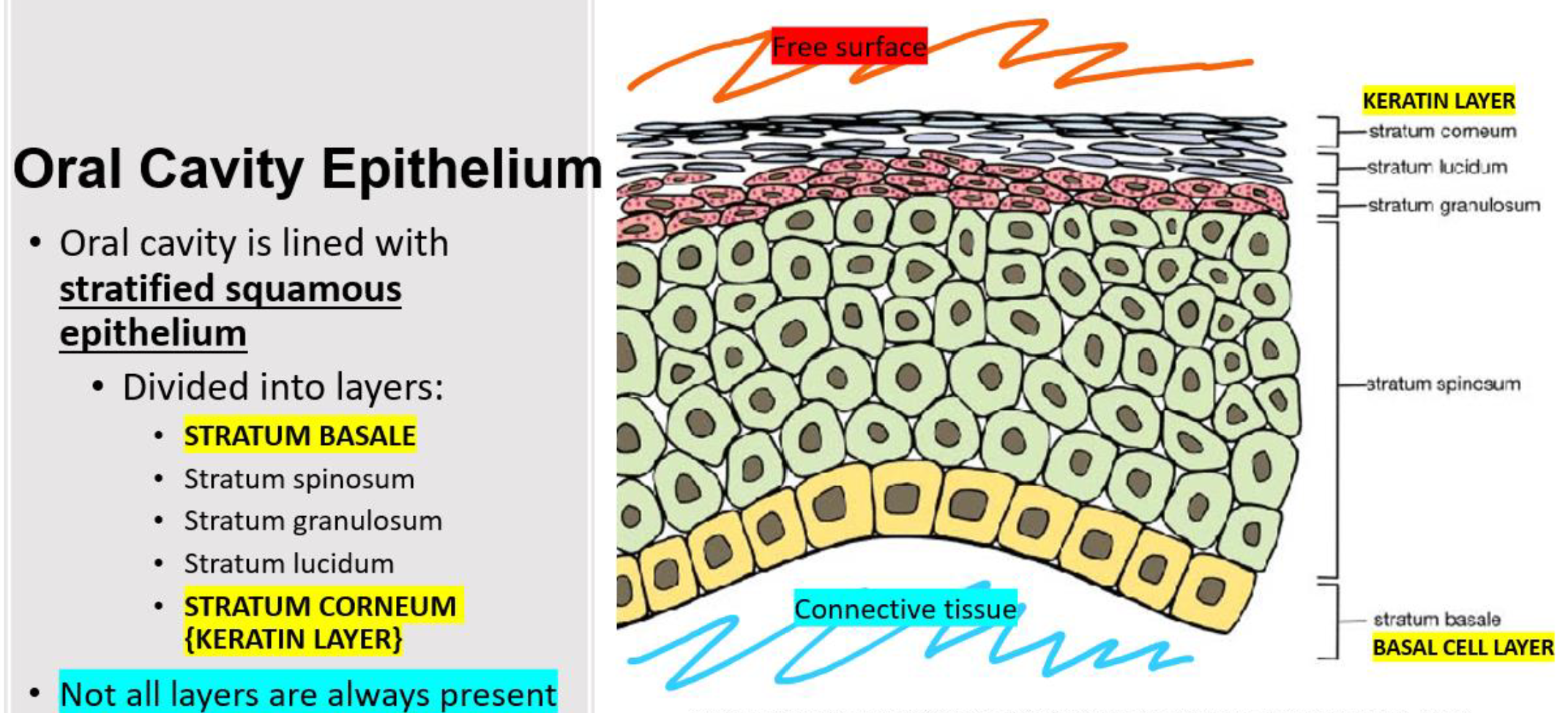
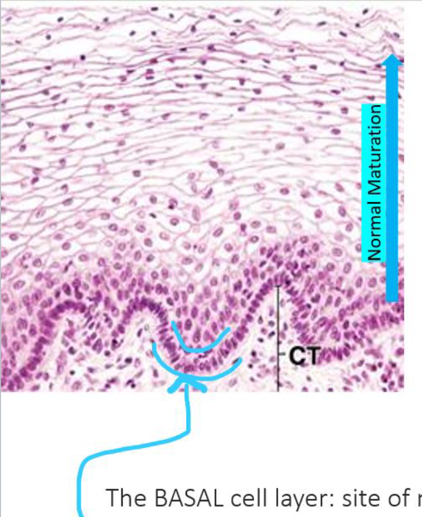
Basal Cell Layer
The Basal Layer in the cell:
Site for normal cytosis for cell replenishment in the oral cavity
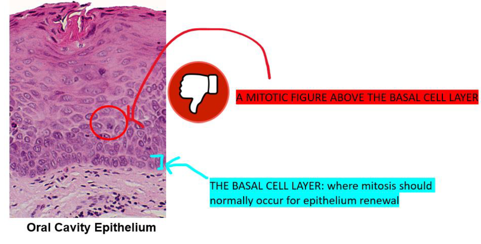
Epithelial Dysplasia
Pre Cancerous Condition
Occurs if cell growth is occurring above the cell layer
Where are the Keratinized vs Non Keratinized Sites in the Oral Cavity
Simple Cuboidal Epithelium
Single layer of square (cube-shaped) cells
Allows for secretion and absorption (or reabsorption)
Forms protective linings
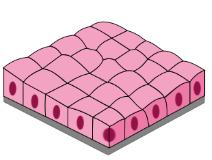
Simple Cuboidal Epithelium Examples (3)
Examples:
Thyroid follicles
Kidney tubules (within nephrons)
Ducts draining salivary glands (smaller ducts)
Thyroid Gland Diagram
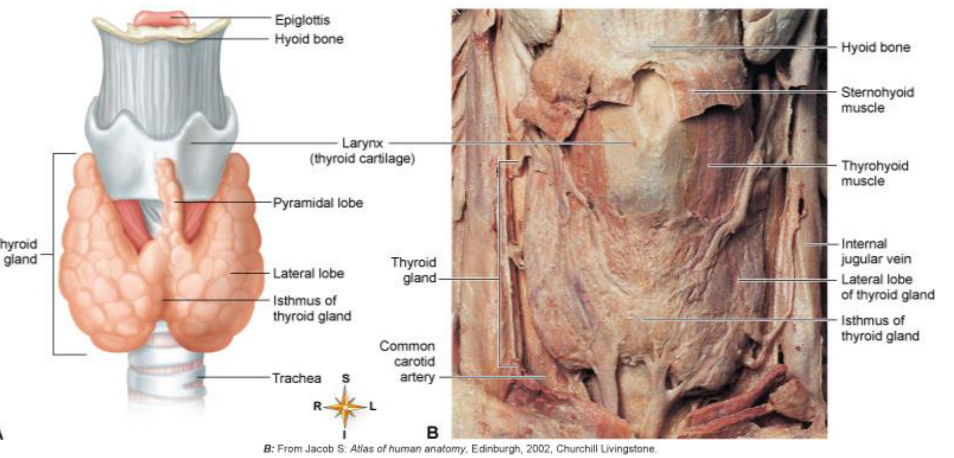
Gross Anatomy of the Thyroid Gland
The thyroid gland is located anterior and lateral to the trachea, just below the thyroid cartilage (Adam's apple).
It consists of two (sometimes three) lobes (one on each side of the trachea).
The lobes are connected by a bridge of thyroid tissue called the isthmus, which is located just below the cricoid cartilage.
The thyroid gland is barely palpable.
The thyroid gland weighs 10 to 20 grams in normal adults.
Microanatomy of the Thyroid Gland
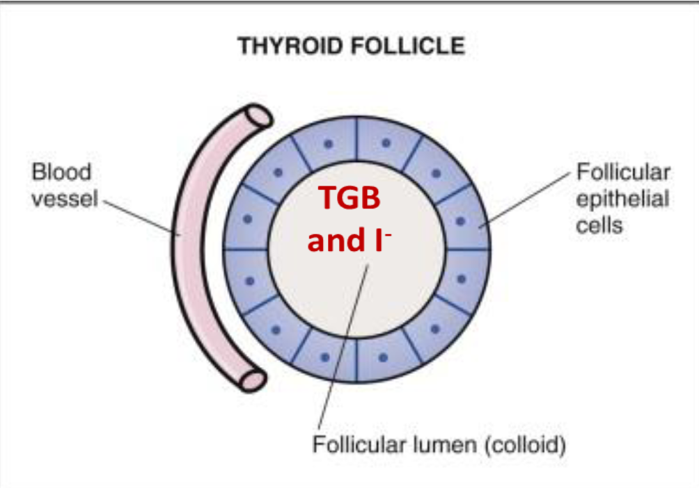
Microscopically, the thyroid is composed of spherical follicles, each composed of a single layer of follicular cells surrounding a lumen filled with colloid
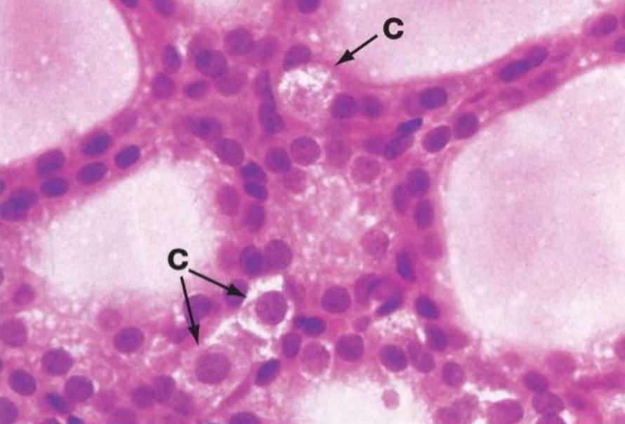
Thyroid follicles are the sites of
THYROID HORMONE (T3 and T4) SYNTHESIS
Nephrons
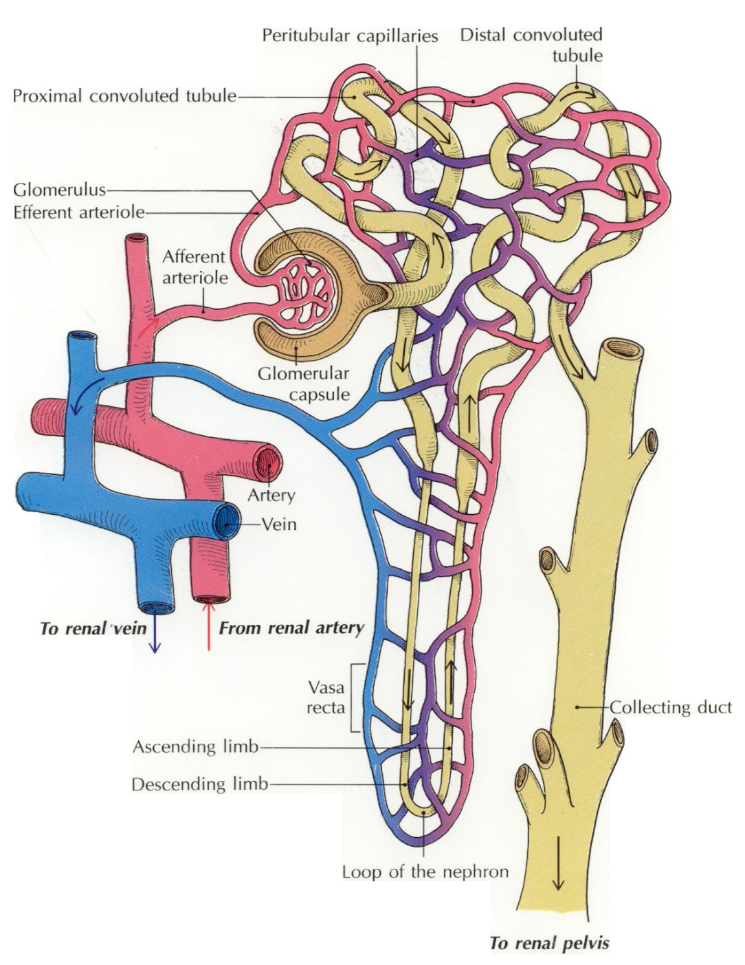
The functional units of the kidney are the NEPHRONS
Each kidney contain approximately 1 million nephrons
Nephrons themselves are NOT cells……but rather they are made up of different types of cells
Nephron Diagram
Nephron Structure
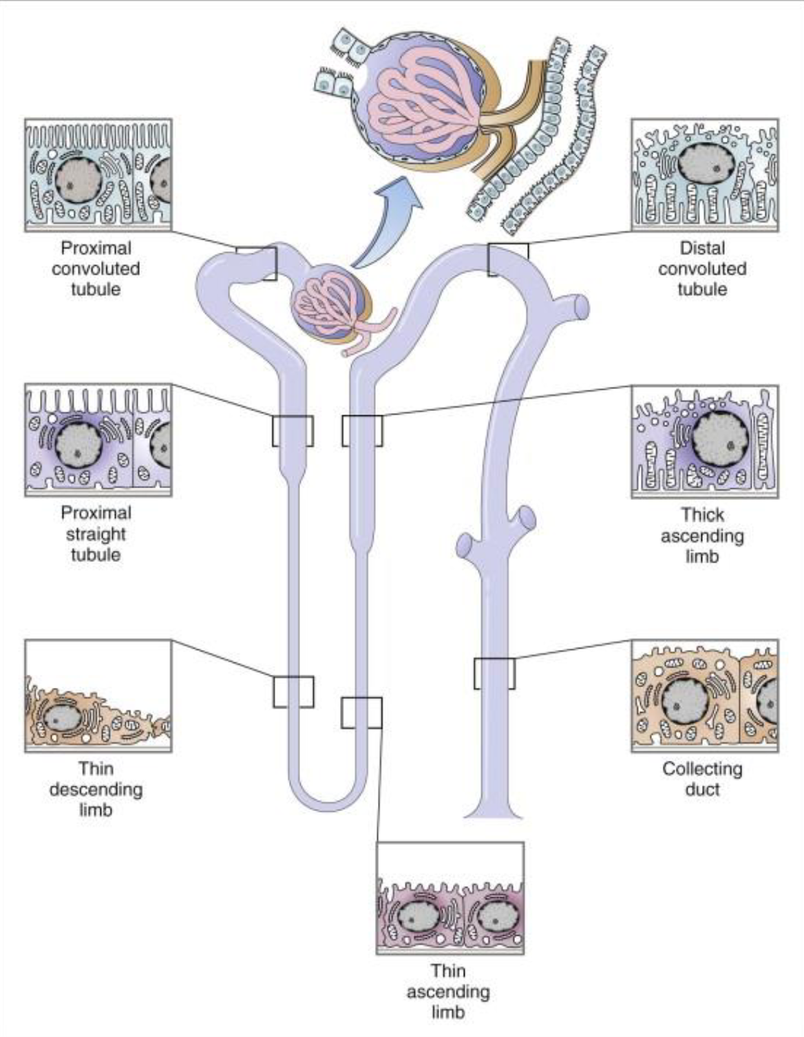
PCT – Tubular reabsorption:
putting a filtered substance back into the blood
.
Notice the MICROVILLI on the cells lining the PCT
.
DCT – Tubular secretion:
Removing a substance from the blood and putting into the kidney tubule
Nephron Structure Diagram
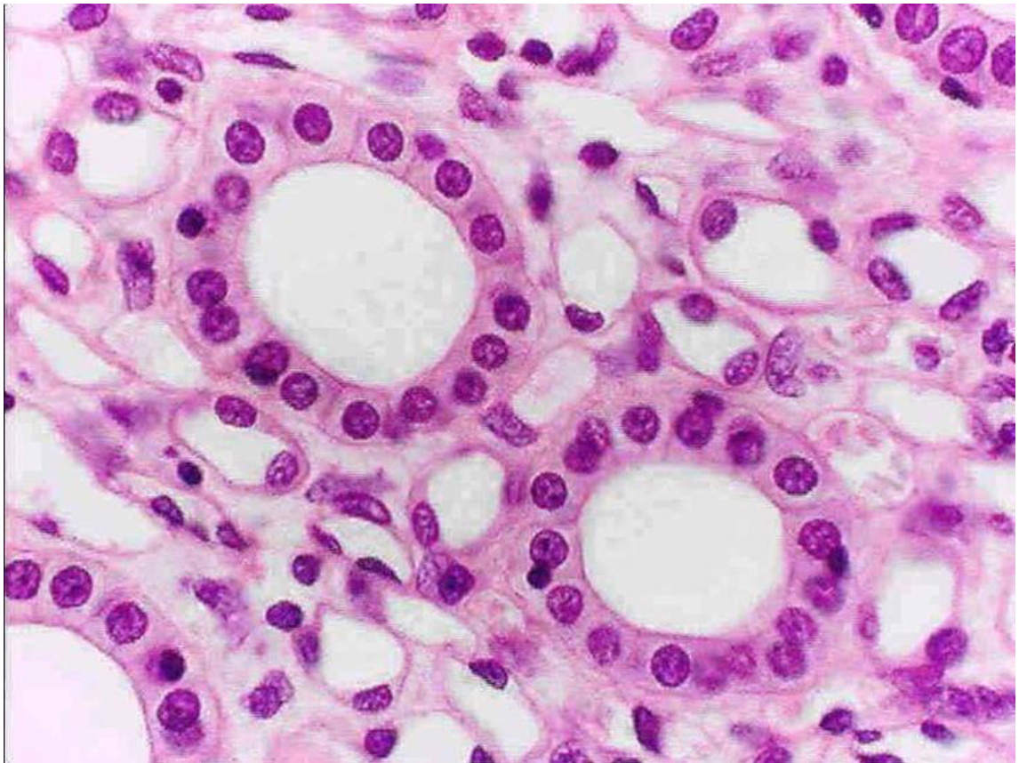
Simple Cuboidal Epithelium in Nephron Tissue Photo
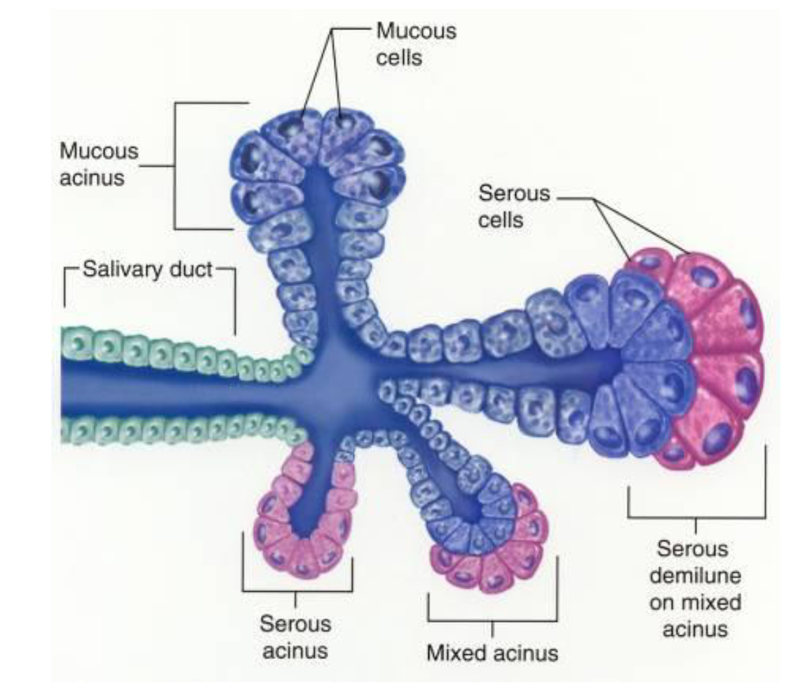
Salivary Gland Histology (3)
Acini are classified as:
• serous
• mucous
• mixed
Salivary Gland Diagram
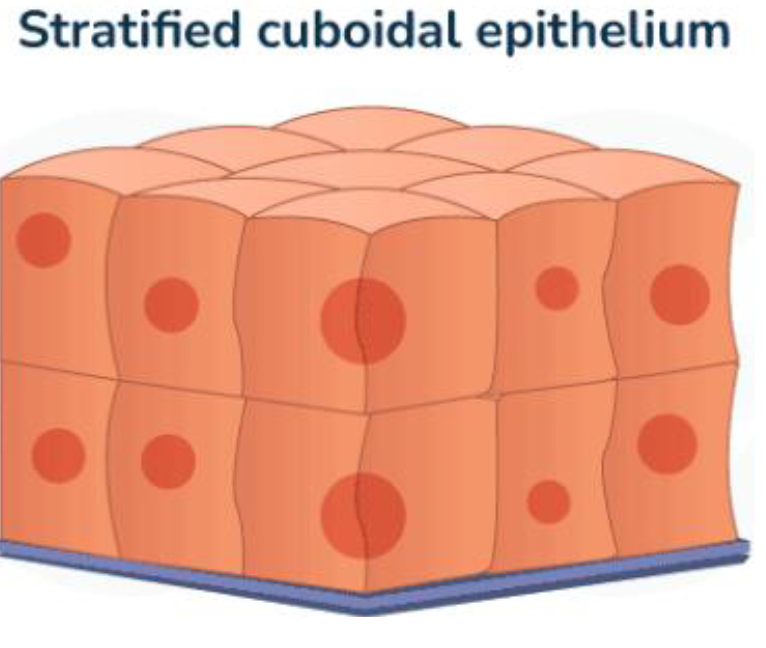
Stratified Cuboidal Epithelium
Multiple layers of square (cube- shaped) cells
Forms protective linings
Stratified Cuboidal Epithelium (3)
Examples:
• Parts of the male urethra
(penile urethra)
• Ducts draining sweat glands
• Ducts draining salivary glands
(larger ducts)
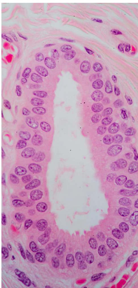
Male Urinary Tract Cellular Photo (Stratified Cuboidal Epithelium)
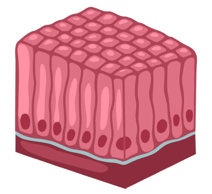
Simple Columnar Epithelium
Single layer of tall cells
Allows for secretion and absorption; forms protective linings; sensory transduction
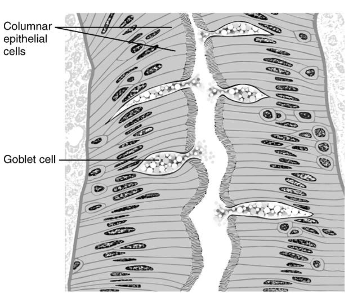
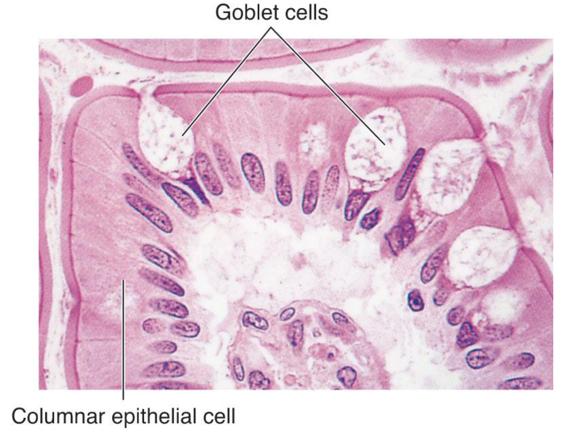
May contain GOBLET CELLS and may have MICROVILLI
Simple Columnar Epithelium Examples
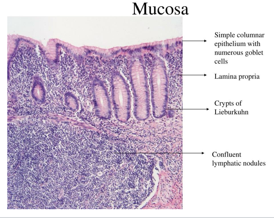
Examples:
• Linings of stomach, small intestine, large
intestine, gall bladder
• Hair cells in the cochlea
Lining of Small Intestine
Lining of Large Intestine
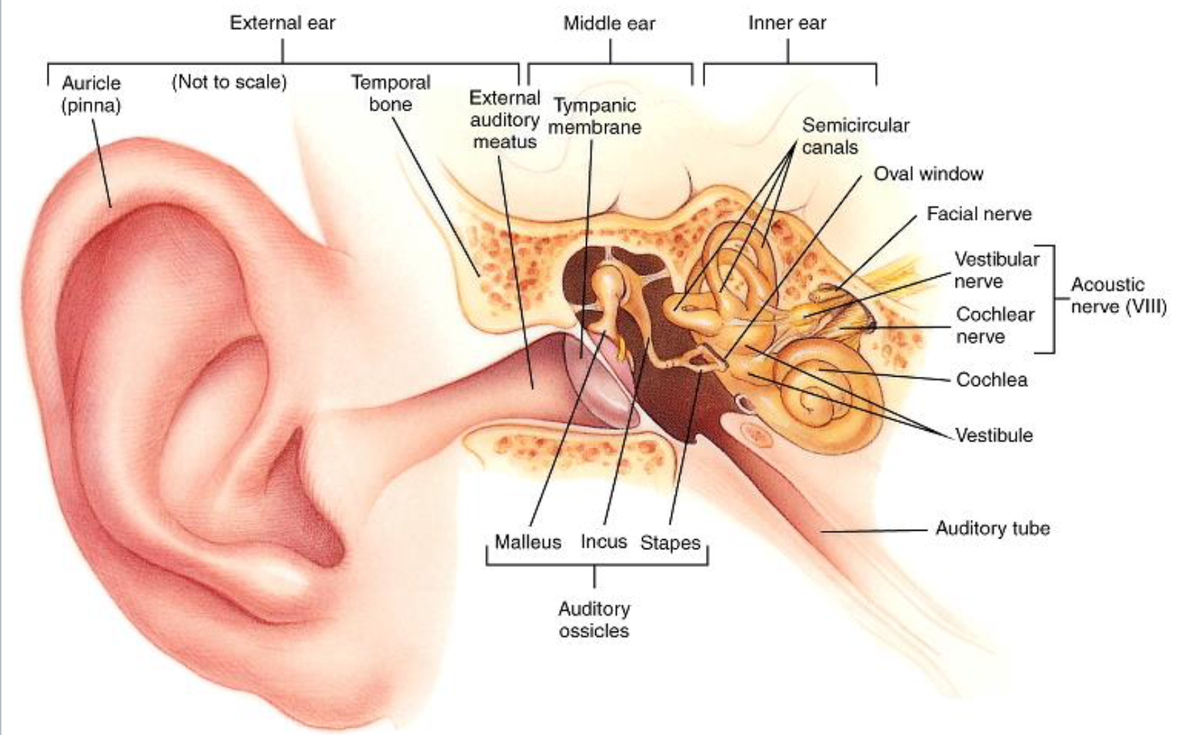
Anatomy of the Inner Ear
Inner ear:
Cochlea
Spiral organ of Corti
Semicircular canals
Vestibule (utricle and saccule)
CN VIII (dgagTrachol Nerve)
Anatomy of the Ear Diagram
3 spiral ducts of the Cochlea
3 Spiral Ducts:
Scala vestibuli
Scala tympani
Scala media or cochlear duct
Anatomy of the Cochlea
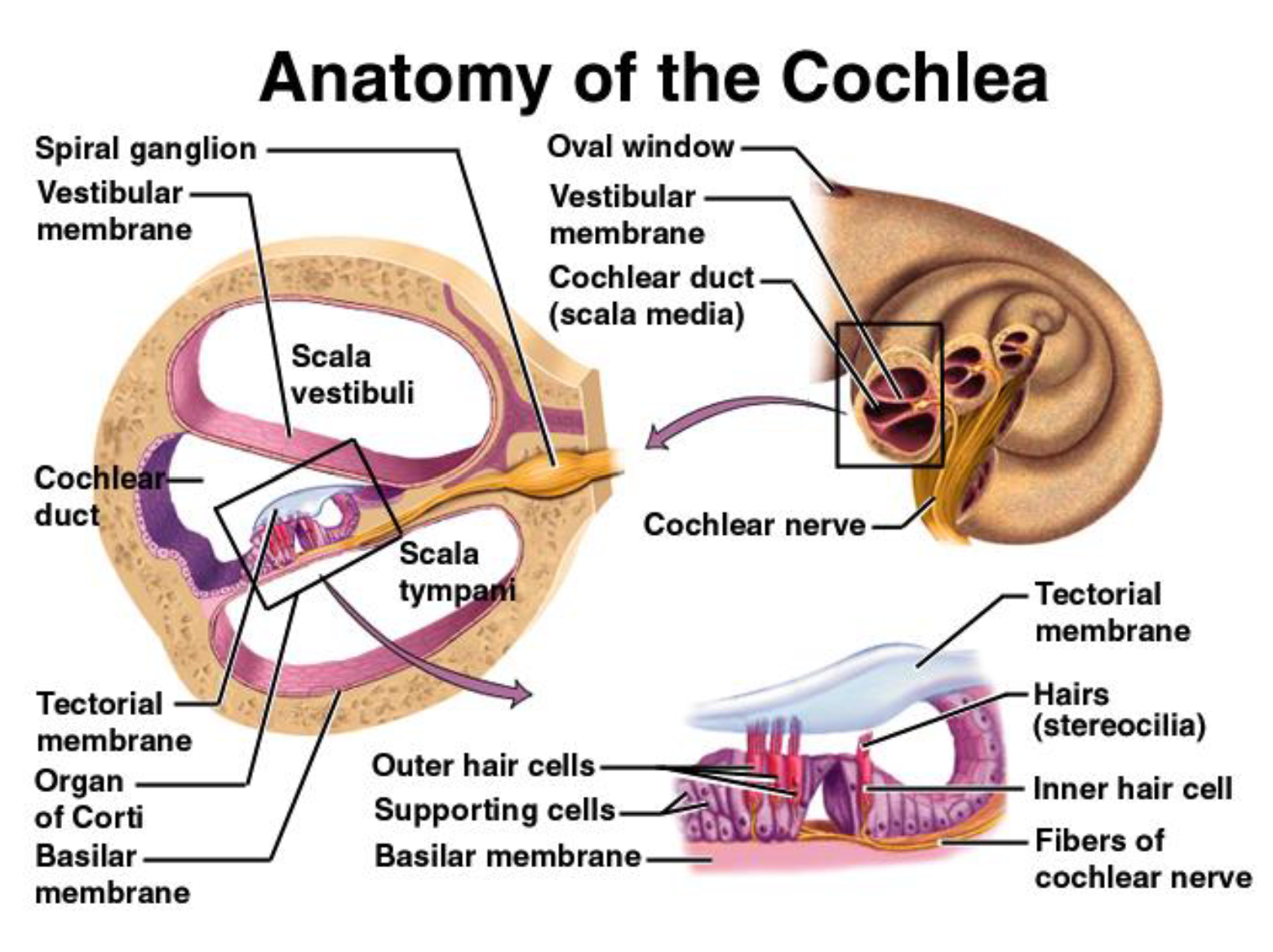
The cochlear duct is separated from the scala vestibuli by the:
vestibular membrane and
from the scala tympani by the basilar membrane.
The cochlea is filled with 2 types of fluid:
Perilymph
Endolymph.
Perilymph
Scala vestibuli and scala typmani; similar to ECF
Endolymph
Scala media; high in [K+] and low in [Na+] compared to ECF
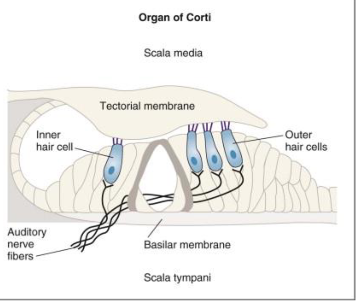
Organ of Corti → Unique property
Only cells with the long hair cili (Put right name)
Resting on the basilar membrane is the spiral organ of
Corti which consists of many supporting cells and hair
cells.
Physiology of Hearing Step 1
When sound waves strike the eardrum, it vibrates back & Forth
This rocking motion is transferred to the ossicles, which in turn vibrate.
As the stapes rocks back & forth against the oval window
Its sets the perilymph in the scala vestibuli into a similar back & forth motion
Physiology of Hearing Step 2
Pressure waves descend through the flexible vestibular membrane, then through the endolymph of the cochlear duct, and finally through the basilar membrane.
Physiology of Hearing Step 3
Back to spiral organ
The tips of the hairs are embedded within and firmly bound to the tectorial membrane above the spiral organ of Corti
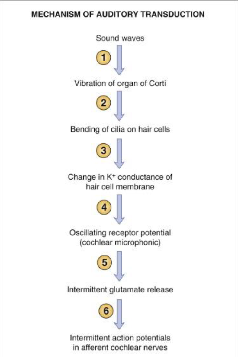
Physiology of Hearing Step 4
As the basilar membrane
vibrates, the bending of the
hair cells is transduced into
electrical signals that travel via
CN VIII.
Mechanism of Auditory Transduction
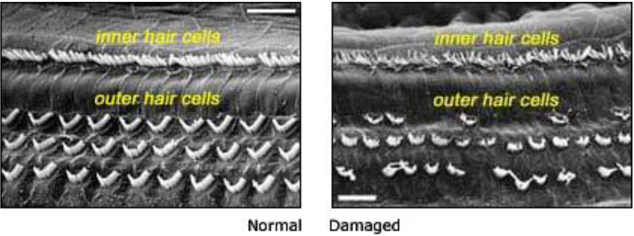
Hair Cell Damage Diagram
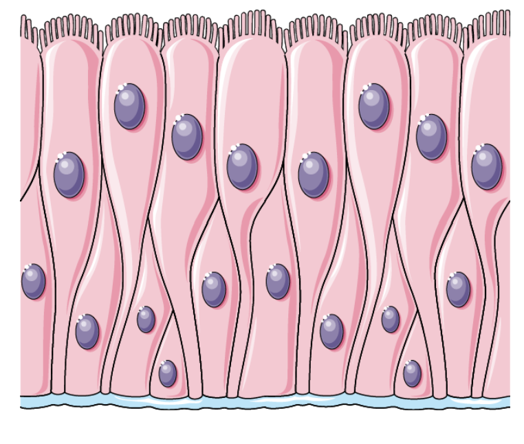
Pseudostratified Columnar Epithelium
Single layer of cells that appears to be stratified due to the varying heights of the cells and placement of their nuclei
.
Allows for secretion, forms protective linings
Usually contains CILIA and GOBLET CELLS
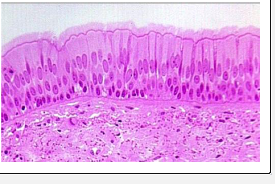
Pseudostratified Columnar Epithelium Examples
Examples:
• Linings of trachea, bronchi, non-
respiratory bronchioles
CONDUCTION ZONE
moves air
• warms air
• moistens air
• filters air
• airway resistance
RESPIRATORY ZONE
gas exchange
Conduction Zone vs. Respiratory Zone
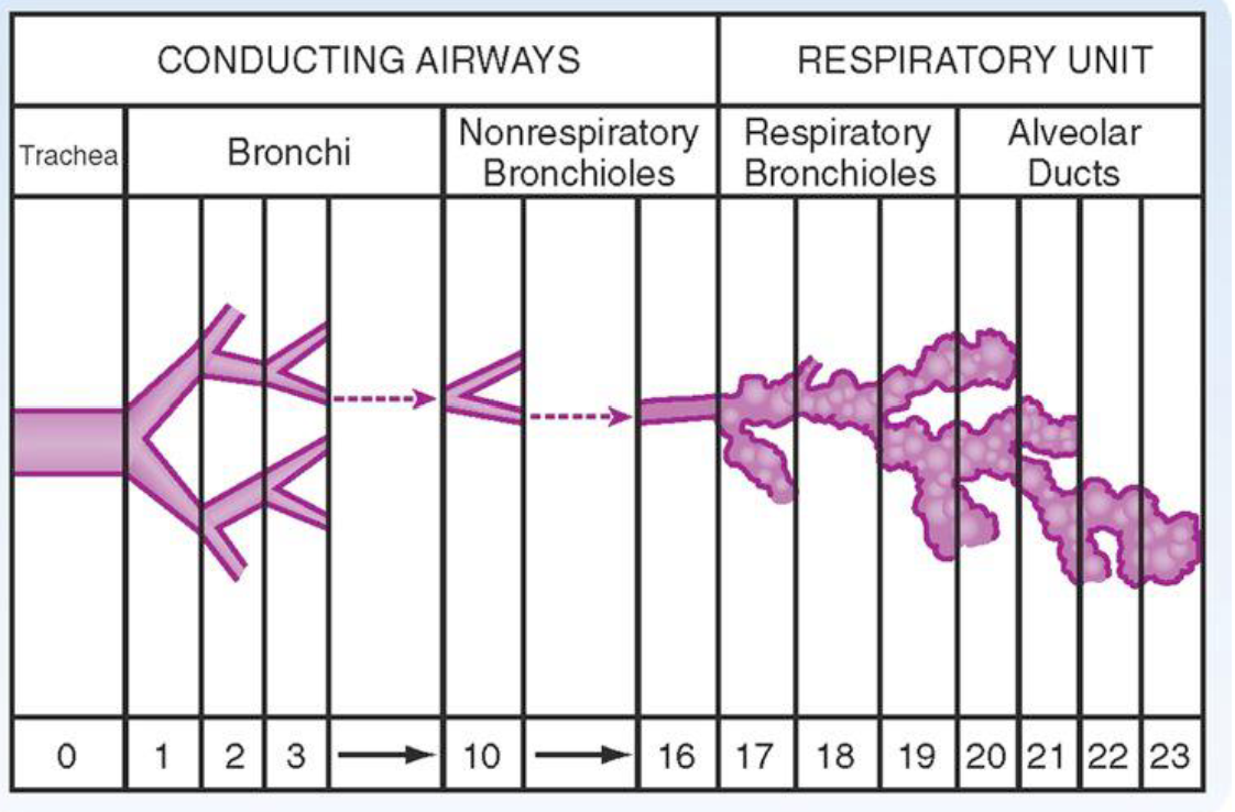
Conduction Zone vs. Respiratory Zone (Smooth Muscle, Cilia and Cartilage)
Goes Aways
Cartilage → After Bronchi
Cilia → After Respiratory Bronchioles
Smooth Muscle → After Alveolar Ducts
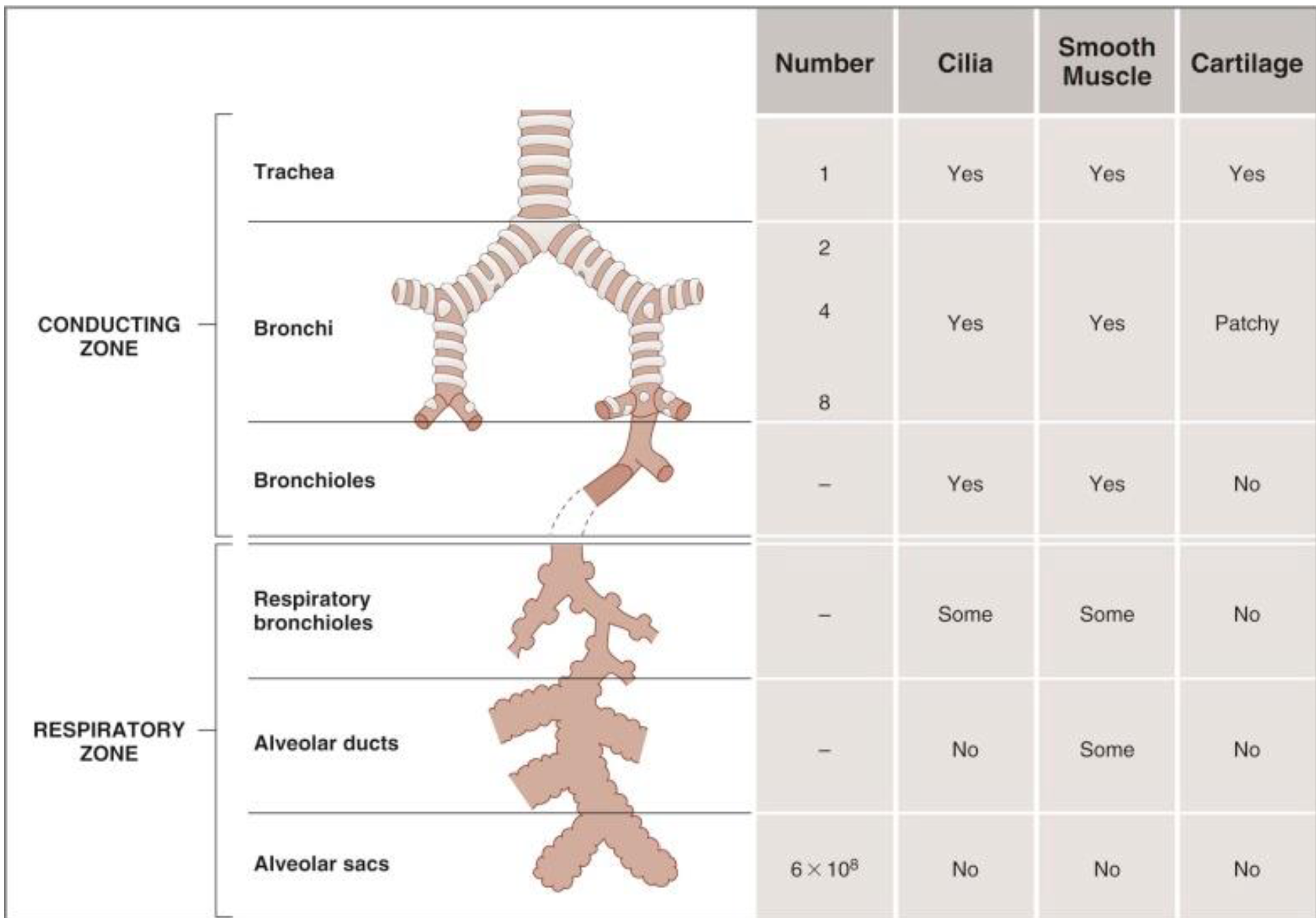
Respiratory Epithelium or Mucosa
From the trachea down to the non- respiratory bronchioles
Transitional Epithelium
Multiple layer of cells that have different shapes with great distensibility
Characterized by its ability to change shape and thickness depending on the degree of distention
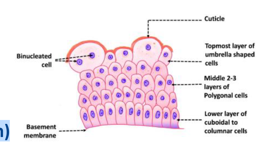
Transitional Epithelium Examples
Examples:
• Linings of ureters and urinary bladder (urothelium)
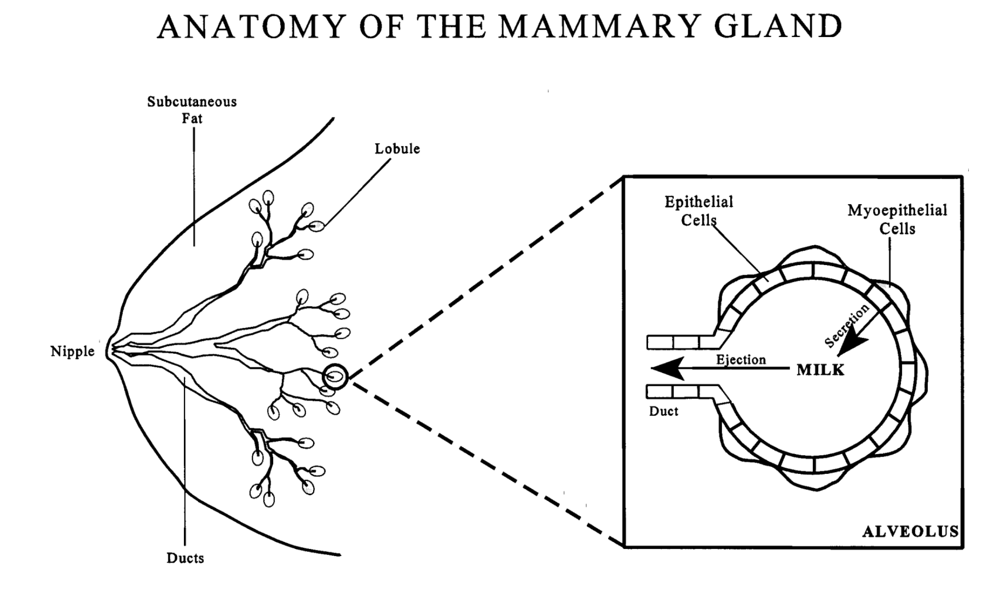
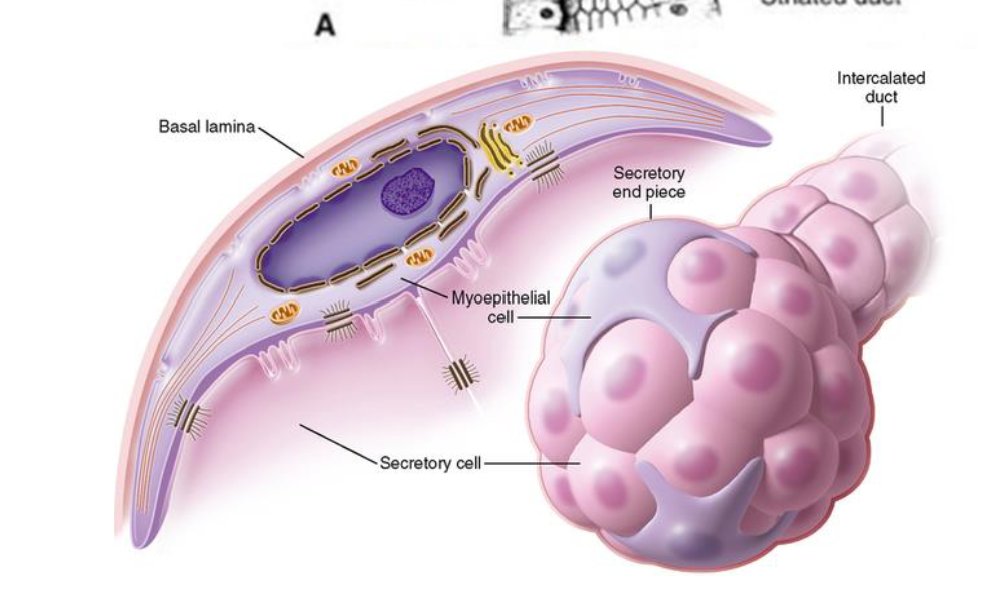
Myoepithelium (Myoepithelial Cells)
Myoepithelial cells are found in various glands – mammary, lacrimal, salivary and sweat
Hybrid of epithelial and smooth muscle cells
.
Have the ability to contract and promote glandular secretion in response to hormones and neurotransmitters
Myoepithelium Diagram
Posterior Pituitary Hormones (2)
Oxytocin
Lactation
Process of Milk Ejection
1) Stimulation of the nipple through suckling produces a neurohumoral reflex that causes secretion of oxytocin.
.
2) In turn, oxytocin causes contraction of the myoepithelial cells of the mammary alveoli & ducts and the ejection of milk.
Diagram of Milk Ejection