ASCP 2024 - Cytology - GYN
 Call Kai
 Learn
Learn Practice Test
 Spaced Repetition
 Match
Match Flashcards
 Knowt Play
1/24

There's no tags or description
Looks like no tags are added yet.
Name | Mastery | Learn | Test | Matching | Spaced | Call with Kai | Chat |
|---|
No analytics yet
Send a link to your students to track their progress
25 Terms
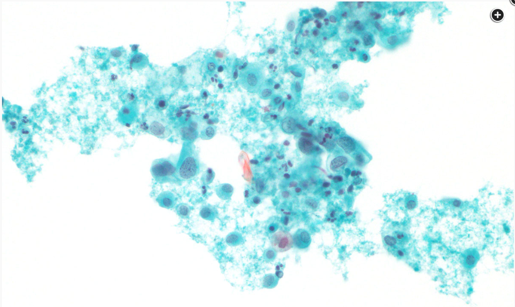
What is the squamous abnormality seen on this Pap smear?
HSIL (with features suspicious for invasion) - dispersed dysplastic cells with high N:C ratio + irregular nuclear membrane seen in background of granular debris suggestive of tumor diathesis
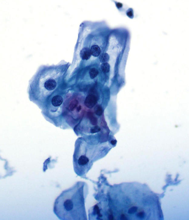
What is the diagnosis and recommended follow-up for this pap smear from a 21F
LSIL; repeat pap in 1 year - hyperchromatic, irregular nuclei, perinuclear halos
Retention period for cytopathology reports
10 years
Retention period for GYN cytopathology glass slides
5 years
Retention period for non-gyn glass cytopathology glass slides (including FNA)
10 years
HPV vaccine (Gardasil) offers protection against which HPV types
HPV 6, 11, 16, 18
Which HPV types cause vulvar condylomas
HPV 6, 11
Which HPV types cause 70% of cervical cancer
HPV 16, 18
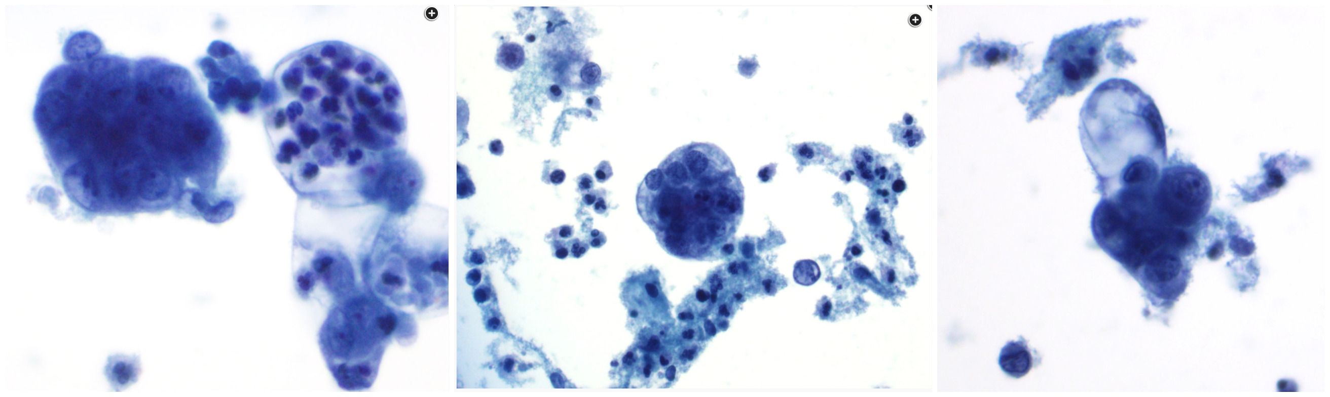
Diagnosis from this Pap smear from 58F
Adenocarcinoma - vacuolated cytoplasm (some w PMNs) + enlarged nuclei w granular chromatin distribution + round/slightly irregular nuclear contours + variable prominent nucleoli arranged in small clusters displaying nuclear overlap; background w granular debris + inflammatory cells; degree of cytoplasmic vacuolization favors glandular origin (vs squamous)
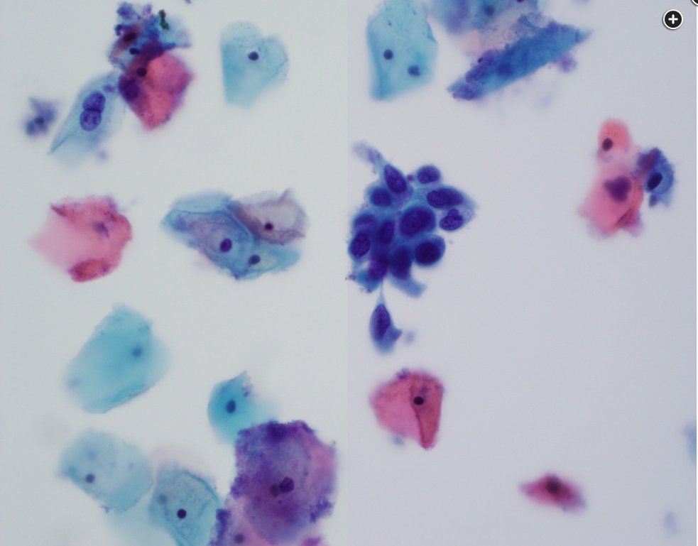
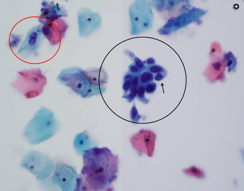
45F pap smear - diagnosis
HSIL
Red circle - LSIL koilocytic change
Black circle - HSIL - cells can be smaller than LSIL, higher N:C ratio, nuclear hyperchromasia, nuclear irregularity
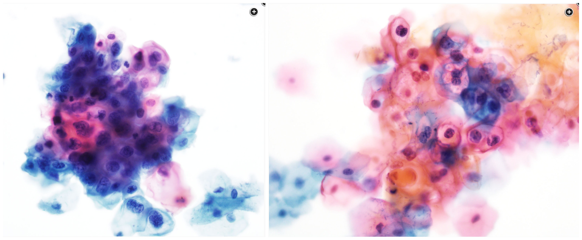
50F pap smear - diagnosis
LSIL - squamous cells w nuclear enlargement + nuclear contour irregularity + hyperchromasia + koilocytes (sharply demarcated cytoplasmic cavities, only seen in LSIL)
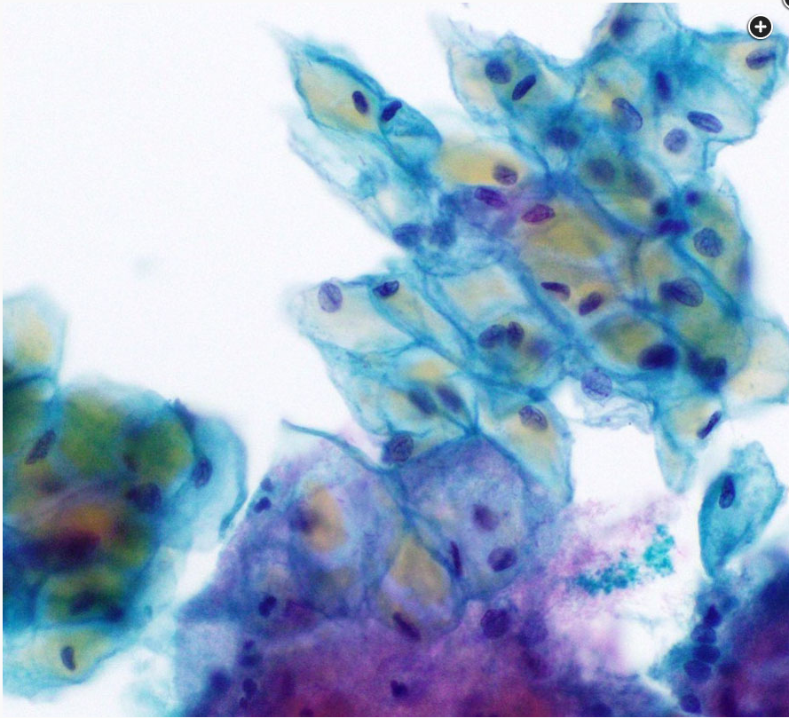
Which characteristic cell is seen in this pap smear
Pseudo-koilocytes - glycogen in squamous cells can have a yellow refractile/cracked appearance and can mimic koilocytic changes
Bethesda classification recommends reporting presence of endometrial cells in a pap smear starting at what age
45 years or older
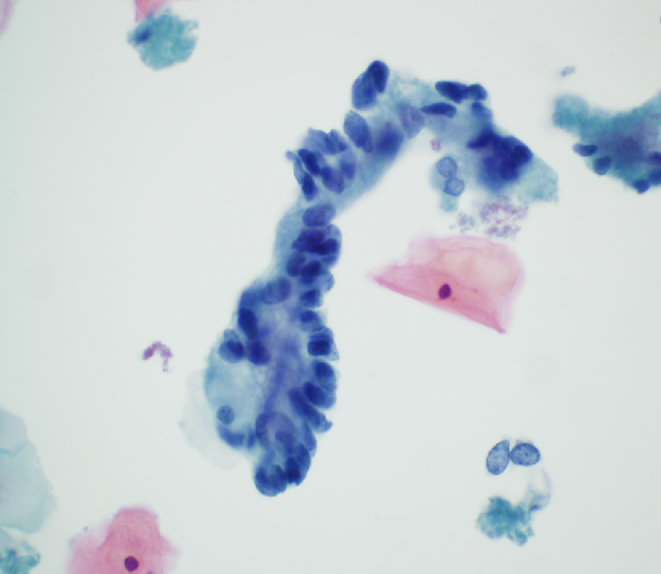
35F pap smear - the finding is best classified as what
Atypical endocervical cells - glandular cells with columnar morphology in a “picket fence” arrangement w significant nuclear crowding and overlap; nuclear enlargement + pleomorphism + more hyperchromatic than surrounding intermediate squamous cells
Which cervical cancer type is most likely to arise from an HPV-independent mechanism?
Adenocarcinoma, gastric type
Which HPV strains is each cervical cancer associated with:
SCC
Adenocarcinoma, usual type
Small cell neuroendocrine carcinoma
SCC - HPV16
Adenocarcinoma, usual type - HPV16 and 18
Small cell neuroendocrine carcinoma - HPV18
Which two mutations are associated with cervical adenocarcinoma, gastric type?
STK11 and p53
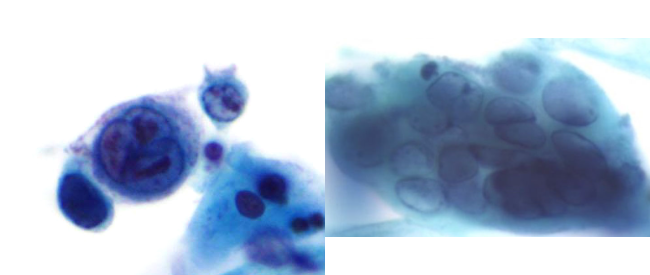
Pap smear: the cells are most consistent with
HSV
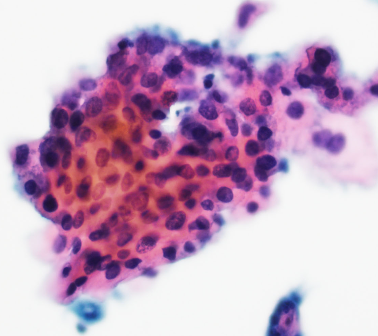
Pap smear: Which HPV subtype is most likely associated with this lesion
HPV18 - HSIL
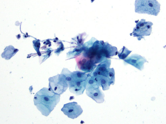
What is the clinical finding depicted in the image typically associated with?
Diabetes mellitus - Candida infection
ASC guidelines for cervical cancer screening (start, end, methods)
Start: 25 years-old with HPV test
End: 65 years-old with a series of negative prior paps
HPV test q5 years if all tests normal from 25-65
HPV + pap co-test q5 years
Pap alone q3 years
What % of women will develop a persistent infection (>2 years) after an acute high-risk HPV infection?
10%
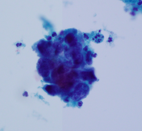
Pap smear: The lesion on the image is best classified as:
Adenocarcinoma, NOS - 3-dimensional groups with overlapping cells with high N:C ratio, nuclear pleomorphism w 3D structure (specific for adenocarcinoma as SILs don’t show this degree of 3D architecture)
Nucleoli are most commonly seen in squamous cells in which condition?
Reactive changes
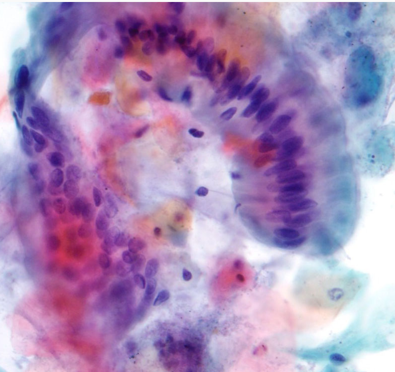
A.nal Pap: how would you report?
Negative for intraepithelial lesion or malignancy - bland columnar epithelium with mucinous features (rectal columnar cells). Background shows benign intermediate type squamous cells and anucleate squames.