BIOS1165 - Embryology
 Call Kai
 Learn
Learn Practice Test
 Spaced Repetition
 Match
Match Flashcards
 Knowt Play
1/58

There's no tags or description
Looks like no tags are added yet.
Name | Mastery | Learn | Test | Matching | Spaced | Call with Kai | Chat |
|---|
No analytics yet
Send a link to your students to track their progress
59 Terms
what are the 4 processes required for normal embryonic development?
Proliferation = ability of a cell to divide which allows for multi-celled organisms
Differentiation = process of turning from one cell type → another e.g early unspecialised embryo → specialised cell
Interaction = between similar / different cell types
Movement = movement of the different processes
what are concepts that help with the molecular regulation of cell identity?
gene expression
protein expression
microRNA
epigenetics
what signalings are involved in regulating cell identity?
Autocrine signaling = cell itself secretes molecules that act back onto itself (local effect)
Paracrine Signaling = one cell secretes a factor → acts on a nearby cell = changes its function / identity (local effect)
Hormonal Signaling = occurs further away, secreted from one area and can affect another area
whats a morphogen?
small molecule (protein) / ligand >> binds to receptor >> downstream signalling >> create a cellular response
Could also act as an inhibitor = no downstream signalling
what. are morphogen gradients?
the morphogen gradient = tells the cell how to behave

in a blastocyst, what process forms it and what does it consist of?
Formed by process called compaction
Consists:
outer trophectoderm cell layer = creates the placenta
Inner cell mass = creates embryo
Zona Pellucida = protects embryo
what are the 2 main steps that occur in post implantation development?
Trophectoderm cells bury into the uterine lining = trophoblast
The more cells we have, the more hcG is produced > required for placental development
Embryo has to adhere, attach, invade the lining (D8-9)
Hypoblast = forms the yolk sac = provides nutrients to embryo before placenta
Epiblast = further differentiates into all cells of the body
Amniotic Cavity = where the amniotic fluid fills in
what is gastrulation and the 3 main steps?
forms the definitive germ layers
Forms the primitive streak > gets longer over time (~1.5mm)
The primitive ectoderm cells undergo gastrulation
Forms the 3 Definitive Germ Layers which will form parts of the body
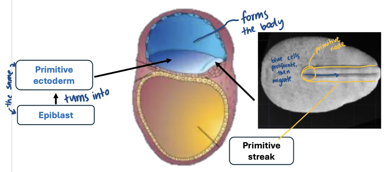
what 3 layers does the primitive ectoderm form? and what are these 3 definitive germ layers forming?

what are these areas called?

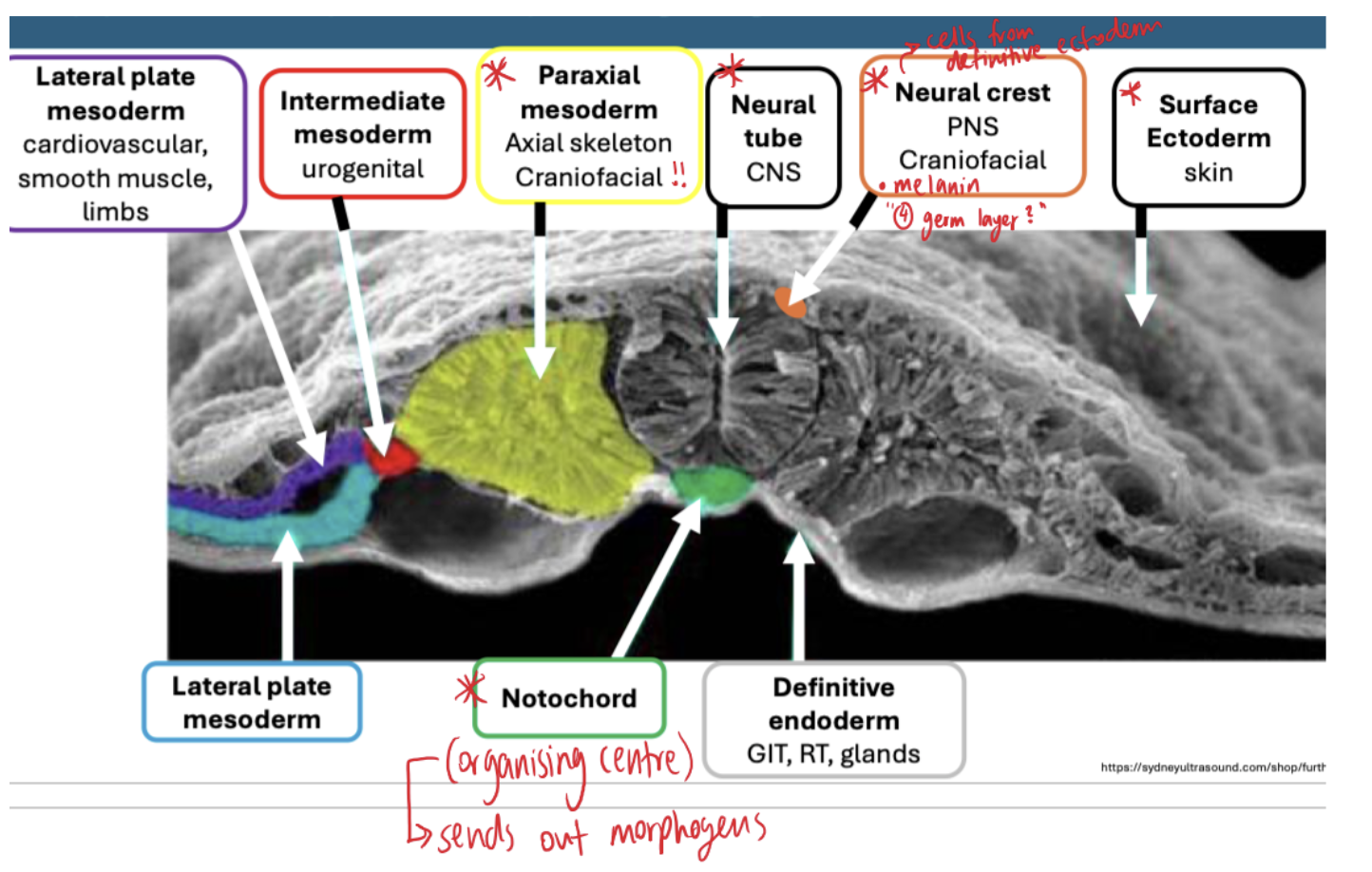
what is the oropharyngeal membrane? prechordal plate? neural plate? notochord? primitive node? primitive streak? cloacal membrane?

oropharyngeal membrane = forms mouth
prechordal plate: organising centre = influences for brain // forms the head, face, pharyngeal
neural plate: formed by thickening of definitive ectoderm = forms the CNS
notochord: from the mesoderm, will send signals to specialised spinal cord and influences the neural plate // forms vertebral discs
primitive node = end of primitive streak
cloacal membrane = forms the anus

what is neurulation and its steps?
formation of the nervous system
Neurulation requires morphogen signals from organisers (prechordal plate, notochord)
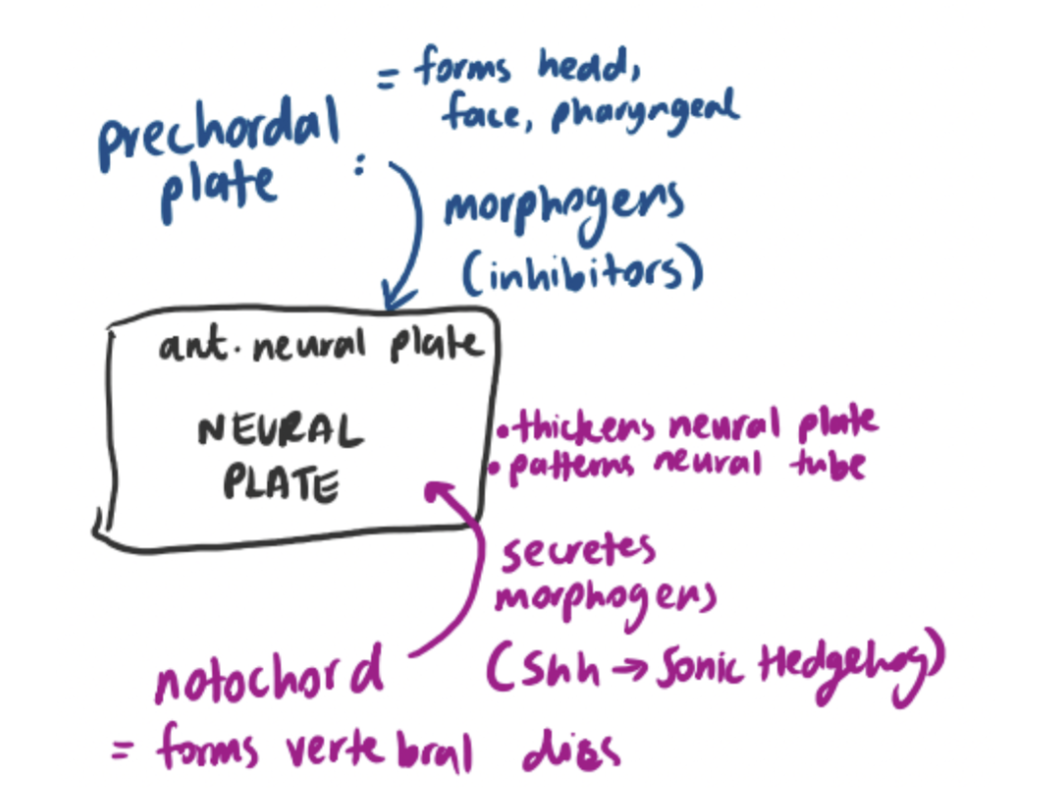
Neural plate derived from definitive ectoderm > ready to undergo neurulation by day 19
Neural Tube Formation

what are the patterning of brain key parts?
primary brain vesicles (3-5 wk) = forms neural tube etc
secondary brain vesicles (5wk) = forms cerebrum, spinal cord etc.
how does the patternign of the spainal cord occur?
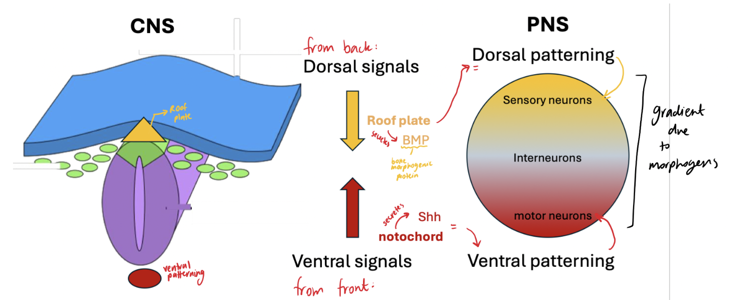
what are neural crest cells? cranial neural crest cells? neural tube?
Neural crest cells — come from dorsal end of neural tube—migrates→ mesenchymal cells
Cranial neural crest cells (subtype): originate from the cranial end of the neural tube = migrates to pharyngeal arches→ bones, cartilage, connective tissue of face, teeth, peripheral neurons, glia of cranial nerves
neural tube = cells have tight connections = NOT migratory
a hollow embryonic structure that develops into the central nervous system, including the brain and spinal cord.
what is somitogenesis?
forming somites
Derived from: paraxial mesoderm
Somites = → blocks of mesoderm (that forms middle structures: blood, muscles, cartilage, bone…)
38 pairs: cranial, occipital, cervical, thoracic, lumbar, sacral, coccygeal regions
Partially give rise to some vertebrae
Forms: axial skeleton (ribs / vertebrae) - bone and cartilage // skeletal muscle

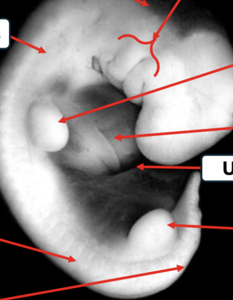
label 4 wk embryo with its somites:

what are pharyngeal arches?
Five paired, bilateral (on both sides) embryonic structures
It counts 1,2,3,4,6 → the 5th one disappears / doesn’t develop for humans
Located on: lateral surface of developing neck / head
Appears as “bulges”
Forms: structures of the face, nasal cavity, mouth, larynx, pharynx, neck
Bone / cartilage
Muscle / connective tissue
Nerves
Glands
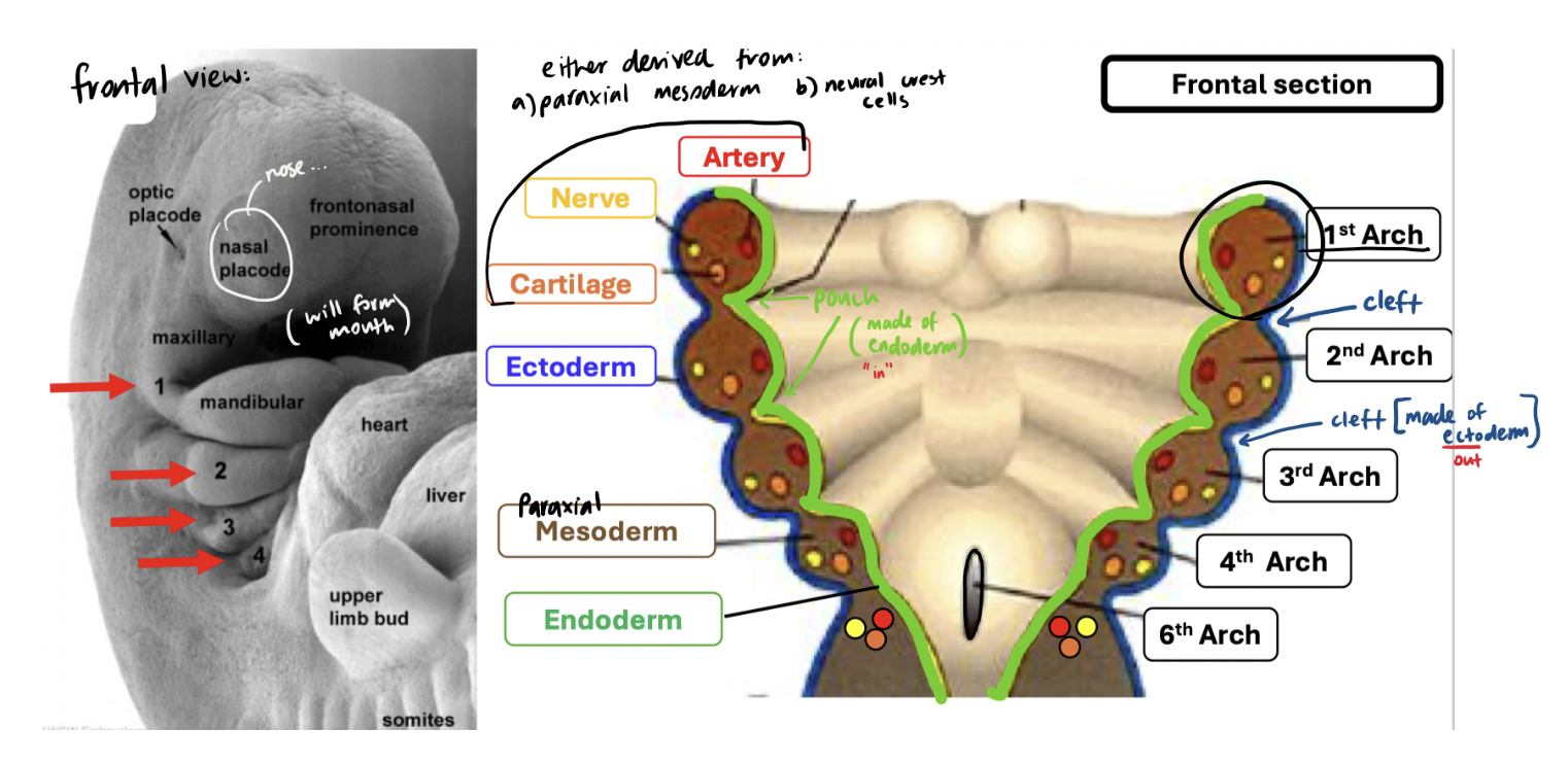
what does the neural crest cells form?
forms the PNS which includes the cranial nerves
what pharyngeal pouches create what?
pouch 1 = middle ear / eustachian tube
pouch 2 = tonsils
pouch 3+4 = parathyroid glands + thymus
what is pharyngeal arch 1 involved in?
muscles of mastication / trigeminal nerve / hearing bones + cartilage
what is pharyngeal arch 2 involved in?
facial expression muscles / facial nerves / anchorage
what is pharyngeal arch 3 involved in?
inspiration muscles, glossopharyngeal nerve, swallowing + speech bones / cartilage
what is pharyngeal arch 4 + 6 involved in?
sound + swallowing muscles / vagus nerve / voice control /+ airway support
what are the nasal septum / nasal alae / nasal bridge / philtrum / upper lip / lower lip formed by?

what is: spina bifida, anecephaly, celft lip, cleft palate
Spina Bifida | Anencephaly |
= improper caudal closure of the neural tube 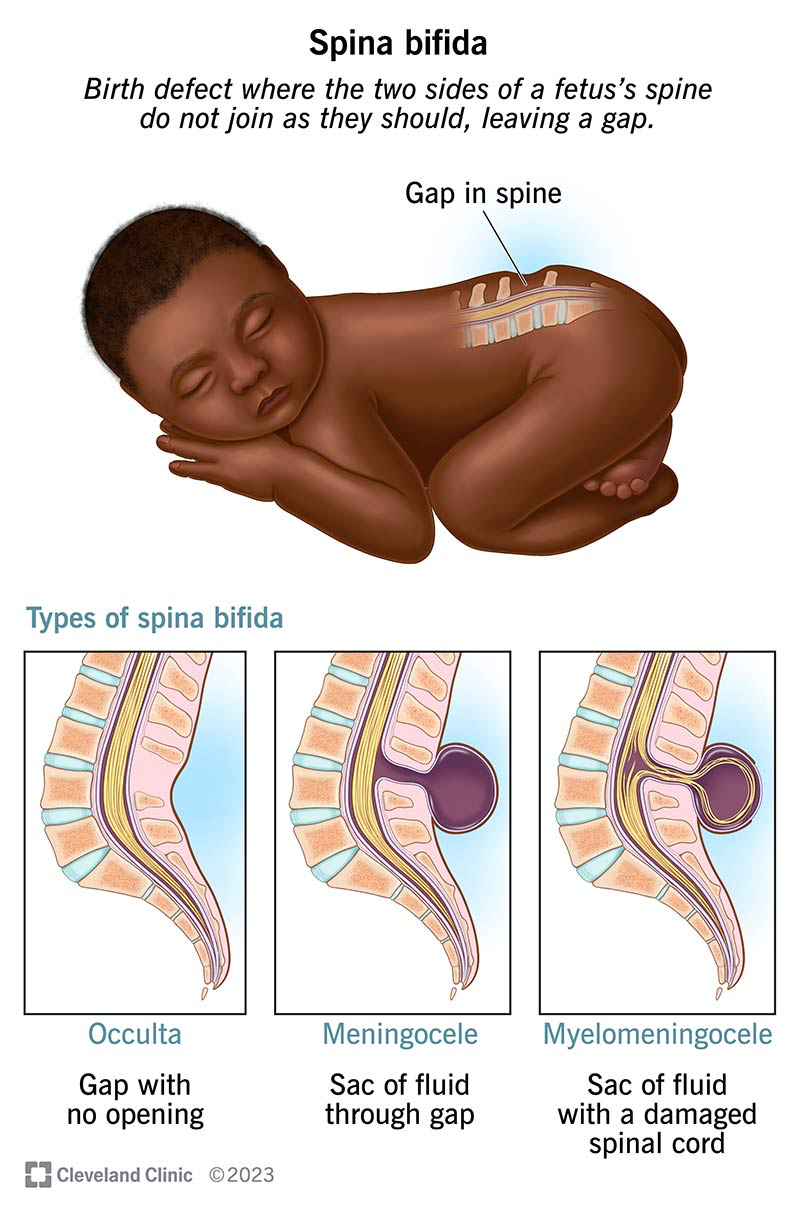 | = improper cranial closure of neural tube 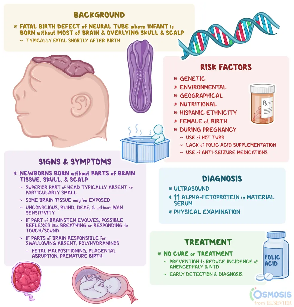 |
Facial Clefts
incomplete fusion of facial structure during development
Cleft Lip: | Cleft Palate: |
Failure of fusion between lateral or medial nasal processes and the maxillary process | Failure of fusion between the primary palate and palatal shelves |
Surgery: 2-3 months post birth | 9-18 months post birth |
Cleft Palate
Primary Palate | Secondary Palate |
|
|
what are submucous clefts? macrostomia?
Submucous Clefts
Involves hard / soft palate
Appears intact, but there are deficits in bone + muscle
Macrostomia
Large mouth → failure of the maxillary and mandibular processes to form
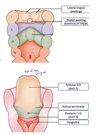
what pharyngeal arches are responsible for what parts of the tongue?
1st Pharyngeal Arch = responsible for anterior ⅔
2nd, 3rd, 4th Pharyngeal Arch = responsible for posterior ⅓
explain the timeline of tongue development:
Week 4: the lateral lingual swellings = they proliferate + enlarge = overtake the medial swelling (tuberculum impar) = will give rise to the tongue
The neural crest cells derived menschyme in Arch 1 = helps proliferate
Week 5: rapid development of lateral lingual swellings = grow over the medial swelling + begin to fuse at the midline
Week 6: tongue structure is recognisable
Skeletal muscle (derived from the occipital somite) = have voluntary control over it
Connective tissues + nerves (derived from the neural crest cells)
Epithelium (derived from surface ectoderm)
‼Cell death = separates tongue from underlying tissue + leaves behind the frenulum
Week 8: formation of basic tongue structure
CRITICAL for taste, perception, swallowing, chewing, speech
what pharyngeal arches are responsible for tongue innervation?
Tongue Innervation (derived from the neural crest cells in the pharyngeal arches):
Pharyngeal Arch 1:
Touch / Pain / Temp: CN V3 = trigeminal nerve (lingual branch)
Pharyngeal Arch 2: Taste: CN VII = facial nerve
Pharyngeal Arch 3:
Touch / Pain / Temp / Taste: CN IX = glossopharyngeal nerve
Pharyngeal Arch 4: vagus nerve (for palatoglossus nerve)
Motor: CN XII = hypoglossal nerve —> from occipital somites
what are two problems that are related with tongue development:
Anklyglossia (Tongue-Tied) | Cleft Tongue (Bifid Tongue) |
|
|
what pharyngeal arches give rise to the larynx?
4 and 6
what is the development steps for the larynx?
Week 4: Laryngotracheal groove → gives rise to opening of trachea
By Week 4: it gets bigger = call it laryngeal orifice
Endoderm cells (outside) will migrate → form the vocal cords
Week 6:
Mesoderm cells from Arch 4 + 6 → forms swellings around the laryngeal orifice to form cartilage + muscles
Epiglottic swelling → will form epiglottis
Arytenoid swelling → will form arytenoid cartilage
arch 4 = epiglottis, thyroid cartilage, cricothyroid muscle
arch 6 = cricoid cartilage, cuneiform cartilage, corniculate cartilage, arytenoid cartilage, laryngeal muscles
Endoderm cells → forms the lining of the larynx
Neural Crest Cells → forms the vagus nerve
Week 12: the larynx is fully developed = lined with endoderm derived epithelium

what pharyngeal arches give rise to the lungs?
4 and 6
what are the developmental steps for the lungs?
Week 5: Trachea & Lung are formed from the lung bud of the primitive foregut
The bifurcation of the lungs are shaped by morphogens
Week 5-16:
~16 divisions to reach terminal bronchiole
Respiration NOT possible
Fetuses born cannot survive
Week 16-23:
Terminal bronchioles divide → primitive alveolar sacs developed
Some respiration possible IF premature birth, BUT require a lot of support
Week 24 - birth:
More alveoli develops
Epithelium lining is thin enough = allows respiration
Surfactant production begins → creates enough surface tension to prevent alveoli collapse, and allows them to open up
If baby is born before 24 weeks, they need an injection as they CAN’T produce surfactants
Have a higher chance of survival ~ 50%
Birth - 8 year old
Lung development continue until 8 years of age, esp because in utero there is no access to oxygen // getting oxygen + removes CO2 through placenta
⅓ of fully formed alveoli present at birth
Babies first breath opens up the system → then ready to breathe
Respiratory bronchioles, terminals, alveolar ducts continue to increase in number

what is oesophageal atresia and trachoesophageal fistula?
Causes:
improper differentiation + elongation of the esophagus = termination of the pipe
Fistula = opening in the wrong area as they are opening on either side
Problem: can’t send food down to stomach / have food will result in food going in the wrong pipe
Treatment: surgery

what are the embryonic origins for these parts of the ear?
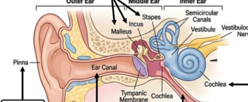
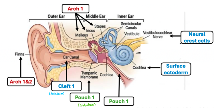
what are the steps for the development of the ear?
Week 4: (D22-24)
Thickening of surface ectoderm= the cells begin to elongate → simple columnar epithelium cells
Morphogens are working on this area = begins to invaginate to form a otic placode
*
D28: Otic Pit formed (the invagination goes so deep)
Week 5:
D30: The otic pit pinches off to form an otic vesicle (otocyst) → closer to the neural tube

Week 6:
Otocyst → develops into the membranous labyrinth
The cochlear portion (cochlear duct) → gives rise to the saccule + cochlear duct
Vestibular portion (saccule) → gives rise to semicircular canals + utricles
Week 7: the cochlear duct makes a turn
Week 8: makes a 1.5 turn
Week 10:
Production of semicircular canals
Production of utricle + saccule = helps with vertical + horizontal motion
2.5 turns of cochlear duct
how does the organ of corti develop?
Differentiation: 8-10 wks
Synaptogenesis: where the synapses of the neurons that connect to the vestibulocochlear nerve (16-20 wks)
Hearing (26-29 wks)
what are the causes of malformation of the middle / external ear + symtpoms?
Malformation of Middle Ear | Malformation of External Ear |
~1/8000 births | Cause: maternal exposure to accutane Symptoms: during 1st trimester, disrupts normal development
|
what is teratology
factors that cause birth defects
what are 2 importatant factors required for a teratogen to cross the placenta?
Molecular Weight (MW)
MW > 1000 = does not easily cross the placenta
MW < 60 = can cross the placenta by simple diffusion or transporter (SHOULD BE AVOIDED)
Most drugs: MW 250–400 (pregnant women have to be careful of what they take)
Hydrophobicity: ability to cross through a plasma membrane
what are key features that determine an embryo’s susceptibility to birth defects?
Conceptus (embryo / fetus when conceived) genotype
Embryos can have the same exposure with varied susceptibility due to their genetic components
E.g Sickle Cell Anaemia → HbAs = protection from malaria // HbAA = susceptible to malaria
Environmental factors
Dose
Timing and length of exposure
what are the weeks for “safe” / “critical period of development” / “less critical”/
safe = 0-2 weeks
critical = 2-8 weeks
less critical = >8 weeks
what happens in the critical period of the development?
ORGAN DEVELOPMENT formation of:
CNS
lower limbs
heart
upper limbs
eyes
ears
lips + midface
palate
teeth
what happens in the less critical stage of development?
maturation of the organ structures
formation of external genitalia
what are 5 main aetiology (causes) of birth defects with examples of each?
Point mutations (7.5%) | Chromosomal Anomalies (6%) | Environmental (5%) | Multifactorial (20%) | Unknown (61.5%) |
Cystic fibrosis Sickle cell anaemia | Trisomy 21 | Alcoholism Rubella CMV Accutane Thalidomide | Familial cleft lip and palate Spina bifida |
what is thaliomide? its cause? symptoms? the critical stage of development affected?
Causes | Thaliomide initially used as a sedative and sleeping tablet
Critical Stage of Development Affected: Limb Development (esp during 20-30 days post fertilisation)
|
Symptoms | Stunted limb development extreme tirdness  |
what is the cause of anticonvulsants like valproate and carbamazepine? and its symptoms?
Causes | Used to: treat epilepsy Critical Development Stage Affected: facial development (occurs earlier in the development)
|
Symptoms | Facial Abnormalities (typical facies)
|
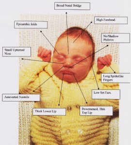
what is the effects of a mother having rubella on her fetus? what are the symtpoms? and risks?
Symptoms | In the Mother: airborne // mild rash + fever In the Conceptus:
|
Risk | 1st Trimester: 25% risk
|
what is the effects of a mother having alcohol while pregnant? and its cause?
Causes | BOTH male and females can be affected by alcohol (the males sperm can also be affected) Impacted Developmental Stage: Facial Development (problems with its fusion) 60% chance of FAS → if alcohol is consumed during pregnancy |
Foetal Alcohol Syndrome |
|
what is the effect of folic acid deficiency while pregnant?
Neural tube defects:
0.2-0.4% pregnancies
INCREASES risk of spina bifida
Folic acid before Pregnancy
reduces risk by 50-70%
Average person needs 0.4 mg/day
5 mg/day for people with epilepsy, diabetes, or family history of NTD
what are the 3 steps followed in drug testing?
preclinical testing (lab testing on cell lines / small animals / large animals)
clinical trials (safety + efficacy)
epidemiology (population based)
what is treacher collins syndrome? its cause? facial abnormalities?
Treacher Collins Syndrome | |
Cause |
|
Facial Abnormalities |
|
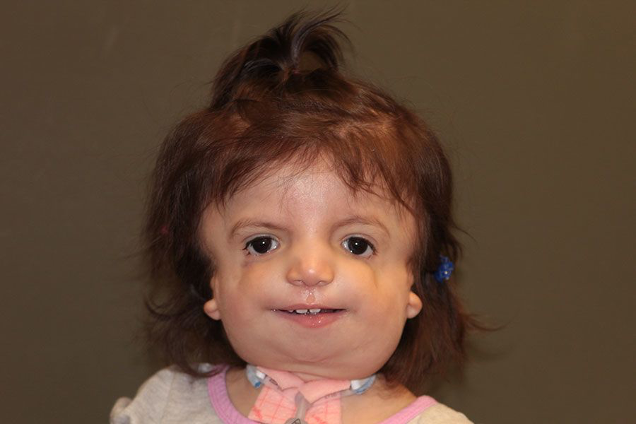
what is the relation of embryological development and treacher collins syndrome? what is its impact on speech etc.?
Embryological Basis | 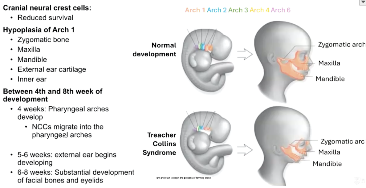 hypoplasia (underdevelopment) of Arch 1 |
Impact of Speech / Swallowing / Hearing |
|
what is goldenhar syndrome? its cause? facial abnormalities? embryological basis?
Cause | Abnormal development of neural crest cells + mesoderm derivatives in pharyngeal arches 1 and 2 = disruption of normal face development |
Facial Abnormalities | Face / Jaw:
Ears:
Eyes:
 |
Embryological Basis |  |
what is noonan syndrome? its cause? facial abnormalitiies? relation to embryology?
Cause | Affects neural crest cell migration + pharyngeal arch development → malformations in craniofacial development  |
Facial Abnormalities |
|
Embryological Basis | Caused by mutations in cell signalling pathways |
what is foetal alcohol syndrome? its cause? facial abnormalities? embryological basis?
Cause | Excessive consumption of alcohol during pregnancy
Toxic by-products of alcohol: negative effect on central nervous, visual, auditory, cardiovascular, renal, skeletal systems |
Facial Abnormalities |
 |
Embryological Basis |  |
what is pierre robin sequence? its cause? facial abnormaliities? embryological basis? effect on speech etc.?
Cause |
|
Facial Abnormalities |
|
Embryological Basis |  |
Effect of speech / swallowing / hearing | Primary management involves surgery to decrease airway obstruction.
|
