BIOM 3200 Midterm 1
0.0(0)
Card Sorting
1/312
Earn XP
Description and Tags
Last updated 8:38 PM on 2/2/23
Name | Mastery | Learn | Test | Matching | Spaced | Call with Kai |
|---|
No analytics yet
Send a link to your students to track their progress
313 Terms
1
New cards
what does Zona glomerulosa do
produces aldosterone. This hormone affects your body in many ways, by: Causing water retention.
Increasing levels of sodium in your intestines.
Increasing levels of sodium in your intestines.
2
New cards
\*Central nervous system is the ________________ and the Peripheral nervous system is everything else
brain and spinal cord
3
New cards
**what is neurophysiology?**
the nervous system function is known as the physiology… with neurons being the minimal functional unit
^^nervous system is made up of discrete individual cells^^
1. how does the location of a nerve cell determine its function?
the nervous system function is known as the physiology… with neurons being the minimal functional unit
^^nervous system is made up of discrete individual cells^^
1. how does the location of a nerve cell determine its function?
the structure & function of a nerve cell is determined by its location in the body
\-it affects physiology bc they all do the same 2 things: 1. conduct action potential (electrical signal) 2. release chemical signals (neurotransmitters)
therefore location changes what those 2 things do aka move arm or make stomach contact etc.
\-it affects physiology bc they all do the same 2 things: 1. conduct action potential (electrical signal) 2. release chemical signals (neurotransmitters)
therefore location changes what those 2 things do aka move arm or make stomach contact etc.
4
New cards
1. what are the 3 functions of the nervous system
hint: what do motor and sensory nerves do
\
1. %%motor nerves%% = control of movement and some functions (2 way = neuron goes in from muscle to brain then one goes back out to muscle to move it)
\
2. %%sensory nerves%% = detection of external stimuli \*one way communication to the brain
\
3. %%circuitry%% (aka integration of neuronal activity and connections) through %%association neurons%%, thoughts emotions, behaviour
\
5
New cards
what are association/interneurons neurons function
\
\
circuitry and connection
neurons within entirely within the CNS responsible for behaviour, thought, emotions etc
integrate message
\
circuitry and connection
neurons within entirely within the CNS responsible for behaviour, thought, emotions etc
integrate message
6
New cards
1. \*what are the 3 components of a neuron
2. what do each 3 do
1. cell body, dendrite, axon
2. cell body aka axon hillock = condenses information and consolidates the signal .“nutritional center” of the neuron where macromolecules are produced.
dendrites= receive info from sensory receptors or other cells and send it to cell body
axons = drivers electrical signals %%from cell body%% to another neuron or an effector organ (muscle)
aka info heads out
7
New cards
neurons perform the function of rapidly moving information by conducting electrical impulses called ______________ from one physical location to another, then converts that electrical impulse to a chemical signal at a __________
action potential
synapse
synapse

8
New cards
the cell body integrates information and the “tail” made up of axons __________ the impulse with the synapse at the end converting it to a chemical signal
conducts
9
New cards
\*sensory/afferent neurons conduct impulses from where to where
from sensory receptors INTO the CNS
peripheral to central
from 5 senses
peripheral to central
from 5 senses
10
New cards
association or interneurons conduct impulses from where to where
located entirely within the CNS and help integrate CNS functions \*ONE NEURON TO ANOTHERR\*
take message, integrate it and sent it on
take message, integrate it and sent it on
11
New cards
\*motor or efferent neurons conduct impulses from where to where
exit \*\*
sensory receptors OUT Of the CNS… to effector organs like muscles or glands)
central to peripheral
sensory receptors OUT Of the CNS… to effector organs like muscles or glands)
central to peripheral
12
New cards
\*somatic motor neurons
reflex and voluntary control of skeletal muscles
* *move hand away* \*
* *move hand away* \*
13
New cards
\*autonomic motor neurons control what
@@involuntary@@ control of smooth muscle, cardiac muscle and glands
2 lines bc sends to multiple organs sometimes
2 lines bc sends to multiple organs sometimes
14
New cards
autonomic motor neurons are further subdivided into sympathetic and parasympathetic … differentiate both
sympathetic = GO
parasympathetic = STOP
parasympathetic = STOP
15
New cards
\*you put your hand on a stove… what happens next (include what type of nervous system, neuron etc)
PNS - hand on stove, receptor (like nerve ending) sensory/afferent neuron goes to
CNS.. association neuron transfers signal to motor neuron which takes it to efferent/motor neuron
PNS - takes hand away , effector
CNS.. association neuron transfers signal to motor neuron which takes it to efferent/motor neuron
PNS - takes hand away , effector
16
New cards
type of neuron: pseudopolar (unipolar)
sensory, 1 process that splits

17
New cards
type of neuron: bipolar
retinal and cochlear , 2 processes, one is a dendrite, one is an axon

18
New cards
\*type of neuron: multipolar
*most common* \*\*\* motor and association, many dendrites but ==one axon==

19
New cards
type of neuron: anaxonic
some CNS neurons , no obvious axon

20
New cards
\*Schwann cells - form myelin sheaths around PNS neuron axons .. what is the purpose
supporting cells IN PNS
\
form myelin sheath = type of protein
insulate so messages → faster
\
form myelin sheath = type of protein
insulate so messages → faster
21
New cards
These gaps in the myelin sheath are known as _____________.__ The successive wrappings of the Schwann cells’ plasma membranes provide insulation around the axon, leaving only the ____________ exposed to produce nerve impulses.
the nodes of Ranvier
22
New cards
Unmyelinated axons are also surrounded by a neurilemma, but they differ from myelinated axons in that they lack the multiple wrappings of Schwann cell plasma membranes that compose the myelin sheath.

23
New cards
\*what do satellite cells do
supporting cells IN PNS
\
support neuron cell bodies w/in ganglia of the PNS
wrap around cell body to protect from environment
\
support neuron cell bodies w/in ganglia of the PNS
wrap around cell body to protect from environment

24
New cards
\*what do oligodendrocytes do
supporting cells IN CNS
just like Schwann cells in PNS aka form myelin sheath around neuron axons
just like Schwann cells in PNS aka form myelin sheath around neuron axons

25
New cards
\*what do microglia do
supporting cells IN CNS
migrate through CNS and phagocytose debris
==-macrophage, pick up debris==
migrate through CNS and phagocytose debris
==-macrophage, pick up debris==

26
New cards
\*what do astrocytes do
help regulate external environment of neurons in CNS
==-connect between everything - regulate== **ex: on blood vessel so it communicates w neuron**
Processes terminate in “end feet” at capillaries, others on neurons (axon, cell body, or dendrite) thus they can influence interactions between neurons and blood
==-connect between everything - regulate== **ex: on blood vessel so it communicates w neuron**
Processes terminate in “end feet” at capillaries, others on neurons (axon, cell body, or dendrite) thus they can influence interactions between neurons and blood

27
New cards
\*what do ependymal cells do
\-line ventricles (cavities) of the brain and spinal cord - in CNS
==keep shape in brain and spinal cord==
==keep shape in brain and spinal cord==

28
New cards
All axons in the PNS (myelinated and unmyelinated) are surrounded by a continuous living sheath of Schwann cells known as the ___________. The axons of the CNS, by contrast, lack it (Schwann cells are found only in the PNS).
neurilemma
\-good for nerve regeneration x
\-good for nerve regeneration x
29
New cards
\*what is the difference between how supporting cells cover a neuron between the PNS and CNS
PNS= bubble gum around stick, wrapped around a single neuron
\
CNS= wrapped around CLUSTER of neurons
\
CNS= wrapped around CLUSTER of neurons

30
New cards
what is the most abundant glial supporting cell in the CNS
astrocytes
up to 90% of nervous tissue
up to 90% of nervous tissue
31
New cards
what are glial cells
non-neuronal cells in the central nervous system (brain and spinal cord) and the peripheral nervous system that do not produce electrical impulses.
32
New cards
ASTROCYTES take up K+ from the Extra cellular fluid which diffuses from neurons when they fire… what is the purpose of this
the K+ needs to be reabsorbed to maintain proper ionic environment for neurons
33
New cards
ASTROCYTES can take up neurotransmitter glutamate and transform it to glutamine after they are released by neurons after firing
\
neurotransmitter = carry chemical signals (“messages”) from one neuron (nerve cell) to the next target cell
\
neurotransmitter = carry chemical signals (“messages”) from one neuron (nerve cell) to the next target cell
glutamine can be released back into neurons, which can use it to reform the neurotransmitter glutamate.
34
New cards
ASTROCYTES : the end feet surrounding blood capillaries take glucose up from the blood then do what w it
metabolize it to lactate, then release it for use as an energy source by neurons, which metabolize it aerobically into CO2 and H2O for production of ATP
\
\-neurons need help picking up ex: converted glucose to use for energy and to make neurotransmitters
\
\-neurons need help picking up ex: converted glucose to use for energy and to make neurotransmitters
35
New cards
astrocytes are needed for the formation of synapses in the CNS … what do synapses do
communicate between neurons
36
New cards
astrocytes regulate neurogenesis in the adult brain.. how is this helpful
needed for stem cells to differentiate into both glial cells and neurons
37
New cards
astrocytes help w formation of the BBB and release neurotransmitters like glutamate, ATP, adenosine etc that can do what
stimulate or inhibit activity of neurons
38
New cards
capillaries in brain do not have PORES between adjacent endothelial cells (cells that make up the walls of the capillaries) instead they are joined by…
tight junctions made by astrocytes
39
New cards
\*BBB imposes very strict control of what can move from the blood plasma into the brain .. with **astrocytes** making up the barrier and altering function
what can move through
\
causes trouble w drugs getting through
what can move through
\
causes trouble w drugs getting through
non polar O2 and CO2 , organic molecules including alcoho and barbiturates
polar molecules
LIPID soluble molecules
polar molecules
LIPID soluble molecules
40
New cards
who had numerical synesthesia
Daniel Tammet
41
New cards
what was special about Kim Peek
extrodinary memory
absence of corpus calllosum
mega savant
absence of corpus calllosum
mega savant
42
New cards
what was Christopher Langan known for
smartest man alive
43
New cards
what happened to Phineas Gage
\-had pole through left eye
\-pierced prefrontal cortex, made him sociopathic and changed behaviour
\-pierced prefrontal cortex, made him sociopathic and changed behaviour
44
New cards
CNS depressants directly affect brain cells, specifically areas involved in inhibiting behaviours ,, also altered speech, slowed reactions, time fog memory
alcohol gets through
45
New cards
In less 10 seconds, nicotine molecules cross the blood- brain barrier and fit like tiny keys into locks normally activated by acetylcholine neurotransmitter, and increases the levels of several other neurotransmitters ( like ______________ ).
dopamine
46
New cards
Other components in tobacco smoke decrease MAO (monoamine oxidases) activity, the enzyme that is needed to break down _____________
neurotransmitters (dopamine, norepinephrine, and serotonin)
47
New cards
Inhibition of NO through a variety of neurotransmitter receptors including N-methyl-D- aspartate and substance P.
48
New cards
how does rabies affect brain
virus infects brain, causes it to swell, immune cells and antibodies can’t enter the brain
49
New cards
CNS contains brain and spinal cord
PNS contains ___________-
PNS contains ___________-
peripheral nerves and ganglia
50
New cards

\*the efferent nervous system of the peripheral nervous system splits into the somatic and autonomic nervous system…
1\. what do each do hint: in/voluntary ? which parts of body
2\. how many neurons are involved in each. draw the autonomic
1\. what do each do hint: in/voluntary ? which parts of body
2\. how many neurons are involved in each. draw the autonomic
somatic - send axons to skeletal muscles (voluntary control). conduct impulses along a SINGLE axon from PNS to CNS in spinal cord etc
\
autonomic - (involuntary, smooth muscle, heart, gland) TWO neurons , 1st cell body in CNS gray matter (preganglionic neuron) synapses with 2nd neuron called a postganglionic neuron, that has an axon that extends from the autonomic ganglion to the effector organ where it synapses w target tissue
\
autonomic - (involuntary, smooth muscle, heart, gland) TWO neurons , 1st cell body in CNS gray matter (preganglionic neuron) synapses with 2nd neuron called a postganglionic neuron, that has an axon that extends from the autonomic ganglion to the effector organ where it synapses w target tissue

51
New cards
which one out of somatic motor neurons or autonomic motor neurons have ganglia
autonomic motor neurons have it
found in the cell bodies of postganglionic autonomic fibers located in paravertebral, prevertebral and terminal ganglia
found in the cell bodies of postganglionic autonomic fibers located in paravertebral, prevertebral and terminal ganglia

52
New cards
what are ganglia
Ganglia are clusters of nerve cell bodies found throughout the body. They are part of the peripheral nervous system and carry nerve signals to and from the central nervous system
53
New cards
\*match the following to somatic and autonomic motor neurons:
1. either excitatory or inhibitory
2. excitatory only
1. either excitatory or inhibitory
2. excitatory only
1. either excitatory or inhibitory - AUTONOMIC
1. excitatory only - SOMATIC
54
New cards
21\. Following an Action Potential, the following all occur in the postsynaptic neuron (ONE \n INCORRECT): \n A. chemically gated channels open in the postsynaptic neuron dendrites \n B. the presynaptic terminal bouton synapses with the postsynaptic axon \n C. the postsynaptic neuron undergoes inward diffusion of Na+ causing depolarization \n D. localized, decremental conduction of excitatory postsynaptic potential (ESPS) occurs \n E. voltage-gated Na+ and then K+ channels open starting at the axon hilloc
\n B. the presynaptic terminal bouton synapses with the postsynaptic axon
\
should say dendrrite \n
\
should say dendrrite \n
55
New cards
The embryonic brain region called the Prosencephalon develops into these regions (ONE \n INCORRECT): \n A. telencephalon \n B. hypothalamus \n C. suprachiasmatic nucleus (SCN) \n D. frontal cerebral lobe \n E. cerebellum
E. cerebellum
56
New cards
\*type of neuromuscular junction in somatic motor neurons vs autonomic motor neurons
\
hint: think of where the signal is going
\
hint: think of where the signal is going
somatic - specialized motor end plate The somatic neuromuscular junction is the site of communication between motor neurons and skeletal muscle fibres.
autonomic - all areas of smooth muscle cells contain receptor proteins for neurotransmitter
autonomic - all areas of smooth muscle cells contain receptor proteins for neurotransmitter
57
New cards
\*damage to an autonomic nerve makes its target tissue more sensitive than normal to stimulating agents. This phenomenon is called …
denervation hypersensitivity
58
New cards
True or False: somatic motor neurons are fast conducting and autonomic motor neurons are slow conducting … and all fibes involved are myelinated
false! postganglionic fibers in autonomic motor neurons are unmyelinated
59
New cards
\*kinda a hard one… what happens when denervation occurs to somatic and autonomic motor neurons
somatic - paralysis and atrophy to muscles
autonomic - muscle tone and function persist \*but target cells show denervation hypersensivity
autonomic - muscle tone and function persist \*but target cells show denervation hypersensivity
60
New cards
how do the sympathetic and parasympathetic divisions of the autonomic nervous system differ
parasympathtic - rest and digest
sympathetic - fight or flight
\
most organs receive input from both
\
particular locations of their preganglionic neuron cell bodies within the CNS, and in the location of their ganglia.
sympathetic - fight or flight
\
most organs receive input from both
\
particular locations of their preganglionic neuron cell bodies within the CNS, and in the location of their ganglia.

61
New cards
what are the four organs without dual innervation from the sympathetic and parasympathetic nervous system
and what is the nervous system that single-handedly controls them
and what is the nervous system that single-handedly controls them
**only sympathetic**
\-adrenal medullla
\-arrector pili muscles in skin
\-sweat glands
\-most blood vessels
\-adrenal medullla
\-arrector pili muscles in skin
\-sweat glands
\-most blood vessels
62
New cards
1. \*what are the two main neurotransmitters used in the autonomic nervous system
2. what releases them
3. how do they differ
cholinergic neurons = acetylcholine
adrenergic neurons = norepinephrine
\
3. they both bind diff receptors to mediate target organ response
adrenergic neurons = norepinephrine
\
3. they both bind diff receptors to mediate target organ response
63
New cards
for parasympathetic and sympathetic outflow, state whether the pre and post ganglionic axons involved are cholinergic or adrenergic and the order of them from the CNS to the effector cell

64
New cards
true or false: acetylcholine is the neurotransmitter for ALL PREganglionic fibers (both sympathetic and parasympathetic)
true
65
New cards
true or false: acetylcholine is the transmitter released most by sympathetic postganglionic fibers at their synapses with effector cells
false
released most by *parasympathetic* postganglionic fibers
released most by *parasympathetic* postganglionic fibers
66
New cards
the neurotransmitter released most by sympathetic nerve fibers is _____________ so the type of transmission is called _____________
norepinephrine
adrenergic
adrenergic
67
New cards
what is dysautonomia
dysfunction of the autonomic nervous system
can be caused by brain injury, diabetes, genetics, tick bite
can be caused by brain injury, diabetes, genetics, tick bite
68
New cards
what happens after you are bit by a tick
hint: how does it spread
hint: how does it spread
substances in tick saliva disrupt immune response, spirochetes multiply in the skin
immune response causes the bullseye look but *neutrophils dont appear to get rid of infection*
bacteria spread via bloodstream to joints, heart, nervous system
immune response causes the bullseye look but *neutrophils dont appear to get rid of infection*
bacteria spread via bloodstream to joints, heart, nervous system
69
New cards
synesthesia vs chromothesia vs synaesthesia
\
who had chromothesia
\
who had chromothesia
letters or numbers are perceived as colours
music as colours - MOZART
coloured sounds taste sweet
music as colours - MOZART
coloured sounds taste sweet
70
New cards
71
New cards

the cell membrane is just 2 molecules thick, 2 phospholipid molecules thick (phospholipid bilayer)
72
New cards
explain simple passive diffusion and example of what passes through
(there are a few) hint; think ions
(there are a few) hint; think ions
1. small uncharged molecules (relatively lipid soluble) can diffuse through the lipid bilayer (eg. steroid hormones)
2. small charged molecules (ions) can pass through water filled pores
these ion channels tho… some are leaky and ions flow in or out as needed
3. voltage-^^gated^^ ion channels
\*\*integral membrane proteins can act as transporters
73
New cards
active transport requires ATP. explain the Na+/K+ ATPase pump
why is it important
why is it important
moves Na+ out of the cell
moves K+ into the cell
ATP turns to ADP to do this
\*establishes electrochemical gradient
\
The sodium-potassium pump carries out a form of active transport FOR MAKING ACTION POTENTIAL MOVEEEE
moves K+ into the cell
ATP turns to ADP to do this
\*establishes electrochemical gradient
\
The sodium-potassium pump carries out a form of active transport FOR MAKING ACTION POTENTIAL MOVEEEE

74
New cards
why are ion channels especially important in the nervous system
they help produce electrical impulses that transmit info rapidly
\---- voltage gated ion channels open and change the membrane potential of the cell and that’s how electrical signals are conducted in neurons
\---- voltage gated ion channels open and change the membrane potential of the cell and that’s how electrical signals are conducted in neurons
75
New cards
All cells in the body have a potential difference – or voltage – across the membrane. This is called the resting membrane potential….
what is it for neurons
what is it for neurons
\-70 mV on the INSIDE of the cell
76
New cards
what are the locations of Na+ and K+ at the start of action potentials
Na+ starts outside (then rushes in)
K+ starts inside (then rushes out)
K+ starts inside (then rushes out)
77
New cards
the primary function of nerve cells is to receive, conduct and transmit signals in the form of :
action potentials.. which go along the nerves, taking signals from one place to another place
78
New cards
what is an action potential
why do they need to be reamplified along the way and HOW
why do they need to be reamplified along the way and HOW
1. momentary discharges %%(depolarizations)%% of the resting membrane potential caused by a rapid influx of Na+ caused by the opening of sodium ion channels
%%= make positive%%
\
2. they have to travel a long way w/o weakening, so we use voltage-gated ion channels
79
New cards
what are the three steps of ion gating in axons
1. electrical event must be strong enough to trigger the axonal membrane beyond its threshold, if it is it initiates the action potential (aka channel has to get positive enough from sodium coming in) ----voltage gates sodium channels open from stimulus and Na+ rushes into cell and makes it very positive (depolarization).. if its strong enough there is an action potential
1. Na+ channel closes and the permeability of Na+ decreases
2. Action potential trickles into the next channel down axon, now making it depolarized and triggers another action potential by surpassing threshod
Meanwhie old channel cell has to repolarize, which is done by letting K+ diffuse out of the cell to restore the original resting membrane potential Sodium channels of refractory area are inactivated, that’s why action potential can’t flow backwards
\
**K+ is only pumped out of the cell once the action potential is reached** \*\*\*\*\*\* @@__*When the depolarization reaches about -55 mV a neuron will fire an action potential. **__@@
\*\*the Na+ and K+ pumps are constantly working to in the plasma membrane, they pump out the Na+ that entered the axon during an action potential and pump in the K+ that left
\
\

80
New cards
the size of all action potentials is the same bc once the depolarization reaches that -55 mV threshold, it will always fire an action potential
\
*all or none principle*
\
*all or none principle*
81
New cards
1. compare how action potentials flow in non-myelinated axons vs myelinated
2. what does this allow
hint: saltatory conduction
**non-myelinated axons**= the action potential passes smoothly along the axon, and all parts of the membrane are depolarized
**myelinated axons**= action potential jumps between non-insulted nodes by saltatory conduction
allows more rapid movement of the action potentials, and needs less energy to restore the membrane after the action potential has been transmitted \*\*dont need to go through every Na channel
\----- refractory period still exists in these neurons

82
New cards
axons end close to another cell so once the action potential reaches the end of the axon, they stimulate the next cell.. how tho
presynaptic nerve ending releases neurotransmitters that stimulate action potentials in the post synaptic cell
they hop across 10nm space between
they hop across 10nm space between

83
New cards
in the CNS, the 2nd cell is also a neuron
what is it in the PNS
what is it in the PNS
could be a neuron, or en effector cell within a muscle of gland
84
New cards
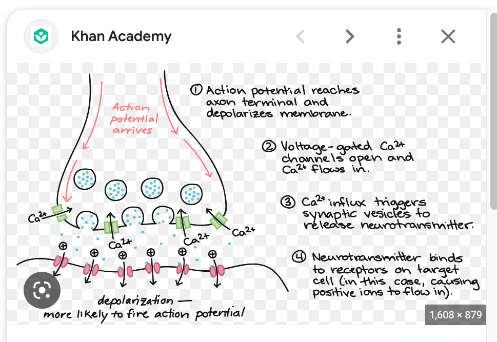
what are the steps of the synapse triggerring the next cell
1. action potential reach axon terminals
2. voltage-gated Ca2+ channels open
3. Ca2+ binds to sensor protein in cytoplasm
4. Ca2+ protein complex stimulated fusion and exocytosis of neurotransmitter

85
New cards
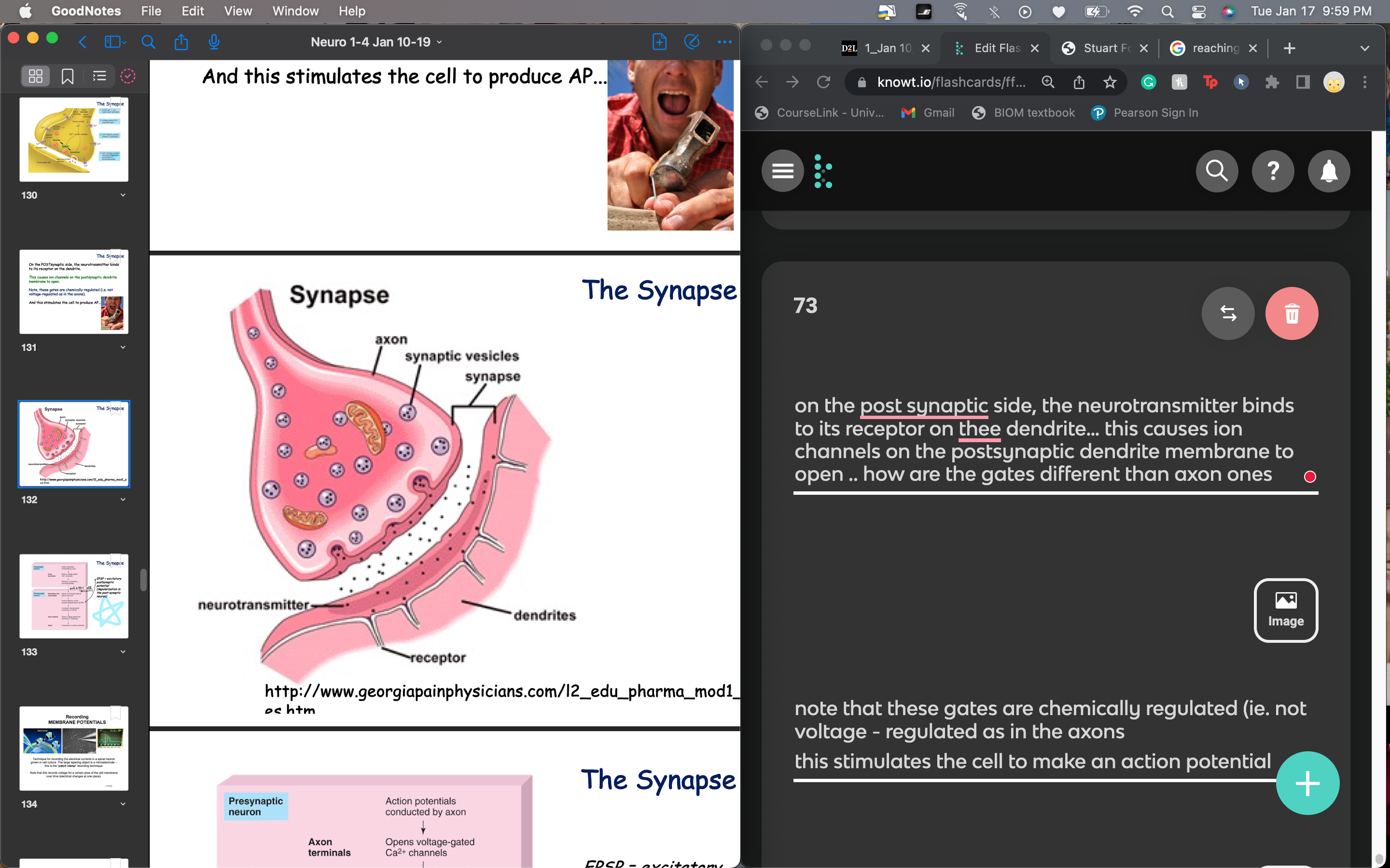
on the post synaptic side, the neurotransmitter binds to its receptor on thee dendrite… this causes ion channels on the postsynaptic dendrite membrane to open .. how are the gates different than axon ones
note that these gates are chemically regulated (ie. not voltage - regulated as in the axons
this stimulates the cell to make an action potential
this stimulates the cell to make an action potential

86
New cards
what are the steps of the action potential in the presynaptic neuron in the axon terminal
action potentials conducted by axon (travel through) and reached axon terminal
voltage gated Ca2+ channels open and Ca2+ flows in
release of excitatory neurotransmitter bc of Ca2+ influc
voltage gated Ca2+ channels open and Ca2+ flows in
release of excitatory neurotransmitter bc of Ca2+ influc

87
New cards
what are the steps of the action potential in the postsynaptic neuron in the next neuron
1. the neurotransmitter bind to receptor proteins on next cell…. ==neurotransmitter receptors are, for the most part, ligand-gated ion channels, that open in response to being bound by the neurotransmitter they are specific for.==
2. Activation of postsynaptic receptors leads to the opening or closing of ligand gated channels in the cell membrane. (chemically)
3. inward diffusion of Na+ causes depolarization (EPSP = excitatory postsynaptic potential aka depolarization in the postsynaptic neuron…… because the membrane potential moves toward the threshold required for action potentials)
4. localized, decremental conduction of EPSP… less and less effective propagation of an impulse due to a progressive decrease in membrane potential
at axon hillock : 5. opens voltage-gated Na+ channels then K+ channels.. aka moving action potential
6. at AXON - conduction of action potential
88
New cards
how to record membrane potentials
patch clamp recording technique - record electrical currrent in spinal neuron grown in cell culture using microelectrode
\
*voltage for a certain area of the cell membrane over time so it changes in one place*
\
*voltage for a certain area of the cell membrane over time so it changes in one place*
89
New cards
myotonia are ?
what are they caused by
\
hint; fainting sheep
what are they caused by
\
hint; fainting sheep
neuromuscular disorders characterized by delayed relaxation of skeletal muscle after voluntary contraction or electrical stimulation
\*problem in muscle
can be caused by mutations in muscle Cl- channel
channel gates do not open properly
repolarization delayed, several Action Potentials fire instead of just one
\*problem in muscle
can be caused by mutations in muscle Cl- channel
channel gates do not open properly
repolarization delayed, several Action Potentials fire instead of just one
90
New cards
describe information flow from dendrites to axon
1. dendrites collect electrical signals
2. cell body integrates incoming signals and generates outgoing signal to axon
1. axon passes electrical signals to dendrites of another cell or to an effector cell
91
New cards
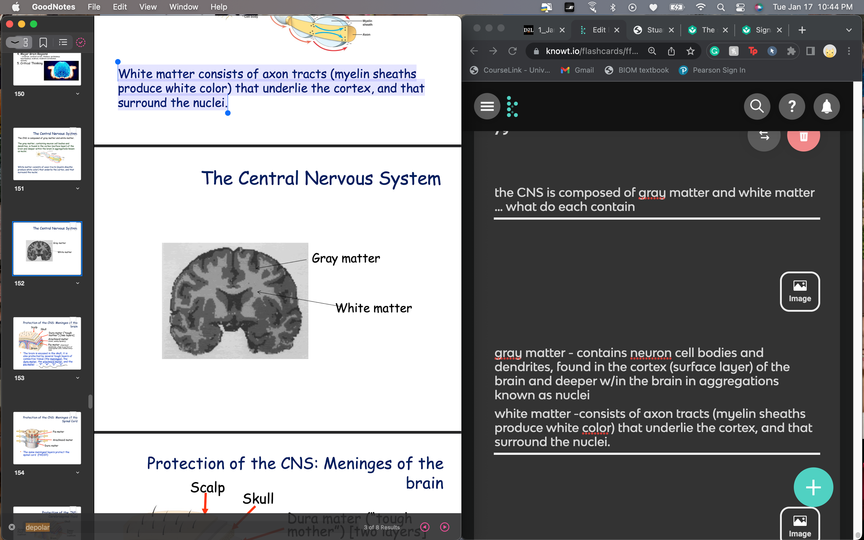
the CNS is composed of gray matter and white matter … what do each contain
gray matter - contains neuron @@cell bodies and dendrites@@, found in the cortex (surface layer) of the brain and deeper w/in the brain in aggregations known as nuclei
white matter -consists of @@axon@@ tracts (myelin sheaths produce white color) that underlie the cortex, and that surround the nuclei.
white matter -consists of @@axon@@ tracts (myelin sheaths produce white color) that underlie the cortex, and that surround the nuclei.

92
New cards
neurotrophin =Any of a family of autocrine regulators secreted by neurons and neuroglial cells that promote axon growth and other effects. Nerve growth factor (NGF) is an example.
what does NGF do
what does NGF do
NGF is required for the maintenance of sympathetic ganglia, and neurotrophins are required for mature sensory neurons to regenerate after injury
Neurotrophins regulate the survival and differentiation of adult neural stem cells in parts of brain for memory and learning
Neurotrophins regulate the survival and differentiation of adult neural stem cells in parts of brain for memory and learning
93
New cards
explain the P.A.D.S in terms of protection of the skull… P.S these same meningeal layers protect the spinal cord!
P - Pia matter (innermost membrane, clings to surface of brain/spinal cord, follows every fold
A - arachnoid mater (spider/webby)
D - dura mater (tough mother and 2 layers)
S - skull and scalp
A - arachnoid mater (spider/webby)
D - dura mater (tough mother and 2 layers)
S - skull and scalp
94
New cards
in addition to the skull and meninges, the brain is protected by two fluid cushions that give some protection for the brain against head traumas….
what are the two cavities and
where are they located
\
P.S the cavities are filled w cerebrospinal fluid
what are the two cavities and
where are they located
\
P.S the cavities are filled w cerebrospinal fluid
outer cavity - superior sagittal sinus (SSS) sits right under dura mater
inner cavity - subarachnoid space - space between arachnoid and pia mater
inner cavity - subarachnoid space - space between arachnoid and pia mater
95
New cards
what does the cerebrospinal fluid found in brain cavities and spinal cord do
provides protection and is similar to blood plasma
CSF tap can be used to examine for signs of disease
CSF tap can be used to examine for signs of disease
96
New cards
there are 31 pairs of spinal nerves split into 6 sections
\-each nerve is a mixed nerve composed of sensory and motor fibers, packed together but they are separated near the attachment of the nerve to the spinal cord
\-each nerve is a mixed nerve composed of sensory and motor fibers, packed together but they are separated near the attachment of the nerve to the spinal cord
cranial
cervical
thoracic
lumbar
sacral
coccygeal
cervical
thoracic
lumbar
sacral
coccygeal
97
New cards
the spinal cord extends from the brain stem to the pelvic region, ending right before the end of the vertebral column…. how do spinal nerves enter or leave the spinal cord
they enter and leave in between the vertebrae
98
New cards
explain the link between the spinal cord and peripheral nervous system
interneurons also communicate w one another along the length of the spinal cord
an afferent sensory stimulus can be translated up or down the spina cord by the interneurons (afferent = peripheral to CNS)
\
interneurons are the ones in between - they connect spinal motor and sensory neurons \*exclusive to CNS
an afferent sensory stimulus can be translated up or down the spina cord by the interneurons (afferent = peripheral to CNS)
\
interneurons are the ones in between - they connect spinal motor and sensory neurons \*exclusive to CNS
99
New cards
how to tell lower motor neuron damage when doing a myotatic stretch reflex eg: knee-jerk reflex
with lower motor neuron damage - the reflex will be diminished
100
New cards
how to tell upper rmotor neuron damage when doing a myotatic stretch reflex eg: knee-jerk reflex
with upper motor neuron damage, reflex will be exaggerated