Introduction
1/11
There's no tags or description
Looks like no tags are added yet.
Name | Mastery | Learn | Test | Matching | Spaced |
|---|
No study sessions yet.
12 Terms
There are thousands of different types of microorganisms:
useful microorganisms for humans, like commensal microorganisms or microorganisms used by humans. Among them we remember the normal flora (Staphylococcus epidermidis e E. coli), in agriculture (bacteria capable of fixing atmospheric nitrogen), in the food industry for dairy products (Lactobacillus) bakery products (Saccharomyces cerevisiae) alcoholic beverages (Saccharomyces cerevisiae, Acetobacter), for methane production (methanogenic bacteria) and for biotechnology (antibiotics, human insulin, interferon, vaccines, transgenic plants and animals).
microorganisms that are infectious diseases agents, like Meningitis, Tuberculosis, Malaria, Papilloma virus, sexually trasmitted disease.
Bacteria can be classified in base of:
shape
temperature they can grow at
gram stain test
ability to live related to oxygen
nutrition
biochemical charachteristics
Classification based on shape
Cocci have a spherical shape and may be individual, in pairs (diplococcuss), chain (streptococcus), cluster (staphylococcus) or groups of eight cells in a cubic space (sarcine)
Bacilli have a rods shape and may be individual, in pairs (diplobacillus), chain (streptobacillus)
Spirilla have a spiral shape and are usually single
classification based on temperature
Cryophilic or psychrophilic bacteria develop around 15-20°C but can also multiply at 0 °J, in some cases, even at -7 °C. Their habitat is represented by the oceans and the Antarctic regions; they are also able to grow in chilled and frozen foods.
Mesophilic bacteria prefer temperature between 20-40°C. Human and animal pathogens are mesophilic and have adapted to the body temperature (37°C) even for fever (40°C). It is important to emphasize that mesophilic do not grow at refrigeration temperatures, and because the microorganism responsible for food alterations are mesophilic, the low temperatures for the preservation of foods are explained. Low temperatures generally slow the development of bacteria without killing them. Freezing does not kill most of the bacteria present in biological samples or in foods: when the temperatures return to the optimum, the microorganisms begin to multiply. The ability to withstand at very low temperatures is favoured by the presence of the capsule.
Thermophilic bacteria growth at temperatures above 40°C. The habitats from which such bacteria can be isolated include hot springs, tropical soils, water heating systems, and the warm currents of some oceans. The temperature range of this group has recently been raised to 90°C, as it has been shown that some bacteria have grown in a hot spring at that temperature. It is important to emphasize that the thermophilic bacterium proteins differ from the others because they are not denatured at high temperature. The thermostability of these proteins is intrinsic, depending on the composition and sequence of amino acids responsible for the appearance of strong bonds such as covalent disulfide bridges, and many hydrogen bonds and other weak bonds that stabilize the structure of proteins and enzymes.
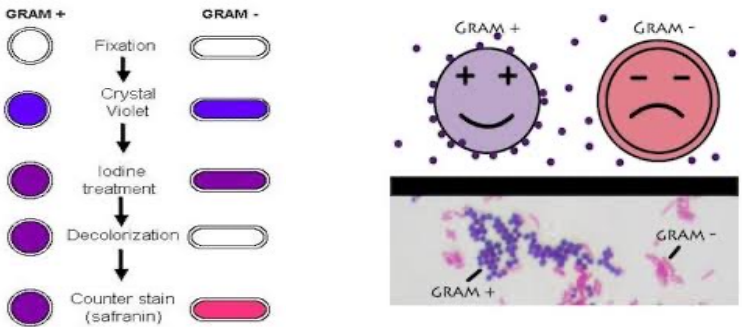
Gram stain test and how to perform it
Gram stain test that allows to divide the bacteria in Gram+ and Gram-. Gram staining differentiates bacteria by the chemical and physical properties of their cell walls. The bacteria are first stained with crystal-violet dye and then with an iodinated solution (Lugol's liquid); then washed with ethyl alcohol or acetone and counterstain with (safranin or fuchsine). The bacteria that lose the first stains are called Gram-, those who retain Gram+.
classification based on oxygen levels
Aerobic if they can survive and grow only in the presence of oxygen (the most common cause of clinical infection) or anaerobic bacteria that does not require oxygen to grow.
Obligate anaerobic bacteria are microorganisms that can only live in the absence of oxygen. Intestinal bacteria are a type of anaerobic bacteria defined oxygen- intolerant bacteria, in fact, if they encounter oxygen, they stop not only to multiply but die in a very short time.
Facultative anaerobic bacteria are microorganisms that can also survive in the absence of oxygen without being affected, but they growth better in the presence of this element. Microaerophilic bacteria can multiply in the presence of air (about 20% oxygen), but unlike the facultative anaerobic bacteria better with very low oxygen concentration (2-18%).
classification based on nutrition
Autotrophic bacteria are organisms that, like green plants, can synthesize their cellular constituents using simple inorganic substances.
The photosynthetic bacteria use light energy to produce useful chemical energy for vital processes.
The chemoautotrophic bacteria use inorganic compounds for energy and to synthesize their cellular constituents. The difference from photosynthesis is that chemosynthesis can also occur in the absence of light.
Heterotrophic bacteria, such as animals, are organisms that can only metabolize compounds already synthesized by other organisms. Most bacteria belong to this group. These bacteria can be further divided in saprophytic bacteria from parasites.
Saprophytic bacteria get food from plant and animal decomposition.
The parasitic bacteria, however, use the metabolism of other animals to obtain the food (internal bacteria) without ever causing obvious damage (see also symbiosis)
Differences between types of luminescence
Luminescence is the generic term that defines the emission of light by molecules without specifying the cause at its origin.
The most famous form of luminescence is that resulting from the absorption of light (photo-luminescence), of which fluorescence is the most common example.
Less commonly known is chemiluminescence. Chemiluminescence represents luminescence resulting from an exergonic chemical process, leading to a product in an electronically excited state, which decays by emitting photons.
In chemiluminescence, bioluminescence is always caused by a chemical process but occurs in living organisms and is catalysed by enzymes. Bioluminescence is widely used in research to reveal microorganisms.
Prokaryotic cell structure
Cell wall, capsule, flagella/pili/fimbriae, cytoplasmic membrane
Structure of the cell wall
the cell wall is made by peptidoglycan, also called murine. It's formed with two polymers, some amino acids and two sugars (NAG: N-acetylglucosamine, and NAM: N-acetylmuramic acid). These two molecules are linked together with a $\beta$-1,4-glycosidic bridge and can be broken by the lysozyme. Then some peptides form a lateral chain: L-alanine, glutamic acid and then two different peptides based on the Gram +(L-Lysine) or Gram - (L-diaminopimeric). The peptides give a rigid structure to the prokaryotic cell, strengthened by the presence of bridges between lateral chains. This bridge is created by transferase, losing the last peptide. In Gram + the bridge is made of 5 peptides (pentapeptide bridge) while in the Gram - the bridge is direct (3rd aminoacid linked to the 4th of another branch). Transferase is also the target of some antibiotics, like penicillin.
Cell wall in G+
The cell wall of Gram + is characterized by several layers of peptidoglycan, with filaments of lipoteichoic and teichoic acids.
Cell wall in G-
In Gram - bacteria, between the inner cell and peptidoglycan there’s a periplasmic space, while outside the peptidoglycan there’s an outer membrane (double layer of phospholipids) where the lipopolysaccharides are linked. LPS are made of three parts:
antigen O, differs for the number of saccharides in different bacteria and differentiates the LPS
lipid A, endotoxin that makes the LPS harmful, also presents an acid chain that contributes to avoid cleavage by the complement system
polysaccharide core, made of KDO (not present in human cells).