Immunology MICI 3115 -Midterm 1
 Call Kai
 Learn
Learn Practice Test
 Spaced Repetition
 Match
Match Flashcards
 Knowt Play
1/417

There's no tags or description
Looks like no tags are added yet.
Name | Mastery | Learn | Test | Matching | Spaced | Call with Kai | Chat |
|---|
No analytics yet
Send a link to your students to track their progress
418 Terms
What are the types of lymphoid tissue?
Primary lymphoid tissue: sites of lymphocyte development and maturation
—Thymus (T and NKT) and bone marrow (B and ILC)
Secondary lymphoid tissue: site of lymphocyte activation —Lymph nodes, spleen, and various mucosal-associated lymphoid tissues (MALT), such as gut-associated lymphoid tissue (GALT) Hub of activation for T and B cells
Tertiary lymphoid tissue: sites of lymphocyte activity
—cutaneous-associated lymphoid tissue (CALT)
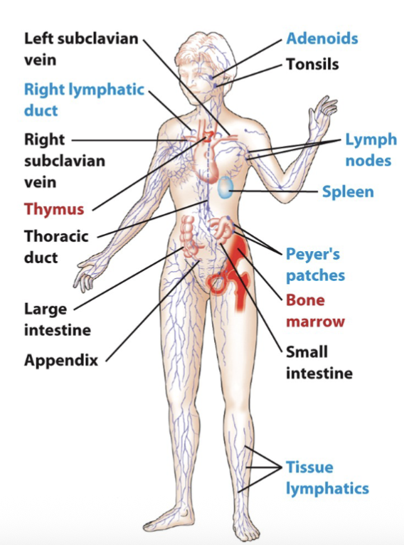
What are the secondary lymphoid tissues?
Lymph nodes, spleen, and various mucosal-associated lymphoid tissues (MALT), such as gut-associated lymphoid tissue (GALT); this is where antigen is trapped, providing an opportunity for interaction with mature lymphocytes and antigen-dependent maturation of T and B cells
Contain high endothelial venules (HEV) that permit transit of
immune cells
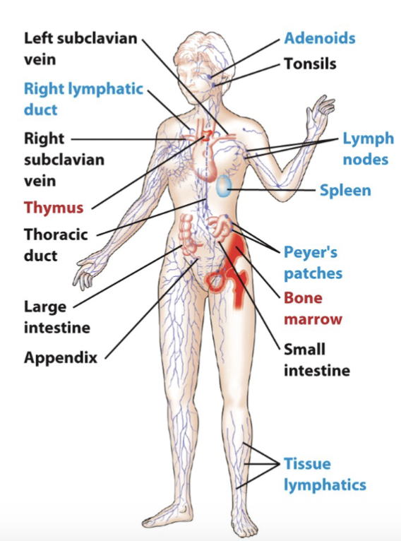
What are the tertiary lymphoid tissues?
Normally contain fewer lymphoid cells than secondary lymphoid organs but can import lymphoid cells during an inflammatory response; e.g., cutaneous-associated lymphoid tissue (CALT)
Can form during inflammatory responses to important lymphoid cells to sites of inflammation.
Distinct from secondary lymphoid tissues because they form during robust/ ongoing immune responses, are less organized, and contain fewer lymphoid cells (normally)
not always present
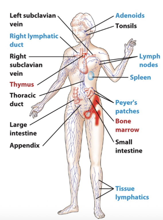
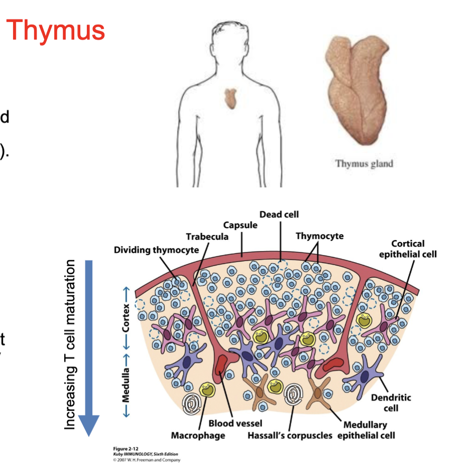
What are the features of the thymus?
Site of T and NKT and some ILC cell maturation
The thymus is a bi-lobed encapsulated organ with lobules, separated by connective tissue strands (trabeculae)
Outermost compartment (cortex) is densely packed with immature,
proliferating thymocytes
Inner compartment (medulla) is only sparsely populated with more mature thymocytes (due to selection)
During maturation thymocytes interact with cortical epithelial cells, medullary epithelial cells, dendritic cells, and macrophages
thymus gets smaller with age
cortex has the most immature T cells
immature T cells that make it through the medulla will be released and finish their development
What are the features of the bone marrow?
Primary lymphoid tissue that is involved in the production of leukocytes
All leukocyte progenitors are produced from hematopoietic stem cells (HSCs)
All immune cells begin their development in the bone marry, but many finish their maturation elsewhere
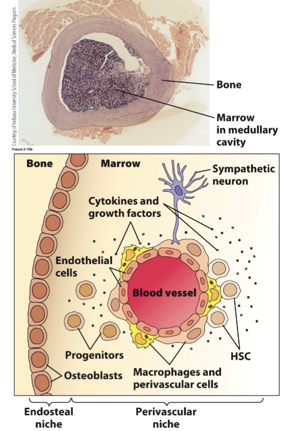
What are the physical components of the bone marrow?
Extracellular matrix: Attachment of stem cells and hematopoietic progenitor cells via adhesion molecules
Stromal Cells (endothelial, perivascular, nerves, macrophages and osteoblasts):
Physical support (scaffold) for the growth of hematopoietic cells. Provide nutrients, produce hematopoietic growth factors and express adhesion molecules that influence differentiation
Hematopoietic growth factors: produced by stromal cells are presented to immobilized stem cells
niche in bone marrow: extracellular matrix, stomal cell + hemopoietic growth factors = able to perform hematopoeisis
all the cells contribute to HSC to grow
What are the features of lymph nodes?
Highly specialized secondary lymphoid tissue
Site of generation of T cell and B cell antibody responses to specific antigen
Provides a site where lymphocytes can interact with antigens and antigen-presenting cells, especially interdigitating dendritic cells
Phagocytosis in the lymph node of particulate matter and microorganisms that enter lymph prevents their entry into blood stream
What are the structure components of lymph nodes?
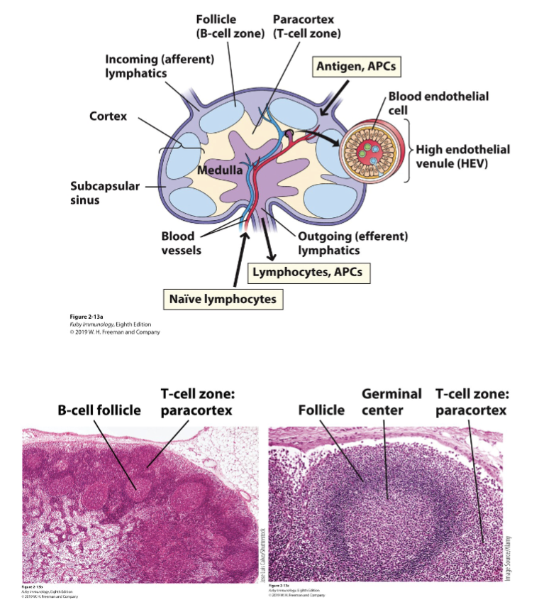
Bean-shaped capsule
Blood supply: Sinuses (subcapsular, cortical medullary), Blood vessels (arterioles, venules, post capillary venules)
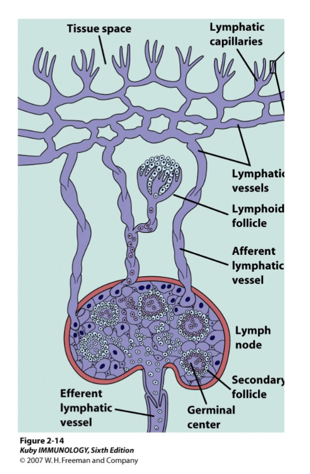
Afferent and efferent lymphatic vessels
Parenchyma (functional tissue):
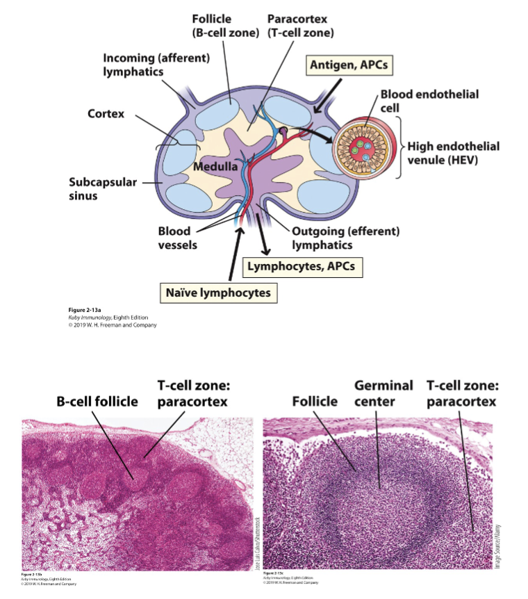
Cortex (lymphoid follicles)
Paracortex
medulla (medullary cords)
Stroma/ Reticular network
What are the afferent and efferent lymphatics?
Lymphatic system vessels
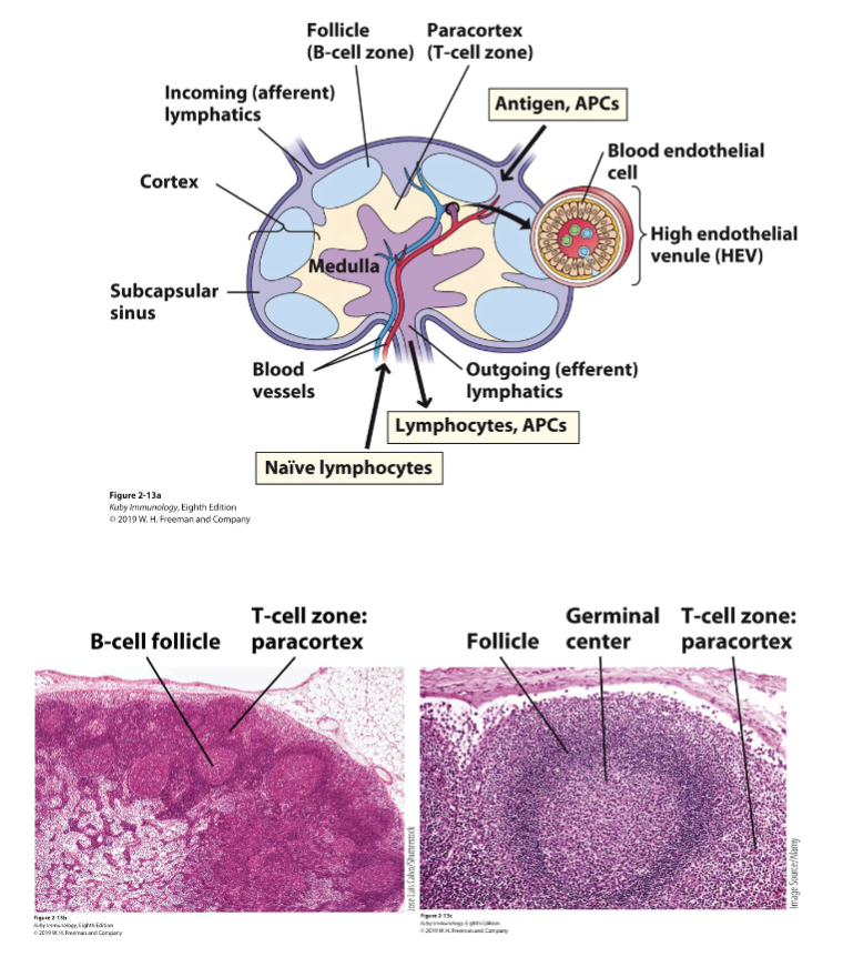
Afferent lymphatics: Arriving at lymphoid tissues
Efferent lymphatics: Exiting lymphoid tissues
Afferent lymphatic vessels bring in lymph fluid containing antigen-carrying dendritic cells, particulate antigen and a few lymphocytes from tissues to regional lymph nodes
Efferent lymphatic vessels take lymph fluid away from lymph nodes via the thoracic duct into venous circulation; carry antibodies secreted by plasma cells and activated/memory T cells and B cells, distributing effector cells and antibodies throughout the body to fight off the infection.
valve system as there is no pumping
some immune cells in the lymphoid follicle
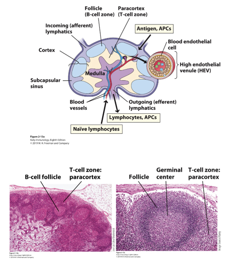
What are the features of the lymph node cortex? What are the features of the paracortex?
Cortex (lymphoid follicles): outer capsule + primary follicles (naïve B, follicular DCs, macrophages) and secondary follicles (activated B cells in germinal centers
Paracortex: T cells + interdigitating DCs
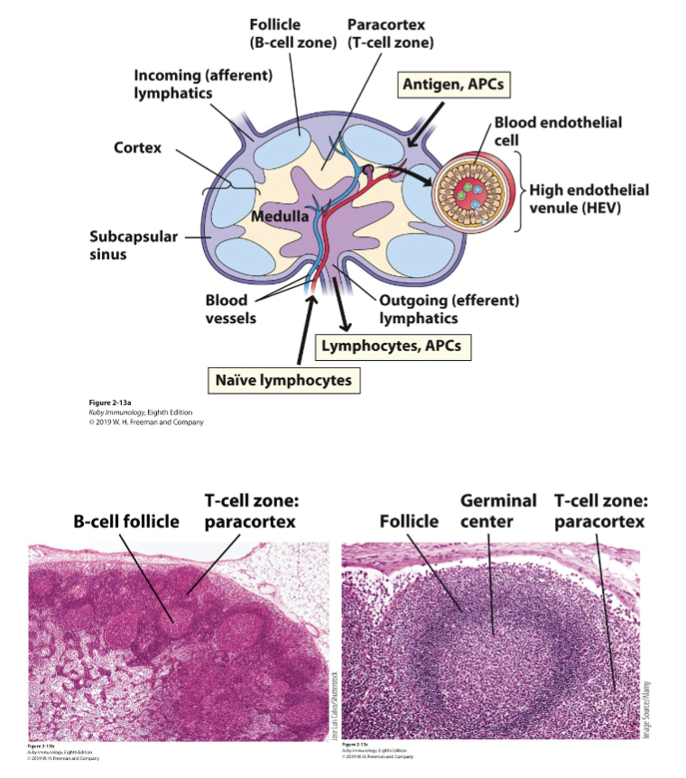
What are the features of the lymph node medulla?
medulla (medullary cords): phagocytic macrophages (eat particulate matter/ microorganism in lymph before it can get into blood), antibody-secreting plasma cells (plasma cells are differentiated B cells), activated/ memory T + B cells (leave lymph node via efferent vessels following activation)
What are the features of the lymph node stroma/ reticular network?
Stroma/ Reticular network: composed of the extracellular matrix, reticular fibres and fibroblastic reticular cells; provides structural support for for lymphocyte support
What are the features of the spleen?
Location where (what happens in the spleen):
-immune responses are mounted against antigens in the blood
-old/defective red blood cells are phagocytosed and recycled
Large ovoid organ situated in upper left quadrant of the abdominal cavity; the spleen is surrounded by a capsule.
where blood bound pathogens get trapped
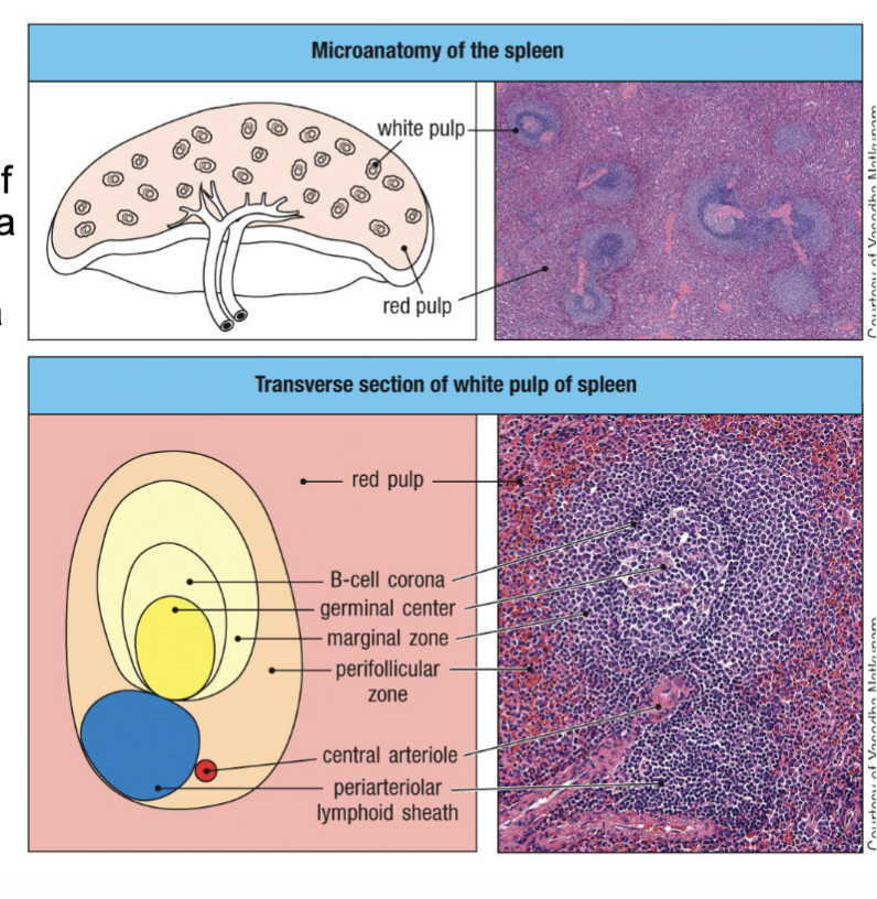
What are the different zones of the spleen?
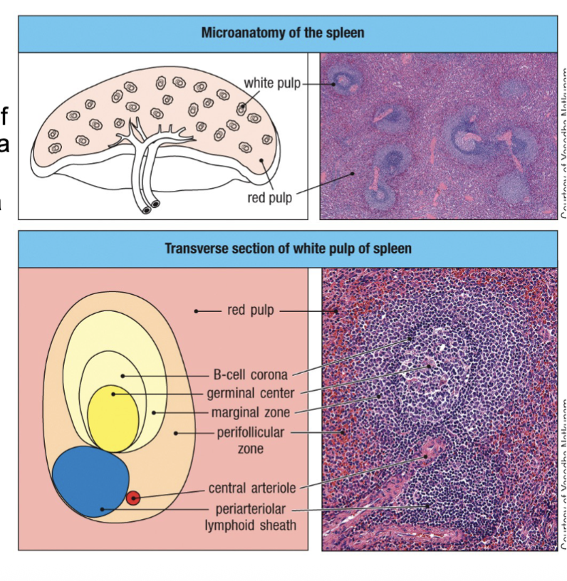
Consists of white pulp and red pulp separated by a marginal zone
-White pulp: Generation of T cell and B cell responses (antibodies) against blood-borne antigens
-Marginal zone: Interdigitating dendritic cells trap blood-borne antigens and transport them to the white pulp
-Red pulp: Defective/old red blood cells and blood-borne pathogens phagocytosed by macrophages
What are MALTs?
Mucosal- associated Lymphoid Tissue
Mucosal sites such as the gut, which has gut associated lymphoid tissue (GALT), have smaller organized secondary lymphoid tissues to trap and present antigen to T and B cells to activate antigen-dependent T and B cell response
What is the role of the lymphatic system?
Connects blood of tissues and the immune system allowing it to get where it needs to.
A network of vessels that collect fluid and lymphocytes that have filtered from the capillaries, through tissues, and ultimately returning them to the bloodstream
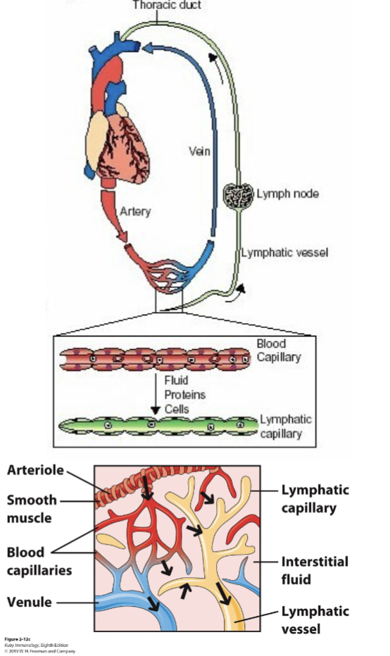
What are the features of the lymphatic system?
leukocytes and antigens are transported and concentrated in lymph nodes to enable their interactions
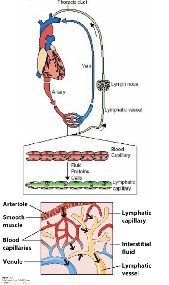
The fluid component of blood (plasma) that leaks out of capillaries is interstitial fluid; most returns to the blood through the capillaries but the remaining fluid, called lymph, is collected by tiny open lymphatic
capillaries
Lymphatic capillaries join and become progressively larger lymphatic vessels, ultimately draining via the thoracic duct into the left subclavian vein back to the blood to return fluid and activated lymphocytes
What are the features of leukocyte abundance in the blood?
Neutrophil- 40-75%
Eosinophil- 1-6%
Basophil- <1%
Monocyte- 2-10%
Lymphocyte- 20-50%
What is homeostasis?
calm state of the cell which ensure the immune cell will not activate until they are needed
What are the two type of immune cells?
Sentinel cells and Circulating/ ready made effector cells
immune cells can be either sentinel cells (stay put/ guard cells) or circulating cells
sentinel in one part can be different in another part of the body, detect and eliminate pathogens
What are the features of sentinel cells?
Cells present (often resident) within tissues with immunologic function
The specific “resident” cells change by location
Function varies by cell types, and include:
-Direct detection and elimination of pathogens
-Recruitment of other immune cells and molecules through release of
cytokines, including chemokines
-Early polarization of immune responses
-Phagocytosis (eating) of pathogens
-Antigen presentation
What are features of Circulating/ready-made effector cells?
Cells present in lymphoid tissues or circulation in the absence of infection
Function varies by cell types, and include:
-Direct detection and elimination of pathogens
-Recruitment of other immune cells and molecules through release of cytokines, including chemokines
-Polarization of immune responses
-Antigen uptake, i.e. phagocytosis (eating), micropinocytosis (sipping)
-Antigen presentation
-Memory
-Antibody Production
What is hematopoiesis?
Hematopoiesis is the formation and development of both red blood cells (erythrocytes) and white blood cells (leukocytes). EVERY leukocyte derives from self-renewing, pluripotent hematopoietic stem cells (HSC).
immune cells come from hematopoiesis (formation of all blood cells)
What are the three lineages of hematopoiesis?
White blood cells
-granulocytes: via granulopoiesis (neutrophils, eosinophils, basophils, mast cells)
-monocytes: via monopoiesis (monocytes, macrophages, dendritic cells)
-lymohocytes: via lymphopoiesis (T cells, B cells, natural killer (NK) cells, ILC, NKT cells)Red blood cells: via erythropoiesis
Platelets: via thrombopoiesis
white blood cells= immune cells = leukocytes
come from pluripotent HSC (self renews)
What are the sites of hematopoiesis?
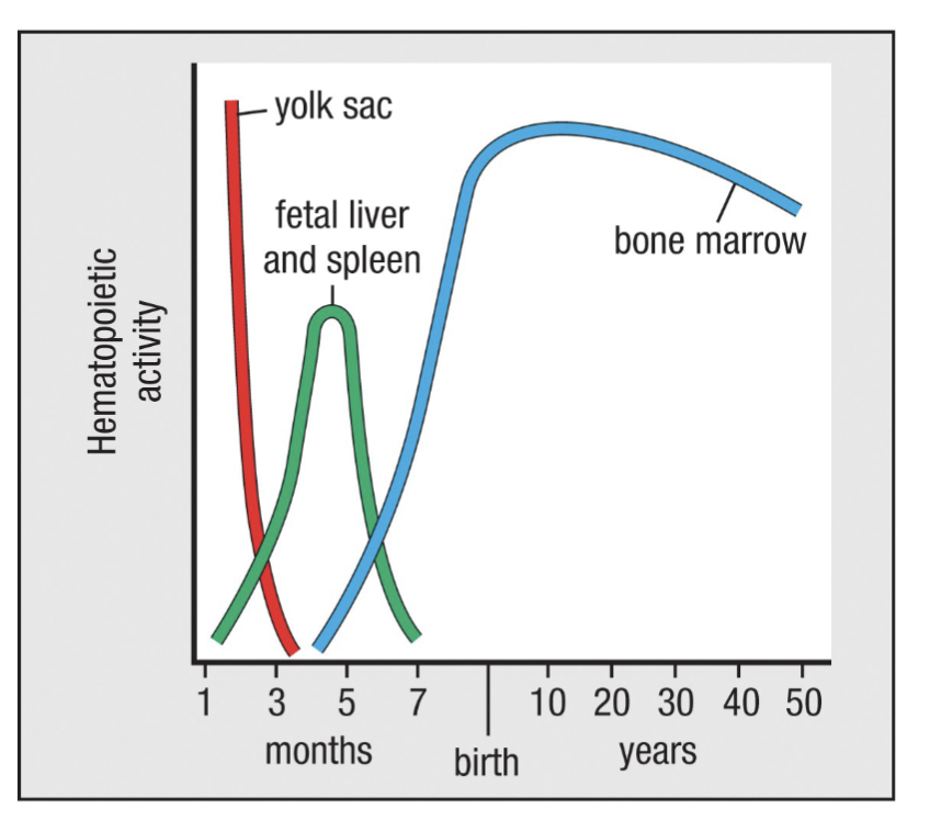
Site according to age
Fetus: 0-2 months-yolk sac, 2-7 months-liver and spleen, 5-9 months-bone marrow
Infants: bone marrow in practically all bones
Adults: vertebrae, ribs, sternum, skull, sacrum, pelvis and end of femurs
adult in bone marrow of long bones + others listed
What are the features of Leukocyte Differentiation and Development
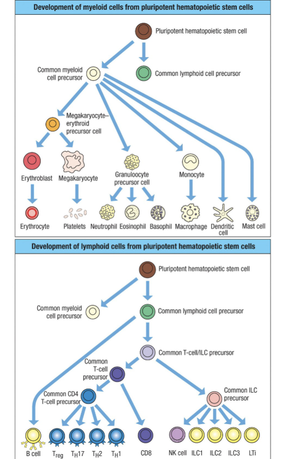
Leukocytes differentiate from hematopoietic stem cells in the bone marrow into two main lineages: myeloid or lymphoid
Within the myeloid lineage are granulocytes (neutrophils, eosinophils, basophils & mast cells) and monocytes
(monocytes, macrophages, dendritic cells)T lymphocytes migrate to the thymus for further maturation
Additional differentiation, maturation and activation-induced differentiation occurs in the periphery and in response to stimulation/growth/tissue-resident factors
pluripotent is the potential to become different things
lymphoid cell precursor gives rise to lymphocytes: innate lymphoid cells (ILC)
How does a developing cell know what to become?
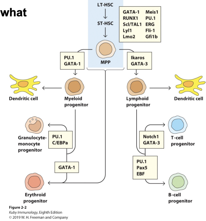
Tightly-regulated transcription factors that progressively specialize cell types through chromatin modification.
Environmental factors trigger transcription factors to turn on or off within cells developing from the HSC, redundancy allows fine tuning of the system
*Ikaros drives differentiation of lymphocytes (shuts down myeloid lineage fate)
*PU.1: low = lymphoid; high = myeloid
*Maintenance of pluripotency in the HSC is controlled by an array of factors
What is Granulopoiesis?
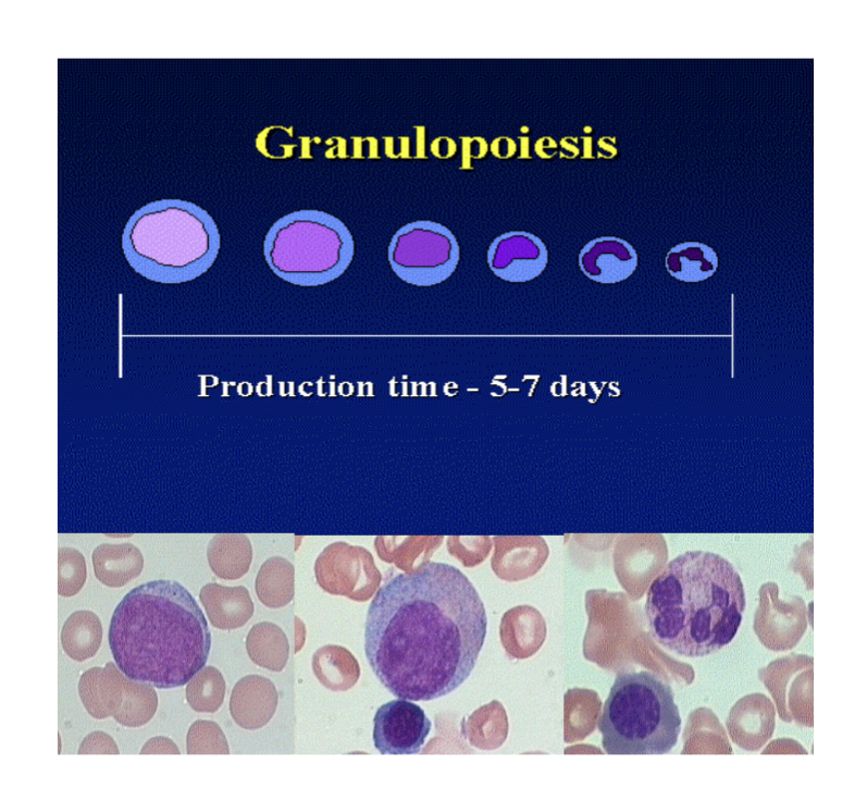
Key regulatory factors for granulopoiesis are:
-Interleukin (IL)-3
-Granulocyte-macrophage-colony stimulating factor (GM-CSF)
-(neutrophils): G-CSF
-(basophils): IL-4
-(eosinophils): IL-4
Maturation characteristics: Nuclear segmentation, acquisition of primary, then secondary granules
Negative feedback inhibition by mature forms
Rate of formation: 1-2 x 10⁹ granulocytes/kg/day
What are the features of granulocytes?
innate immune cells
derived from myeloid progenitor
very early responder to infection, release granule products to damage pathogens and recruit other immune cells
Distinct morphology: segmented nucleus and granules
various granulocytes respond simultaneously
Responses are not antigen specific (allows a quick, generic response to quickly contain infection)
Release products to recruit other immune system cells, including those capable of more specific responses
What are the examples of the granulocytes that release products to recruit other immune system cells?
Neutrophil: Antimicrobial proteins, defensins & lysozymes, direct harm to pathogens
Eosinophil: Cytokines, IL-4,IL-10, IL-13, modulation of adaptive immunity response
Basophil: histamines, vasodilation & smooth muscle activation
listed in order of molecule in granule, examples, function
What are the features of neutrophils?
most abundant leukocyte in circulation,
very short lifespan
first cells recruited to infection sites
respond to infection by: phagocytosis, release of the granule content
Highly responsive to infection – usually first responders to infection, and respond to inflammatory molecules called chemokines to swarm to sites of infection
Increases in circulating PMN (leukocytosis) indicates infection (clinical measurement)
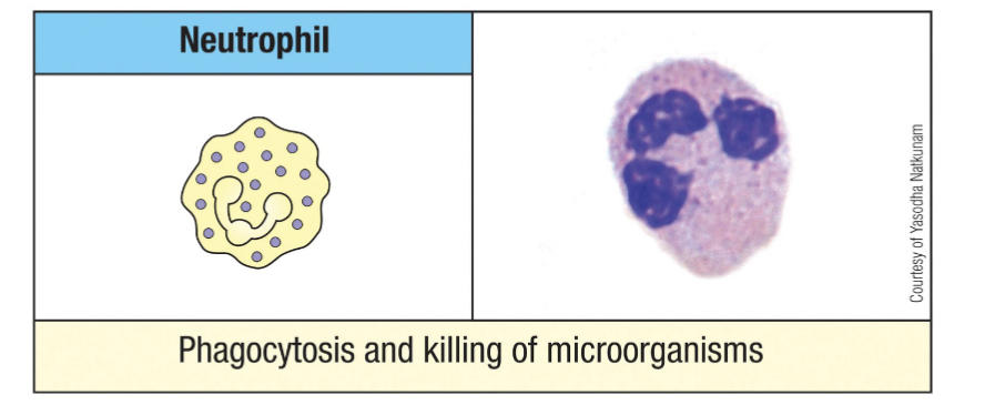
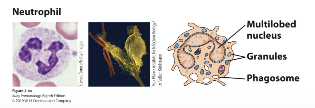
What are the morphological features of neutrophils?
Segmented nucleus (most have 3-4 nuclear segments) connected by tapering chromatin strands
Primary granules (larger, denser; toxic anti-microbial mediators; formed first)
Secondary granules (smaller, contain complement activators and enzymes)
Tertiary granules (phosphatases and metalloproteinases)
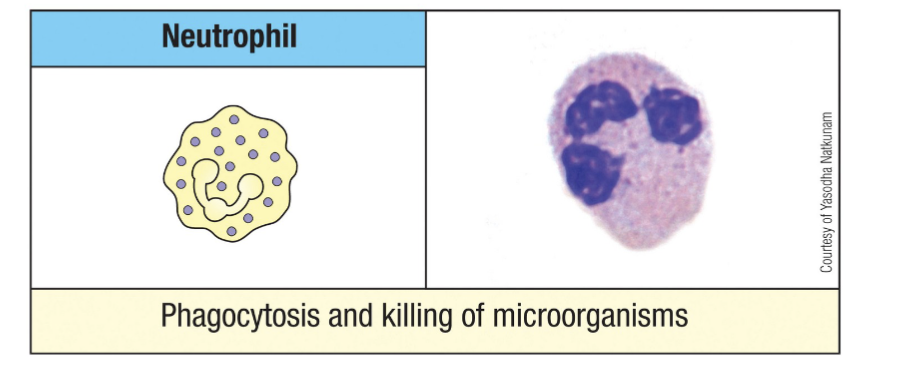
What are the functions of neutrophils?
-Phagocytosis (engulf) bacteria and debris
-Release of chromatin to trap microbes - NETosis
-Secrete proteins to kill bacteria and signal for tissue remodeling
-Assist in shaping the adaptive immune response, when required
-Dead neutrophils accumulate as the major cell type in pus
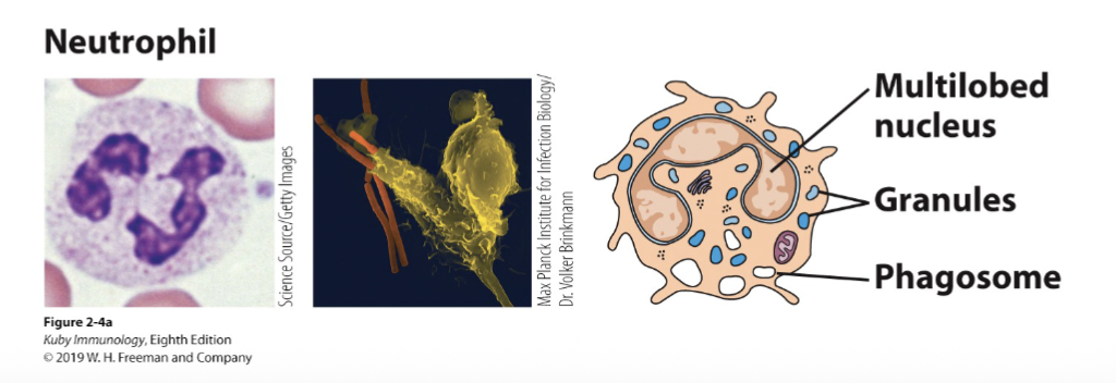
What are the two methods that neutrophils use to kill ingested bacteria?
Oxygen-dependent (mitochondria)
- Reactive oxygen intermediates:
superoxide anion (O2-)
hydroxyl radical (OH)
hydrogen peroxide (H2O2)
hypochlorite anion (ClO-)
-Reactive nitrogen intermediates
nitric oxide (NO)Oxygen-independent (granules)
-Defensins
-Lysozyme
-Hydrolytic enzymes i.e. Collagenase
-Tumor necrosis factor
senses with chemokine receptors moves towards it and engulfs the bacteria
those that say mitochondria are found in the cytoplasm
chemical reactions that disrupt the outer membranes of the element it is attacking
What are the features of eosinophils?
Frequency in circulation: 1-3% of leukocytes (rare circulating cells),
Circulate through the blood and into the tissues
Especially prevalent in the small intestine
innate cells
involved in anti-parasite response, also allergy and asthma
release granule content
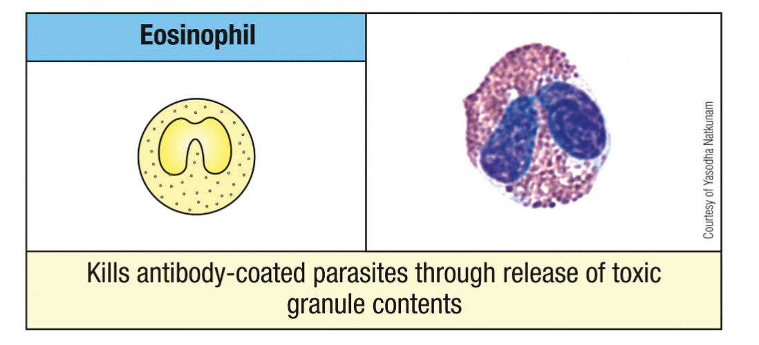
Morphology: segmented nucleus (2 nuclear lobes), spherical granules (eosinophilic granules)
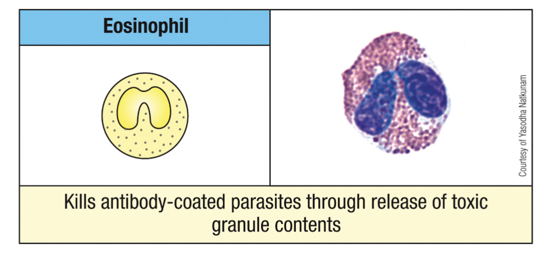
What are the functions of eosinophils?
Involved in anti-parasitic function and also allergy and asthma
Release cytokines to instruct adaptive immune responses; coordinate immune responses, especially against multicellular parasites (i.e.,
worms)
Use lysosomal enzymes and oxygen-radicals like neutrophils
Contain an anti-parasite protein called eosinophil cationic protein (ECP)
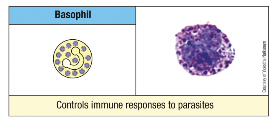
What are the features of basophils?
super rare circulating cells
kills parasites by releasing granule contents
Important for killing extracellular parasites, including multi-cellular worms
Morphology: segmented nucleus (2 nuclear lobes), spherical purple granules (basophilic granules)
similar morphology to mast cells except basophils are much more stationary
Bind circulating antibody/antigen complexes (i.e., tagged pathogens) and release granule contents (i.e., histamines, leukotrienes, prostaglandins and cytokines)
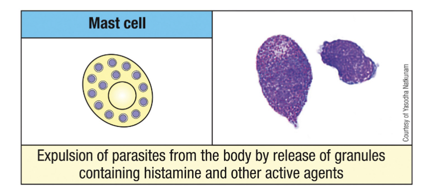
What are the features of Mast Cells?
Produced in the bone marrow and released into blood as immature precursors
two types: mucosa and connective tissue-resident cells
anti-parasite response but also allergy+ asthmas
release granule contents
Important for anti-parasite function and involved in allergic reactions
immature precursor goes into tissue & only there does it mature
Important immune-polarizing cells
Like basophils, they release granule contents (i.e., histamines, leukotrienes, prostaglandins and cytokines)
sentinel version of basophils, very highly granulated cannot distinguish the nucleus
What is monopoiesis?
Key factor: Monocyte-colony stimulating factor (M-CSF)
invade infected tissue and then will differentiate into macrophages and dendritic cells
Maturation Characteristics: Gradual nuclear folding, Acquisition of cytoplasmic granules
Stages in development: Monoblast, Promonocyte, Mature monocyte,
Further differentiation (in tissues) into: Dendritic cells (Granulocyte-monocyte colony stimulating factor: GM-CSF, IL-4) or Macrophages (M-CSF)
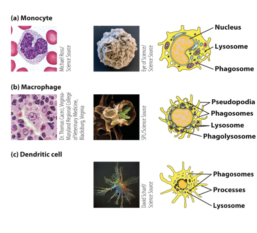
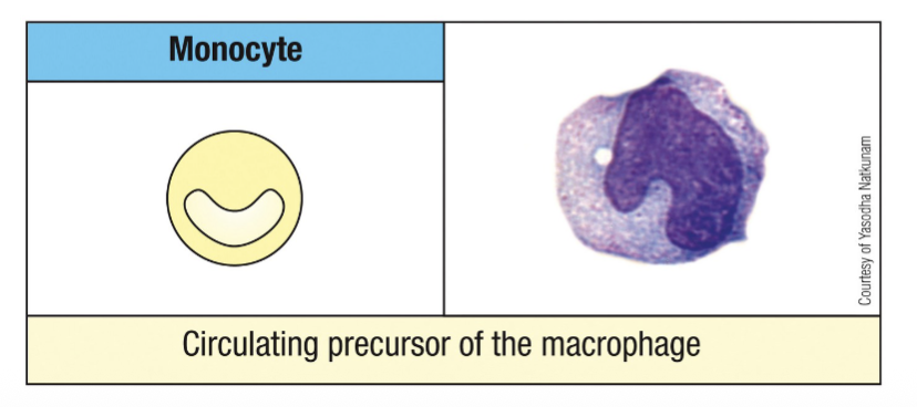
What are the features of monocytes?
Derived from myeloid progenitor
Sentinels to detect infection early in tissues, phagocytose pathogens, and coordinate immune system
Sentinel function – to detect infections and signal immune responses
Monocytes differentiate into macrophages in tissues
Phagocytosis of microorganisms (for immediate and direct immune control)
Phagocytosis and recycling of apoptotic (dead) cells
Killing of ingested microorganisms: oxygen-dependent and oxygen-independent
Recruitment of immune system cells into inflammatory site- Secrete cytokines and chemokines
Present antigen to T cells
What are the features of monocyte differentiation into macrophages?
Changes involved in differentiation:
Increase in size (5-10 fold)
Increased numbers and complexity of organelles
Increased phagocytic activity
Increased levels of hydrolytic enzymes
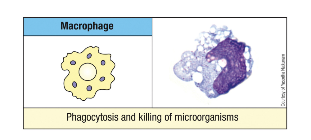
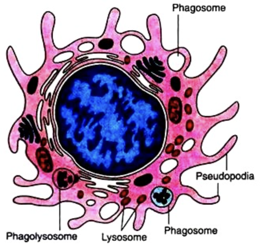
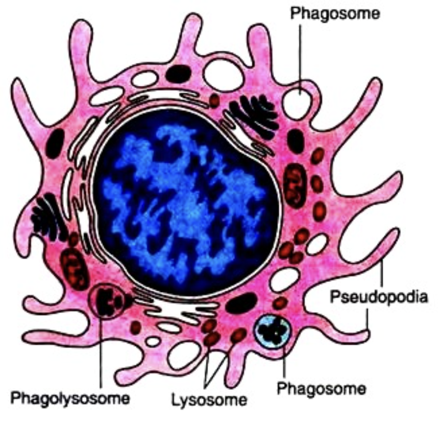
What are the features of macrophages?
circulating monocytes differentiate into macrophages in tissues
macrophages are sentinels & professional phagocytes
also release cytokines
can act as antigen-presenting cells
Macrophages are dispersed throughout the body, traveling by amoeboid movement throughout
tissues
What influences the names of macrophages?
according to location
Alveolar: lung
Histiocytes: connective tissues
Kupffer cells: liver
Mesangial cells: kidney
Microglial cells: brain
Osteoclasts: bone
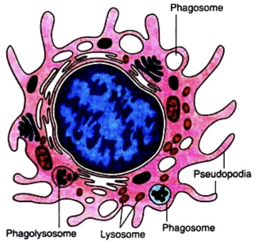
What activates macrophages?
Phagocytosis
Inflammatory Th1 cytokines (i.e. IFN-γ)
Inflammatory mediators
Bacterial components
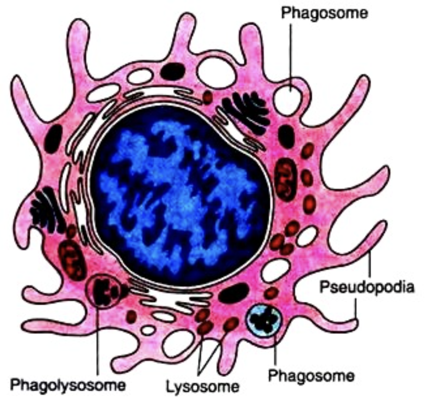
What do macrophages do once activated?
Exhibit enhanced phagocytic activity
Increased killing ability
Increased secretion of inflammatory mediators
Increased migration
Increased ability to activate T cells via antigen-presentation
engulf the bacteria and then it is destroyed via chemical and non-chemical methods
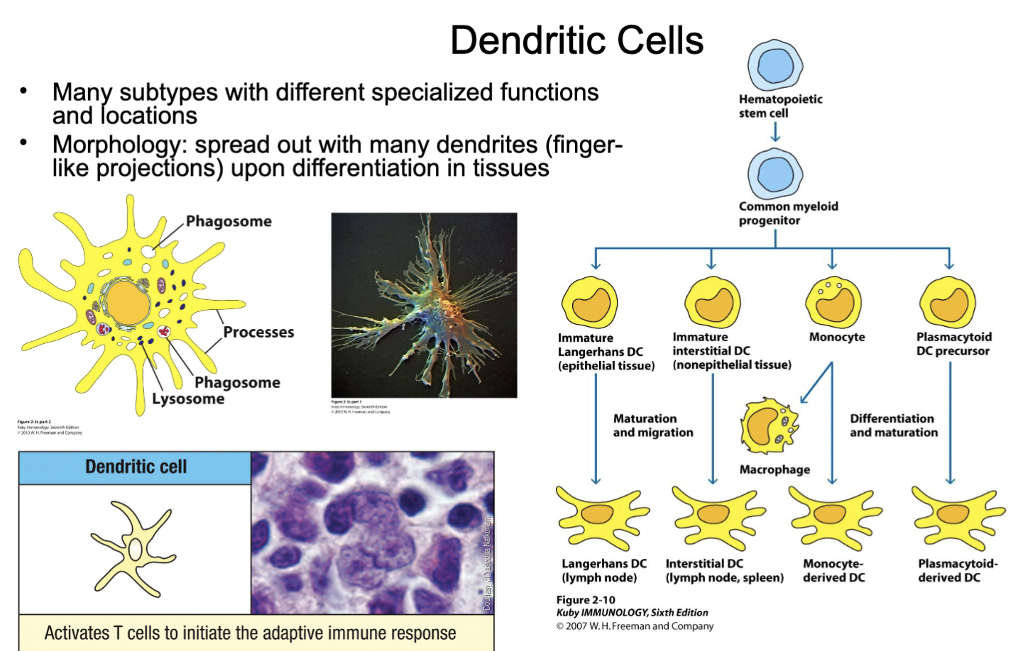
What are the features of dendritic cells (DCs)?
tissue-resident sentinels (mature)
professional antigen presenting cell APC: take up antigen & travel to lymph nodes to activate T cells (adaptive immune cells)
produces cytokines to shape immune response
*uniquely DCs can arise from either myeloid or lymphoid lineage
Many subtypes with different specialized functions and locations
Morphology: spread out with many dendrites (finger-like projections) upon differentiation in tissues
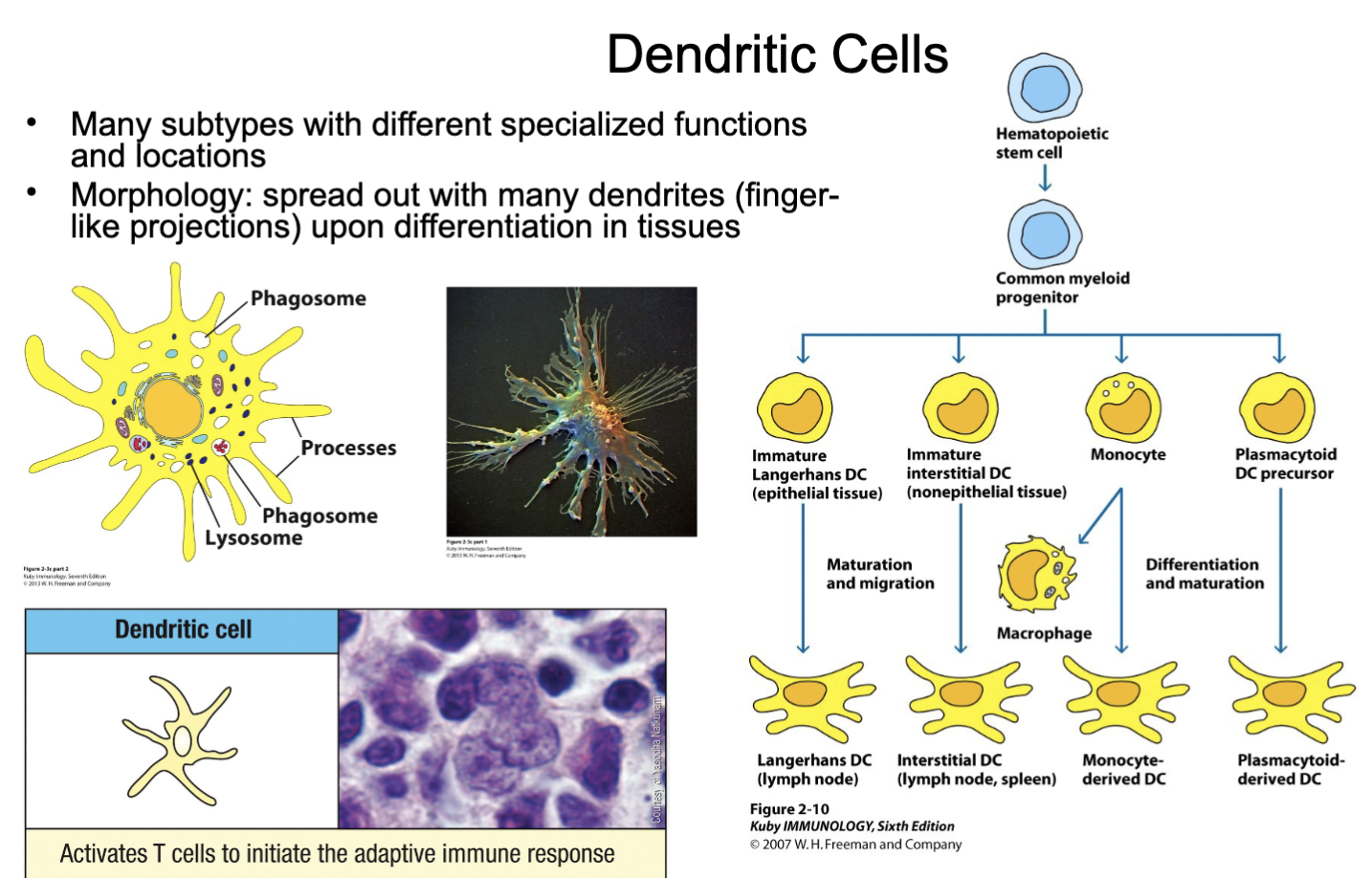
What are the functions of dendritric cells?
Most potent antigen-presenting cell (APC) with ready function
Uniquely, subsets can arise from different lineages (myeloid and lymphoid)
Covered with multiple long membrane extensions like dendrites on nerve cells.
Take up potential antigens by phagocytosis (large eating), pinocytosis (sipping) and receptor-mediated endocytosis
Reside in the tissues and capture antigens from invading pathogens and load the antigen into MHC II and MHC I molecules
Dendritic cells then migrate to lymph nodes and present these antigen to T cells and costimulate T cell activation
Produce cytokines to polarize immune responses
What is lymphopoeisis?
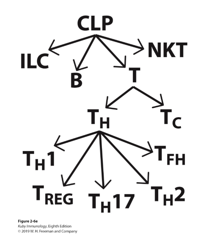
T cells, B cells, NKT cells, NK cells and innate-like lymphocytes arise from same stem cell
Key hematopoietic growth factors (there are many; learn these):IL-3, IL-7, IL-2, IL-4, IL-15
Stages of maturation are defined by surface antigen expression (CD antigens) rather than morphologic features
B cells mature from their progenitors within the bone marrow
NK (natural killer) cells mature from their progenitors and further in the periphery and thymus (one subset)
T and NKT cells develop in the bone marrow but mature in the thymus
Innate lymphoid cells (ILCs) develop in the bone marrow and migrate to tissues. Thymic input for some subsets
What are the features of lymphocytes?
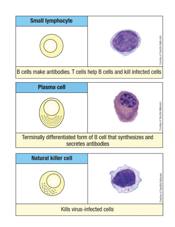
Frequency in circulation: 20-40% of leukocytes
Morphology:
-Naïve (quiescent) T and B lymphocytes
Round nucleus (size of red cells)
Small rim of blue cytoplasm
-Large granular lymphocytes; natural killer (NK) cell, cytotoxic T lymphocyte (CTL), plasma cell (antibody secretion)
Larger cell
More abundant cytoplasm
Large granules containing perforin and granzyme (not plasma cells)
every B cell and T cell have a different receptor
smaller than granulocytes
derived from lymphoid progenitor
make up the adaptive immune cells (T&B) which possess memory & are slower to respond; other lymphocytes like NK cells are faster to respond and function more like innate immune cells
What activates lymphocytes?
by antigen presenting cells APC ex: DC
when the cells are made they are naive
needs 7 days for them to become active, memory cells
What are the features of T cells?
adaptive immune cells which recognize specific antigens and can form memory cells to protect against re-infection
-mature in the thymus
After a T cell begins development and expresses the CD3 complex, it then differentiates into either a CD4+ or a CD8+ T cell
-Further divided into CD4+ and CD8+ T cells
-CD4+ helper T and regulatory T cells
-CD8+ CTL
-CD4:CD8 = 2:1 ratio in healthy blood
-Each exist as naïve, activated and memory cells based on experience and timing with antigen
-Develop in the bone marrow, complete development in the thymus
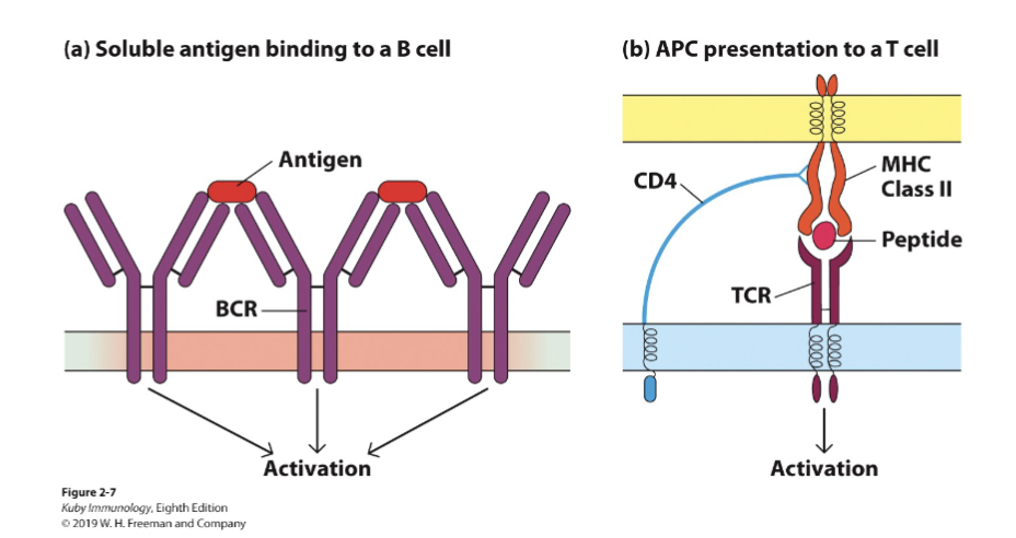
-Express an antigen receptor called the T cell receptor (TCR is a receptor for a peptide presented by MHC1 or MHC2)
main marker is CD3 (if it is there = T cell)
What are the features of CD4+ T cells?
T helper cells
Generals” of the immune response
Help to activate CD8+ T cells, B cells, macrophages and other immune cells; also regulate immune responses
Function by producing a range of different cytokines
Subsets of helper T cells polarize the type of immune response
-CD4+ T Cells are helper cells which produce cytokines to shape immune responses
C4 is helper in regulatory cells (conductor of the orchestra tells the immune system what to, take over after a couple of day)
What are the features of CD8+ cells
Kill virus-infected cells and cancer cells.
Source of cytokines like interferon-γ (IFN-γ).
-CD8+ T Cells are cytotoxic cells which recognize and kill infected & cancer cells,
CD8 main job is to kill infected cells or cancer cells
What are the features of regulatory T cells?
Mostly CD4+
Control immune responses, generally by regulating T cell reactivity
What are the features of B cells?
adaptive immune cells which recognize specific antigens and can form memory cells to protect against re-infection
activated B cells (plasma cells) produce large amounts of antibodies to fight infection
mature/ develop in the bone marrow
B cells, 10-20% (CD19+) if CF19 is present= B cell
Naïve B cells (antigen inexperienced)
Express membrane bound immunoglobulin (antibodies)
Function as APC in secondary immune responses (T helper cells will see it and create a humoral response)
What are the two types of B cells?
Plasma B cells (activated, “antibody factories”)
Memory B cells (maintained after challenge to form memory responses
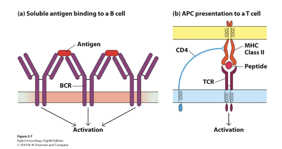
What are the innate-like lymphocytes? What are their features?
-Innate lymphoid cells (ILCs)
-NK cells (CD56+)
-NKT cells
faster response than B and T cells
go around in the body and differentiate between self and non-self
What are the features of Innate lymphoid cells (ILCs)?
Subsets mirror those of CD4+ T cells with polarized function: ILC1, 2 and 3 subsets
Primarily tissue-resident
Major function is cytokine production to polarize immune function
Develop primarily in the bone marrow with thymic input for some subsets
What are the features of natural killer cells (NK cells)? (CD56+)
Discriminate “self” from non-self
Secrete granzyme/perforin to kill target cells
Kill via death receptors
Secrete cytokines to polarize immune responses
sentinel NK cells= Innate lymphoid cells (mostly don’t kill) make cytokines according to their subtype
What are the features of NKT cells?
Express functional T cell receptors that interact with CD1 and conserved glycolipid moieties
Develop in the bone marrow and mature in the thymus
Kill virus-infected and cancer cells
Produce pro-inflammatory cytokines to direct other immune cells
NKT instead of peptides it presents glycolipid moieties
What are the features of the innate immune system?
-Rapid response within hours
-Fixed receptors (received through evolution)
-Limited number of specificities
-Constant during the course of response
What are the features of the adaptive immunity?
Slow response in days to weeks
variable
numerous highly selective specificities
improve during the course of response
What are the default “offense systems” of the immune system
Physical and chemical barriers
Local non-immune cells (i.e. epithelial cells, M cells)
Sentinel cells and molecules
Circulating/ready-made effector cells and molecules
What are the physical structures that interfere with a pathogen’s ability to enter?
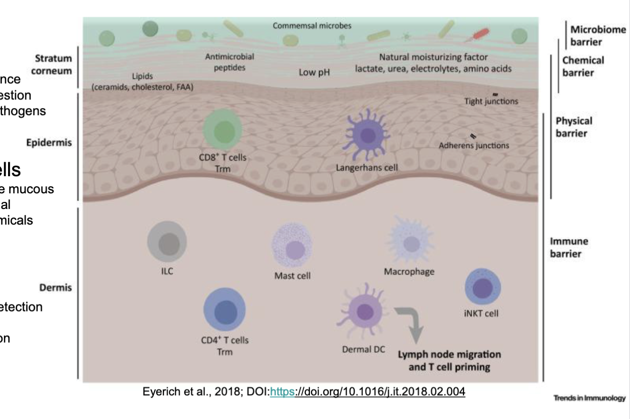
skin (epidermis and dermis)
mucus membranes (epithelium)
How is the skin good at preventing the entry of pathogens?
(epidermis and dermis) is a mechanical barrier impermeable to most
infectious agents
-Epidermis: several layers of epithelial cells with the outermost layer
contains dead cells filled with keratin (waterproofing protein)
type of adhesion in the epithelial cells with tight junctions
dead skin will fall off along with the microbes on it
keratin in the layers (a lot of the bacteria needs water)
How is the mucosal membrane good at preventing the entry of pathogens?
(epithelium) line the interior surfaces of the body and secrete mucus which traps foreign particles such as bacteria and prevents them from adhering to epithelial cells. Foreign particles trapped in mucus are expelled by the mechanical action of cilia, as well as by sneezing, coughing and swallowing. Normal bacterial flora inhibit the growth of many potentially pathogenic bacteria, e.g., gut commensals produce protective peptides.
Gastrointestinal tract: enzymes, normal flora
Respiratory tract: mucus, mechanical action of cilia
Urogenital tract: mucus, fluid flow
What are the barriers to infection found in the skin?
Mechanical: epithelial cells joined by tight junctions, longitudinal flow of air or fluid
Chemical: fatty acids, antimicrobial peptides
Microbial: normal microbiota
What are the barriers to infection found in the gut?
Mechanical: epithelial cells joined by tight junctions, longitudinal flow of air or fluid
Chemical: Low pH, antimicrobial enzymes, antimicrobial peptides
Microbial: normal microbiota
What are the barriers to infection found in the lungs?
Mechanical: epithelial cells joined by tight junctions, movement of mucus by cilia
Chemical: Pulmonary surfactant, antimicrobial peptides
Microbial: normal microbiota
What are the barriers to infection found in the eyes?
Mechanical: epithelial cells joined by tight junctions, tears, nasal cilia
Chemical: antimicrobial enzymes in tears and saliva, antimicrobial peptides
Microbial: normal microbiota
What are the physical and chemical barriers on skin and mucosa (or both)?
-Antimicrobial proteins at epithelial surfaces
-Lysozymes: cleaves peptidoglycans in cell wall of bacteria → lysis of bacteria
-Lactoferrin:
-Secretory leukocyte protease inhibitors (skin, mucosa): blocks epithelial infection by pathogens/ is anti-microbial
-S100 proteins (skin/ mucosa): disrupts membranes of invaders and kills them, also binds to growth factors (Mn & Zn) of pathogens and makes them unavailable
-Surfactant proteins, REGIII proteins antimicrobial peptides at epithelial surfaces: smaller than antimicrobial proteins, kills bac, viruses + fungi by impairing critical components or functions
-Defensins(⍶ + β) (skin/mucosa): disrupts pathogen’s membranes and kills them (bac, fungi, viruses, parasites) secretion of defensins is also induced by PRR signalling
-Cathelicidin(LL-37) (resp. mucosa): disrupt bacteria’s membranes and kills them- secretion also induced by PPR signalling
-Histains (saliva): agains fungi- binds and enter fungal cells and harm it
-Dermcidin (skin, from sweat glands)- antibacterial and antifungal
What are the features of Antimicrobial proteins at epithelial surfaces?
acidic pH of skin- lactic and fatty acids in sweat and sebaceous secretions
What are the features of Lysozymes?
cleaves peptidoglycans in cell wall of bacteria → lysis of bacteria
What are the features of Lactoferrin?
binds iron (taking it from those that use it- mostly respiratory tract/mucosa)
What are the features of Secretory leukocyte protease inhibitors?
skin, mucosa
blocks epithelial infection by pathogens/ is anti-microbial
What are the features of S100 proteins
skin/ mucosa
disrupts membranes of invaders and kills them, also binds to growth factors (Mn & Zn) of pathogens and makes them unavailable
What are the features of Surfactant proteins, REGIII proteins antimicrobial peptides at epithelial surfaces
smaller than antimicrobial proteins, kills bac, viruses + fungi by impairing critical components or functions
What are the features of Defensins(⍶ + β)?
skin/mucosa
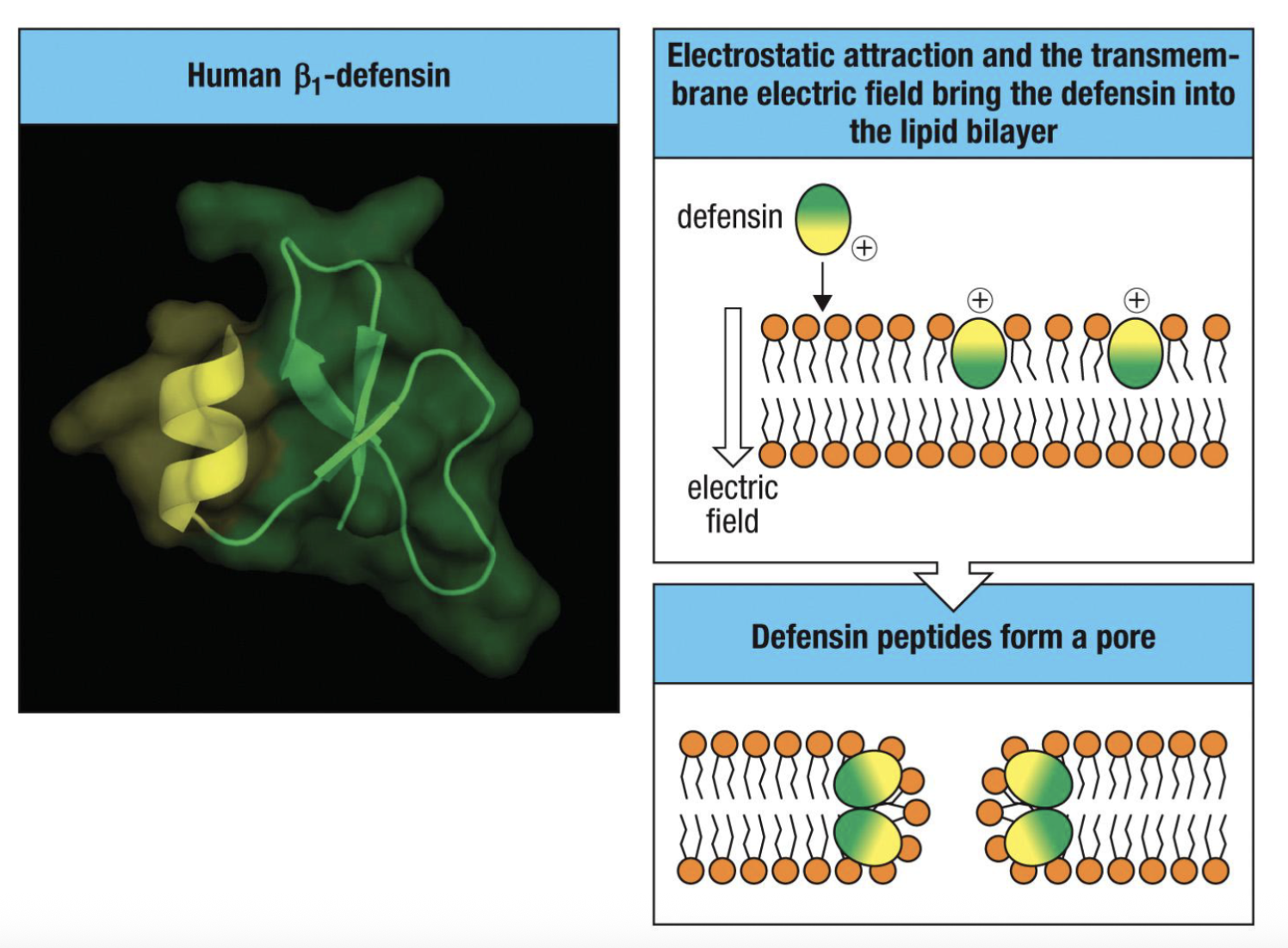
disrupts pathogen’s membranes and kills them (bac, fungi, viruses, parasites) secretion of defensins is also induced by PRR signalling
defensins form a pore and that leads to influx of water and the cell is lysed
What are the features of Cathelicidin(LL-37)
respiratory mucosa
disrupt bacteria’s membranes and kills them- secretion also induced by PPR signalling
What are the features of Histains (saliva)
against fungi- binds and enter fungal cells and harm it
What are the features of Dermcidin?
(skin, from sweat glands)- antibacterial and antifungal
Cellular barriers are a layered set up what is the common set up?
Commensal microflora
a. Maintain balance
b. Help with digestion
c. Crowd out pathogensEpithelial and associated cells
a.Often produce mucous or antimicrobial peptides/chemicalsSentinel cells
a. Tolerance to commensals
b. First line of detection for infection
c. Differ based on location
What are the features of Antimicrobial (host defense) peptides?
Short cationic peptides (typically 29-35 amino acids) with an amphipathic
(has both hydrophilic and hydrophobic parts) structure, constitutively expressedActive against bacteria, fungi and enveloped viruses
Bind to negatively-charged microbial structures and membranes via
electrostatic interaction. (e.g., LPS)Disrupt microbial membrane integrity by introducing bulky hydrophobic
amino acidsInternalized peptides inhibit DNA/RNA/protein synthesis and activate
antimicrobial enzymesKill microbes rapidly (minutes to hours)
As an example: defensins (α + β) are secreted by intestinal epithelial Paneth cells and other epithelial cells; stored in neutrophil granules.
cationic peptides: expressed all the time
interact via + - interactions
Why do defensins not target host cells?
host mammalian bilayer membranes are less attracting for antimicrobial peptides because they are rich in phosphatidilethanolamine, phosphatidilcoline and
sphingomielin and generally neutral in net charge
vs
the bacteria tends to have a negative charge (more attracting)
In order to initiate infection, pathogens must develop a means of getting past the epithelium; this route differs with particular pathogens. What are these different routes?
via the airways (Streptococcus pneumoniae)
via the gastrointestinal tract (E. coli)
via the genitourinary tract (Human Immunodeficiency Virus)
via cuts in the skin (Staphylococcus aureus)
via mosquito bites (West Nile Virus)
After penetration of epithelial barriers, pathogens are detected by innate immune components. What are the components?
Soluble innate immune recognition elements (portions hanging out waiting to encounter the pathogen)
-Mannose-binding lectin; C-reactive protein; complementPhagocytic and other cells
Neutrophils, macrophages, dendritic cells, natural killer (NK) cellsPattern recognition receptors (PRRs) such as Toll-like receptors (TLR) are present on innate immune cells (macrophages, mast cells, dendritic cells) that function as sentinel cells in tissues
Interferons
What are soluble factors?
Various secretions contain substances with anti-bacterial activity; gastric juice - acid; semen - spermine and zinc; milk – antimicrobial peptides, lactoperoxidase; tears, saliva, nasal secretions - lysozyme
Collectins
Complement pathway
What are collectins?
small proteins that are able to kill certain bacteria by cell wall disruption, cause bacteria to aggregate, thereby enhancing phagocytosis, and/or activate complement by the lectin pathway
What is the complement pathway (simple definition)?
A feed-forward cascade that enables killing spontaneously or after antibodies or mannose-binding lectin binds to target cells.
How do interferons play a role in the detection of pathogens once they penetrate the skin?
Interferons→ Type I alpha and beta: transcription is directly induced by PRR signaling these in turn drive the production of type II interferon (gamma).
What are the features of interferons?
Type I: IFN-⍺ and IFN-β
Function: Inhibit viral replication, generate an antiviral state.
Produced by: IFN-⍺ (leukocytes esp. PDC) IFN-β (fibroblasts)
Type II: IFN-ɣ
Function: Enhance killing of virus infected cells, promote Th1 immunity
Produced by: NK cells and T cells
What is the role of Type I interferons? How do they do this?
Inhibit Viral Replication
Induction of type I: IFN-⍺ and IFN-β
Presence of viral RNA or DNA activates IFN-⍺/-β production via stimulation of
pattern recognitions receptors such as TLRs.Interferons then induce antiviral responses in neighbouring cells (paracrine) by binding to IFN-⍺/-β receptors, leading to mRNA degradation and inhibition of protein synthesis, making them refractory to viral growth
pathway of production starts with TLRs
What do Plasmacytoid Dendritic Cell produce at a higher frequency than other cells?
Although many cell types produce small amounts of type I interferons upon viral infection, plasmacytoid dendritic cells produce 100-1000x more type I interferon upon contact with viruses than any other cell type
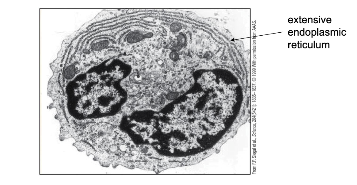
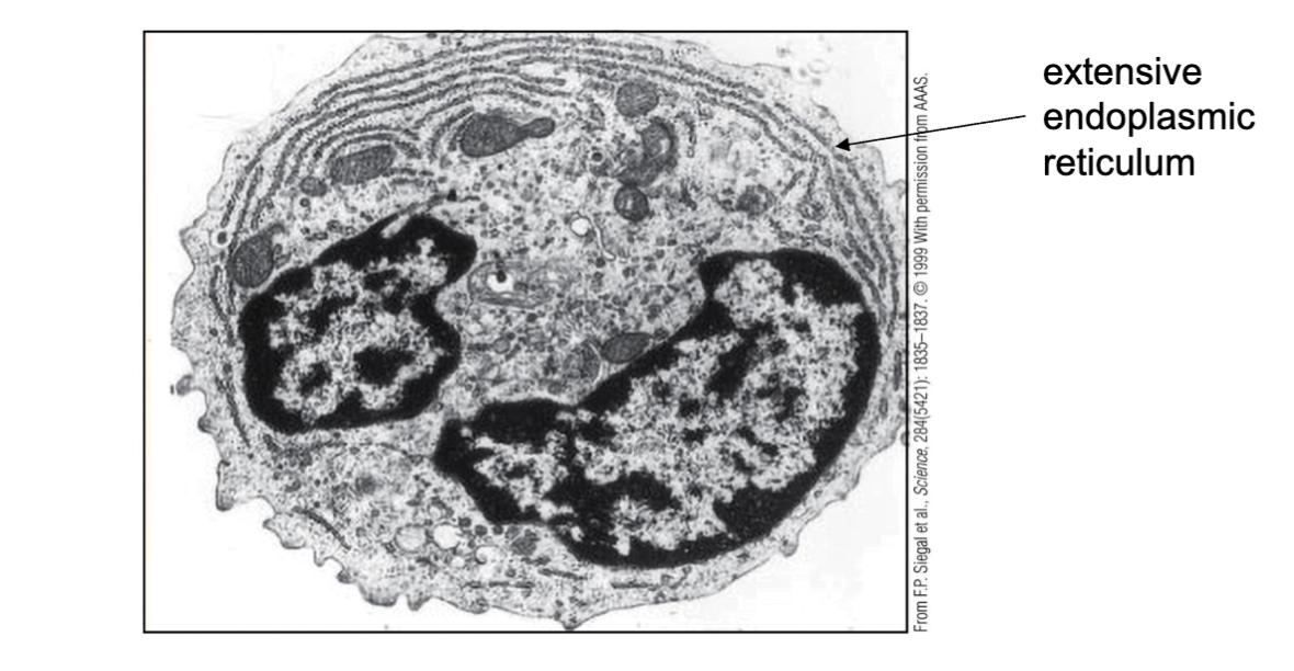
What are the features of Plasmacytoid Dendritic Cells?
Plasmacytoid dendritic cells do not need a productive viral infection in order to upregulate genes coding for type I interferons
Recognition mechanism for viral infection via TLR7/8 that recognize viral ssRNA and TLR9 (CpG DNA) (CpG DNA recognition doesn’t happen in mammalian cells)
Plasmacytoid dendritic cells are important in driving immune responses against viral
infections, e.g. NK cell responses
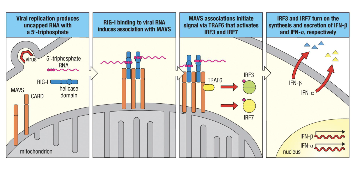
What is the process of: Intracellular recognition of viral nucleic acid by RIG-I
initiates interferon production?
Viral replication produces uncapped RNA with a 5’-triphosphate
RIG-I binding to viral RNA induces association with MAVS
MAVS associations initiate signals via TRAF6 that activate IRF3 and IRF7
IRF3 and IRF7 turn on the secretion of IFN-⍺ and IFN-β respectively
if the virus makes it to the cytoplasm of the cell there are sensors there : most know= RIG-I
5’-triphosphate RNA is not found in our RNA only in viruses
RIG-I will recognize 5’-triphosphate RNA
need to know signalling pathway
What are the cellular responses to infections (induced by actions of soluble innate mediators)?
PRR binding PAMPs
→phagocytosis (soluble innate elements also promote phagocytosis i.e. complement, in addition to PRR binding)
→production of interferons, cytokines, chemokines, anti-microbials
Cytokine production (IL-1,IL-6, TNF) also drives acute phase response (IL-1,IL-6, TNF → hypothalamus→ prostaglandin → fever)
IL-6, TNF also signal HSCs in bone marrow to produce more leukocytes to fight infection
IL-1, IL-6, TNF also signal liver to produce more acute phase proteins (C-reactive protein, serum amyloid A, Fibrinogen, Mannose-binding protein, complement components)
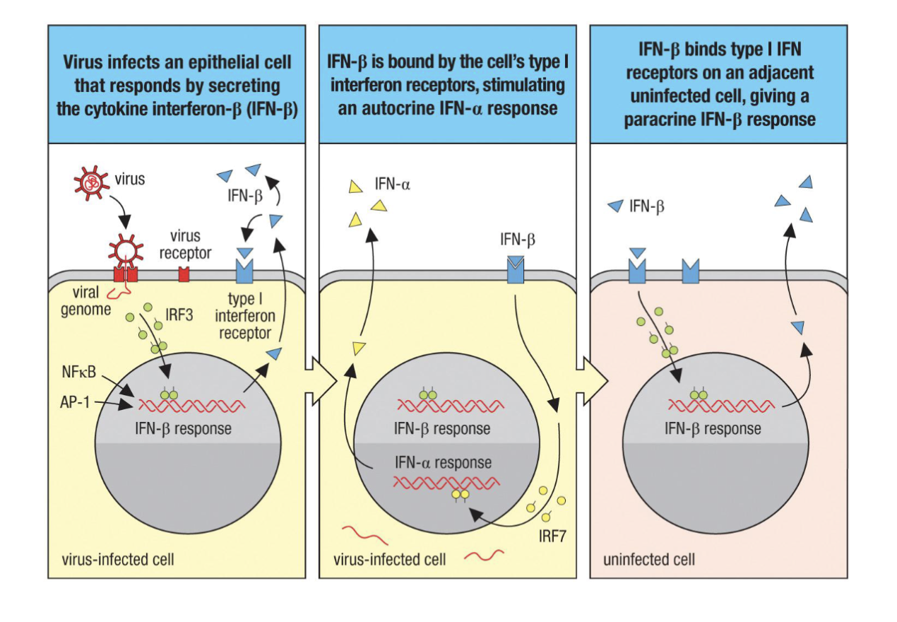
How do Virus-infected cells produce type I interferons and protect adjacent uninfected cells?
Virus infects an epithelial cell that responds by secreting the cytokine interferon-β (IFN-β ).
IFN-β is bound by the cell’s type I interferon receptors stimulating an autocrine IFN-⍺ response
IFN-β bind type I IFN receptor on an adjacent uninfected cell giving a paracrine IFN-β response.
What are the innate initiations of adaptive responses?
Dendritic cell PPRs recognize PAMPs, activating phagocytosis
Dendritic cells migrate to lymph nodes, carrying intact or degraded pathogens
Antigen fragments bound to cell surface MHC proteins are recognized by T cells, with costimulation.
Activated T cells initiate adaptive responses
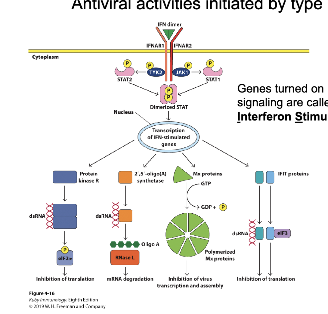
What are the antiviral activities initiated by Type I Interferon?
IFNAR: interferon alpha receptor (1 and 2)
TYK2 & JAK1 are kinases and will phosphorylate the STAT which creates Dimerized STAT
=Transcription of IFN stimulated genes:
kinase R … turns of everything in the cell to stop the virus (good chance it kills the cell, epithelial cell so can be renewed not that big a deal if it dies) ,
2,5’-oligo(A) synthetase which makes Oligo A which will bind RNase L and it will degrade all the RNA in the cell (makes sure to get viral RNA)
Mx protein inhibition of virus transcription and assembly
IFIT protein can recruit and bind elongation factor 3 (stop it from participating in translation) = global inhibition of translation
These products generally inhibit transcription and translation of all genes in the cell; this prevents viral genes from being used