Anatomy 12 | Anterior Thoracic Wall & Mediastinum
1/43
There's no tags or description
Looks like no tags are added yet.
Name | Mastery | Learn | Test | Matching | Spaced | Call with Kai |
|---|
No analytics yet
Send a link to your students to track their progress
44 Terms
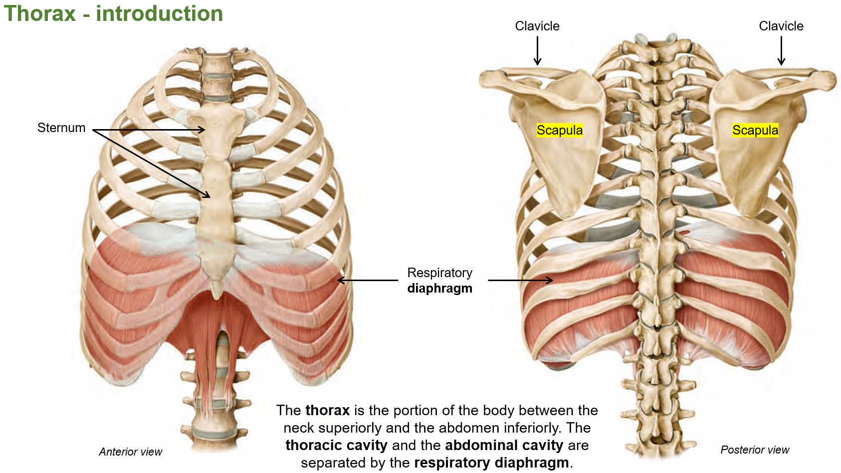
What is the thorax?
Part of the body between the neck and the abdomen. The thoracic cavity and the abdominal cavity are separated by the diaphragm.
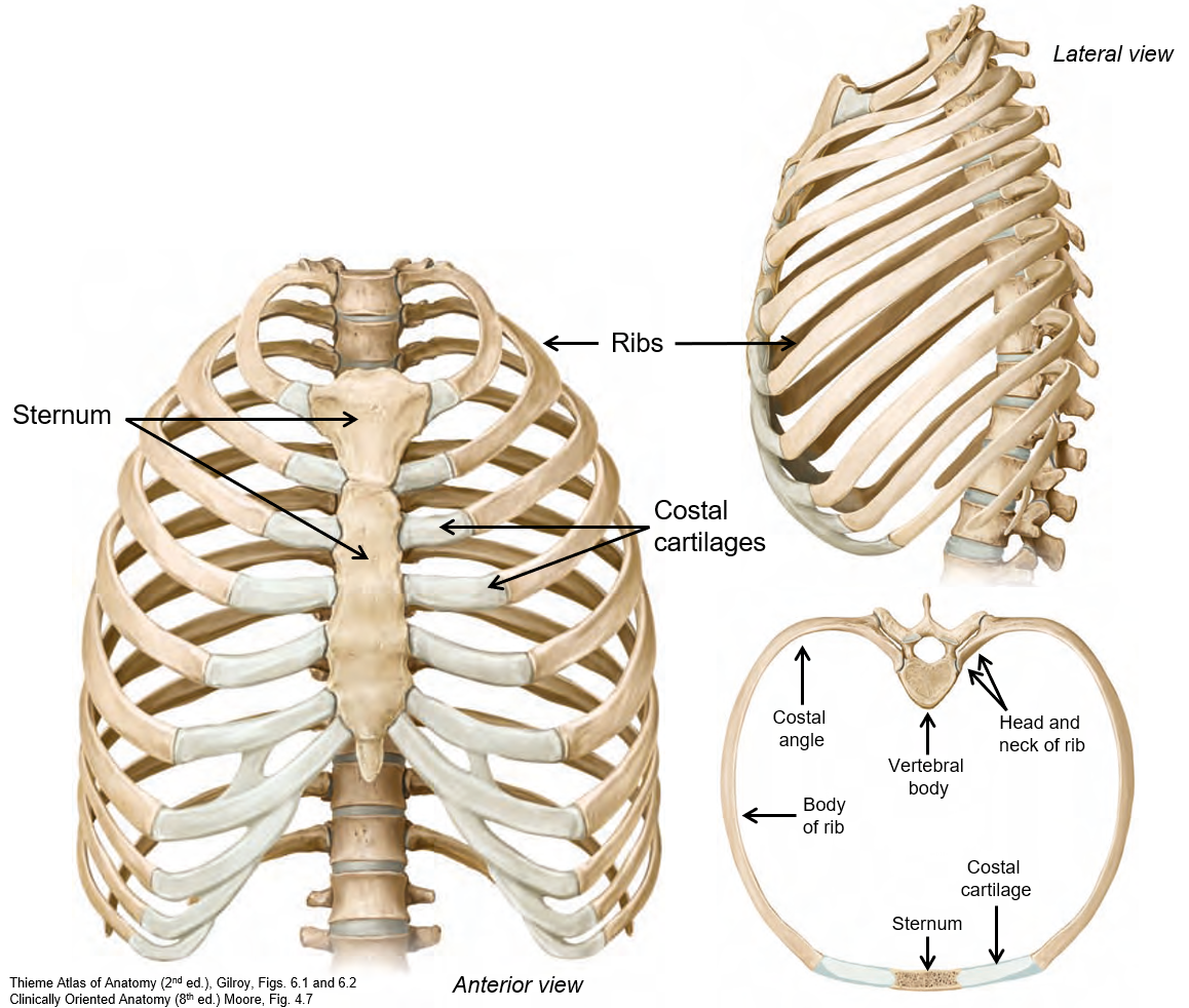
The thoracic cage comprises of?
The ribs, costal cartilages, sternum, and vertebral column. It opens at the thoracic inlet (top) and thoracic outlet (bottom).
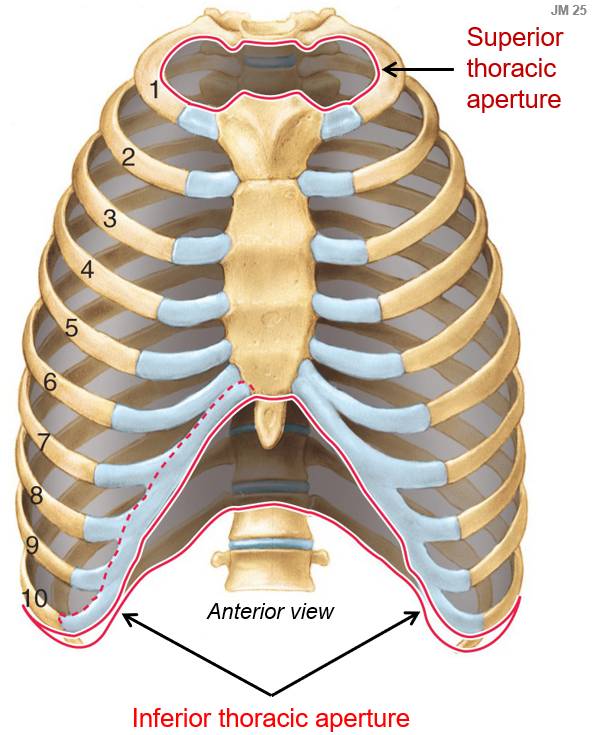
What is the superior and inferior thoracic aperture?
The superior thoracic aperture (thoracic inlet) is the opening at the top of the thoracic cavity that connects to the neck.
The inferior thoracic aperture (thoracic outlet) is the opening at the bottom of the thoracic cavity that connects to the abdomen.
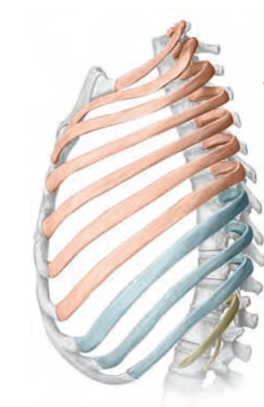
What are true ribs?
True ribs (1st–7th) are ribs that attach directly to the sternum through their own costal cartilage.


How do false ribs differ from true ribs?
False ribs (8th–10th) do not attach directly to the sternum; instead, their cartilage connects to the rib above them.
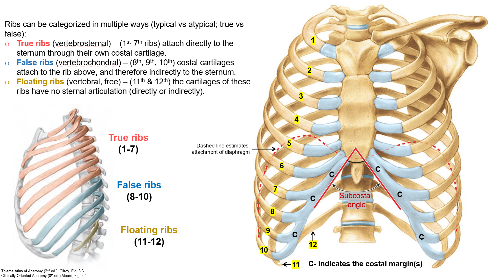

Why are floating ribs called "floating"?
Floating ribs (11th & 12th) do not connect to the sternum at all, making them appear "free" or "floating."

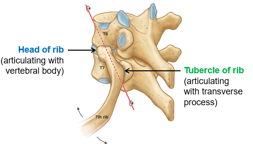
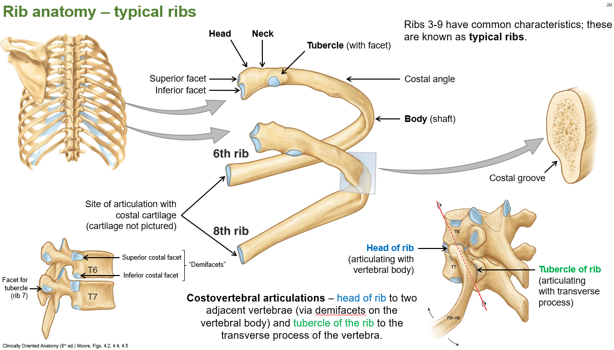
What are costovertebral articulations?
Costovertebral articulations are the joints where the ribs connect to the vertebrae in the spine.
How does the head of the rib connect to the vertebrae?
The head of the rib connects to two adjacent vertebrae through demifacets on the vertebral body.

What part of the rib connects to the transverse process of the vertebra?
The tubercle of the rib connects to the transverse process of the vertebra.
What are typical ribs?
Typical ribs are ribs 3–9, which have common structural characteristics.
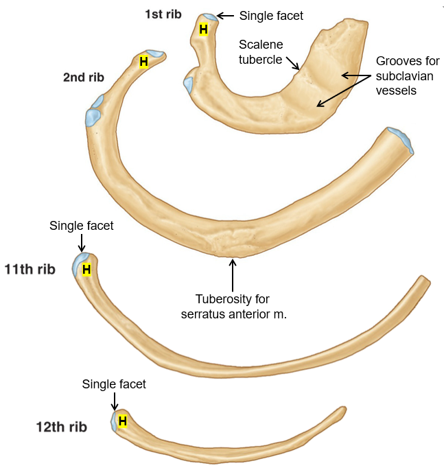
Which ribs are considered atypical?
Ribs 1, 2, 10, 11, and 12 because they have unique features different from typical ribs.
What are the unique features of ribs 1, 2, and 10-12?
Rib 1 – Short, broad, with one facet on its head and grooves for subclavian vessels.
Rib 2 – Has two facets on its head and a rough area for serratus anterior attachment.
Ribs 10-12 – Have one facet on their heads; ribs 11 and 12 are short with no neck or tubercle.
What is the function of the sternum?
The sternum anchors the ribs and protects the mediastinal organs.
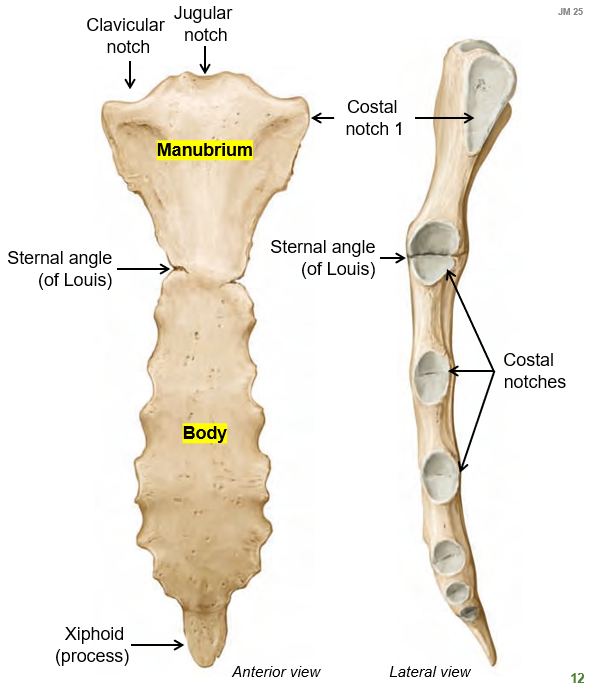
What are the three parts of the sternum?
The sternum consists of
manubrium
body
xiphoid process

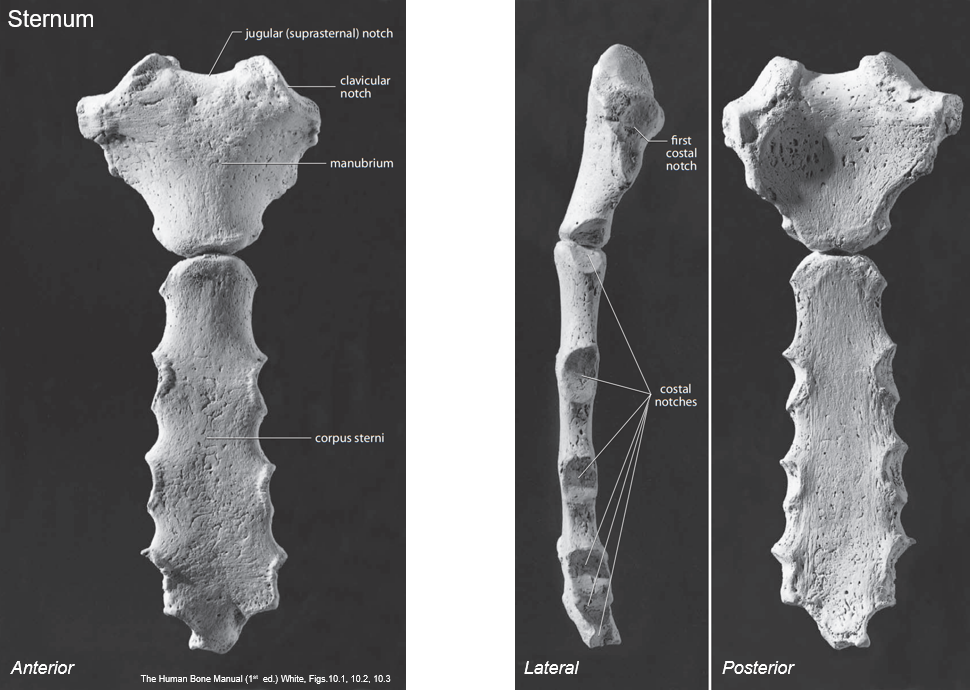
How are the parts of the sternum connected?
By synchondroses, which ossify in middle to late adulthood.
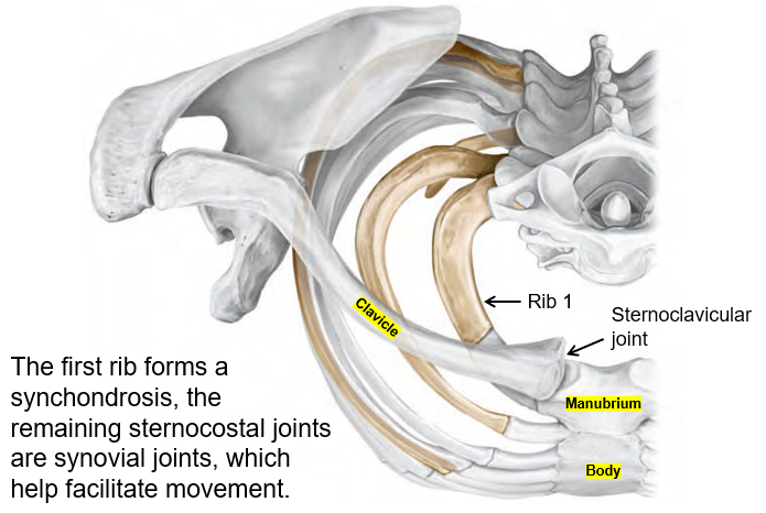
What type of joints are found in the sternocostal region?
The first rib forms a synchondrosis, while the other sternocostal joints are synovial joints, allowing movement.
What is the respiratory diaphragm?
A muscle that separates the thoracic and abdominal cavities. It plays a key role in breathing by contracting and moving downward to increase lung volume
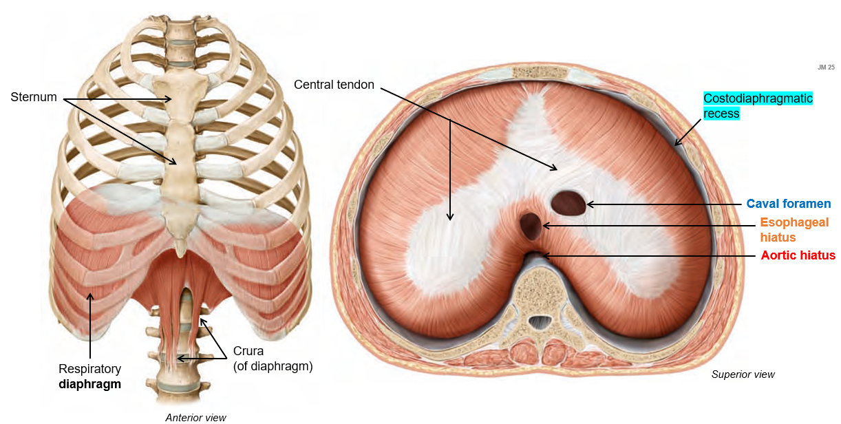
Where does the diaphgram insert?
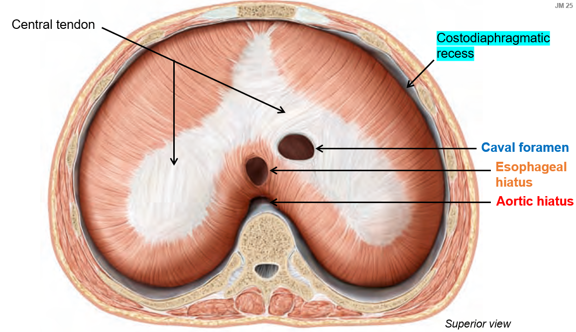
Inserts into the central tendon, a large fibrous structure that helps facilitate its movement during respiration.
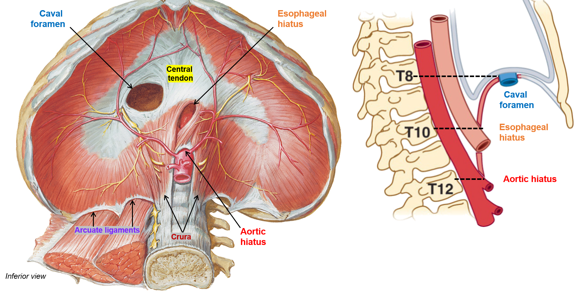

What are the three major openings in the diaphragm, and what structures pass through them?
Caval foramen – Allows the inferior vena cava to pass through, facilitating blood return to the heart.
Esophageal hiatus – Provides a passage for the esophagus and the vagus nerves.
Aortic hiatus – Allows the aorta, thoracic duct, and azygos vein to pass through.

What are the three parts of the diaphragm
Sternal portion – From the xiphoid process.
Costal portion – From the ribs and costal margins.
Lumbar portion – From aponeurotic arches and lumbar vertebrae (L1-L3), forming the left and right diaphragmatic crura.
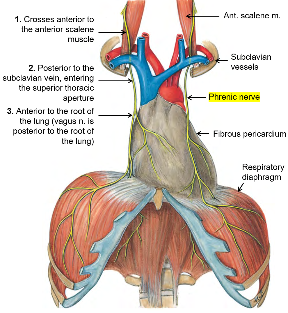
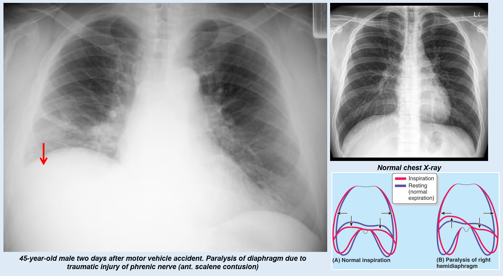
What is the function of the phrenic nerve?
Provides motor innervation to the diaphragm and relays sensory information from the pericardium and parietal pleura.
Which spinal nerve roots contribute to the phrenic nerve?
The phrenic nerve originates from spinal nerve roots C3, C4, and C5. (A derivative of the cervical plexus and some help from brachial plexus)
What is the pathway of the phrenic nerve from the neck to the diaphragm?
The phrenic nerve comes from C3, C4, and C5 and moves downward in front of the anterior scalene (neck muscle).
It passes behind the subclavian vein and enters the top of the chest (superior thoracic aperture).
The nerve moves in front of the lung root, while the vagus nerve stays behind it.
It reaches and controls the diaphragm, helping with breathing.
Mnemonic to remember phrenic nerve innervation?
C3,4,5, keeps the diaphragm alive
What would you expect if you had a lesion to the phrenic nerve?
One side of the diaphragm would not work. Asymmetry
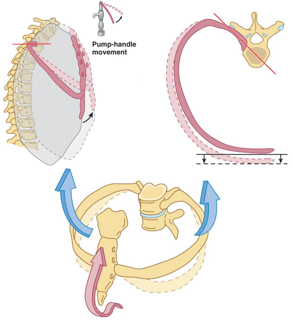
What is the pump handle movement?
Occurs in the upper ribs (2-6), where the anterior ends of the ribs rise, increasing the front-to-back (anterior-posterior) size of the chest during breathing.
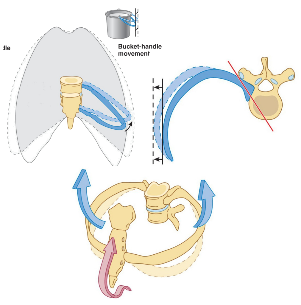
What is the bucket handle movement?
Increases the side-to-side (transverse) dimension of the chest by raising the lateral parts of the ribs during breathing.
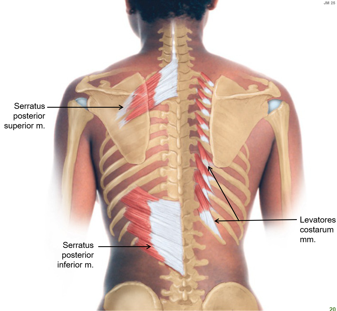
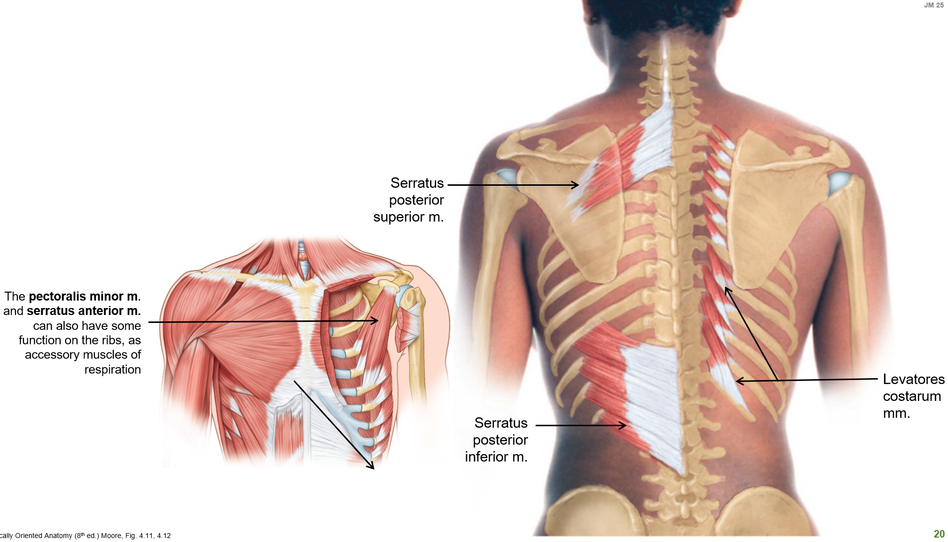
What do the serratus posterior muscles do?
Serratus posterior superior weakly elevates the ribs.
Serratus posterior inferior weakly depresses the ribs.


What do the levatores costarum muscles do?
Help elevate the ribs, assisting in respiration.
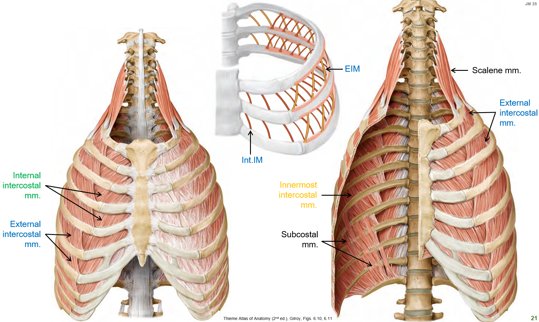
What is the function of the intercostal muscles in respiration?
External intercostal muscles assist with inspiration.
Internal and innermost intercostal muscles assist with expiration.
If the first letter starts with E (external int…) it helps with I (inspiration). If it starts with I, it helps with E (expiration)
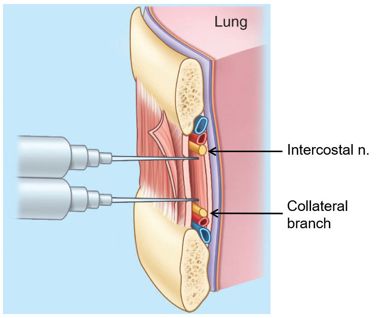
What is the purpose of an intercostal nerve block?
Helps relieve pain from rib fractures or thoracic procedures by numbing the intercostal nerves.
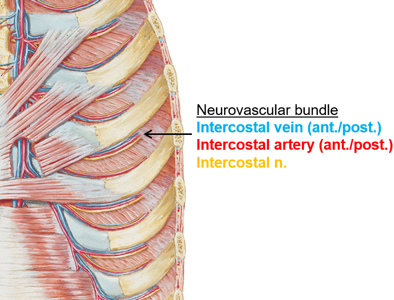
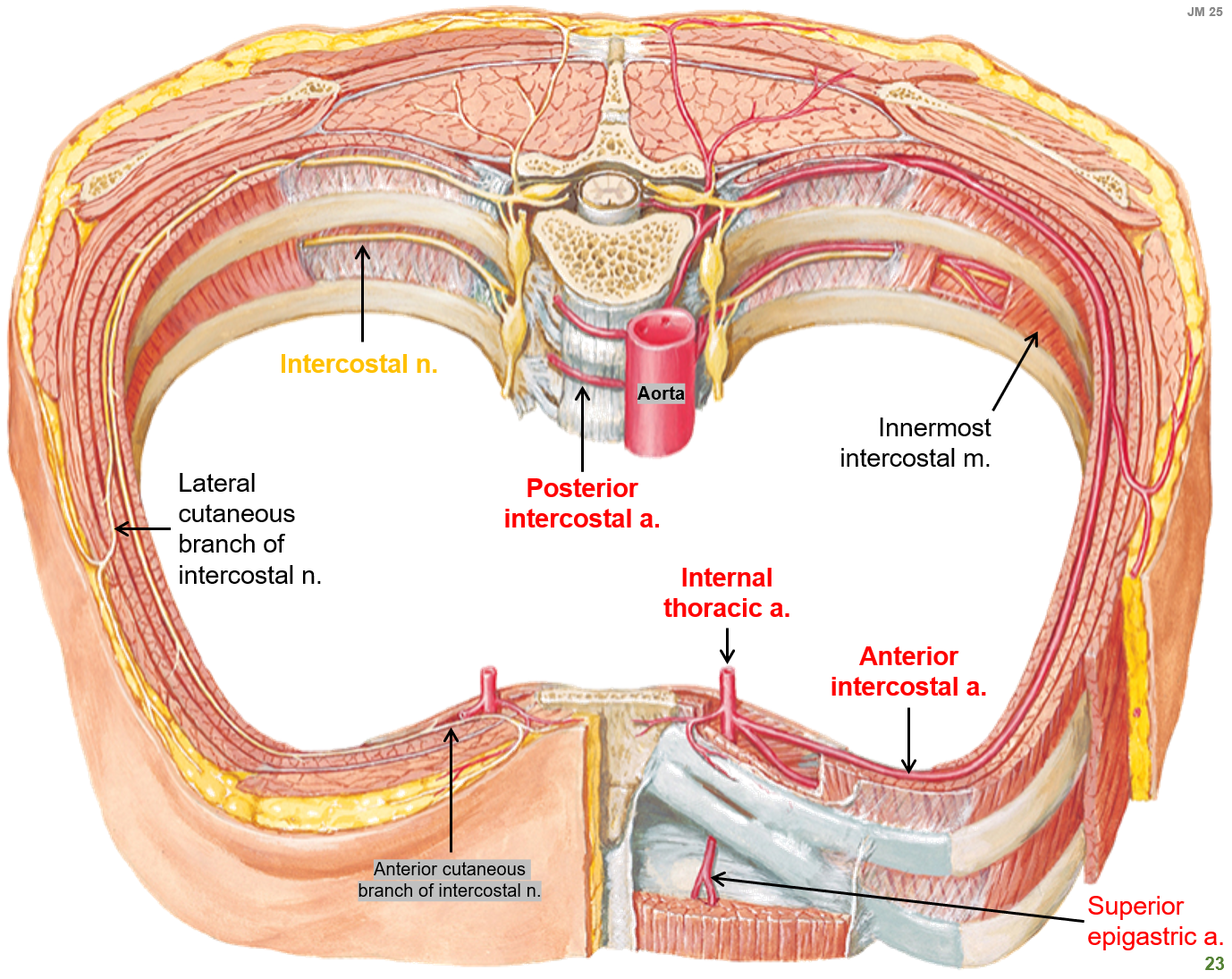
What is the V.A.N. bundle, and where is it located?
V.A.N. bundle consists of a Vein, Artery, and Nerve, which are arranged in the costal groove of each rib.
What is the transversus thoracis muscle?
A thin muscle on the inner chest wall that helps with forced expiration by pulling the ribs down.
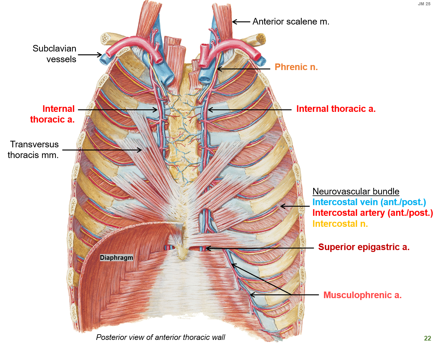
What is the internal thoracic artery and what does it supply?
A blood vessel that runs along the inner chest wall and supplies blood to the thoracic and abdominal walls.

What do the intercostal nerves innervate?
The intercostal muscles (full thickness—deep) at each level and the overlying skin at each level of the thoracic wall.
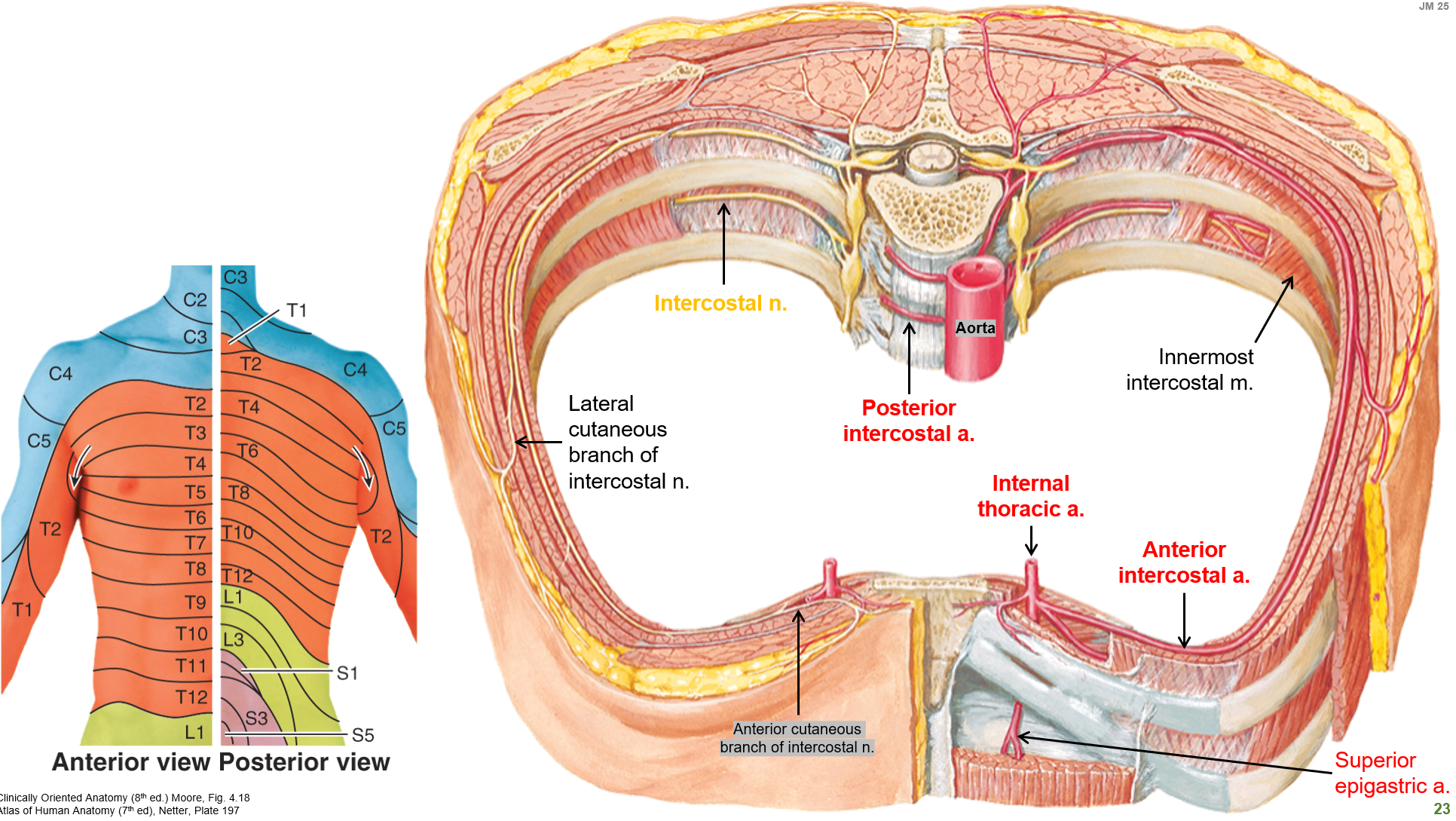
Where are the posterior and anterior intercostal arteries located?
Posterior intercostal arteries branch from the aorta and run along the back of the ribcage
Anterior intercostal arteries come from the internal thoracic artery and run along the front of the ribcage
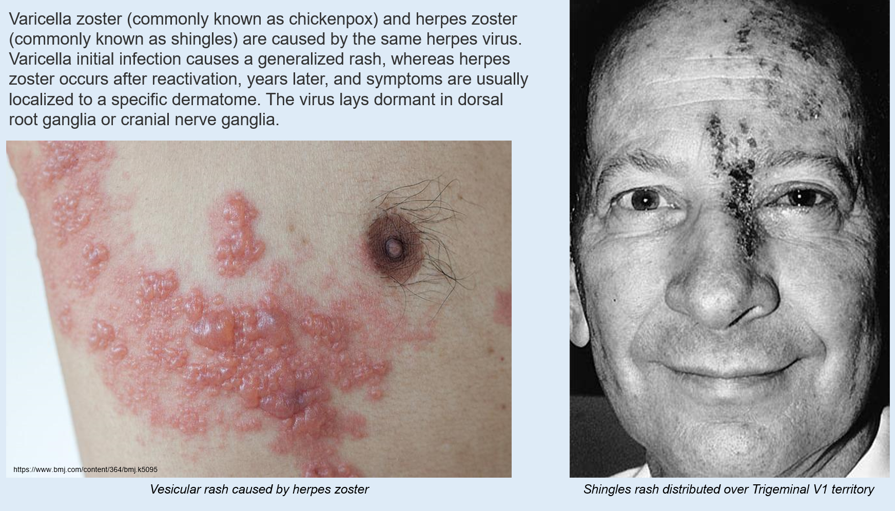
What is the difference between chickenpox and shingles caused by Varicella Zoster virus?
Chickenpox is the initial infection caused by the varicella zoster virus, leading to a generalized rash.
Shingles occurs when the virus reactivates years later, causing a rash in a specific dermatome.

Where does the varicella zoster virus remain dormant?
In the dorsal root ganglia or cranial nerve ganglia until reactivation.
What is a coronary artery bypass graft (C.A.B.G.)?
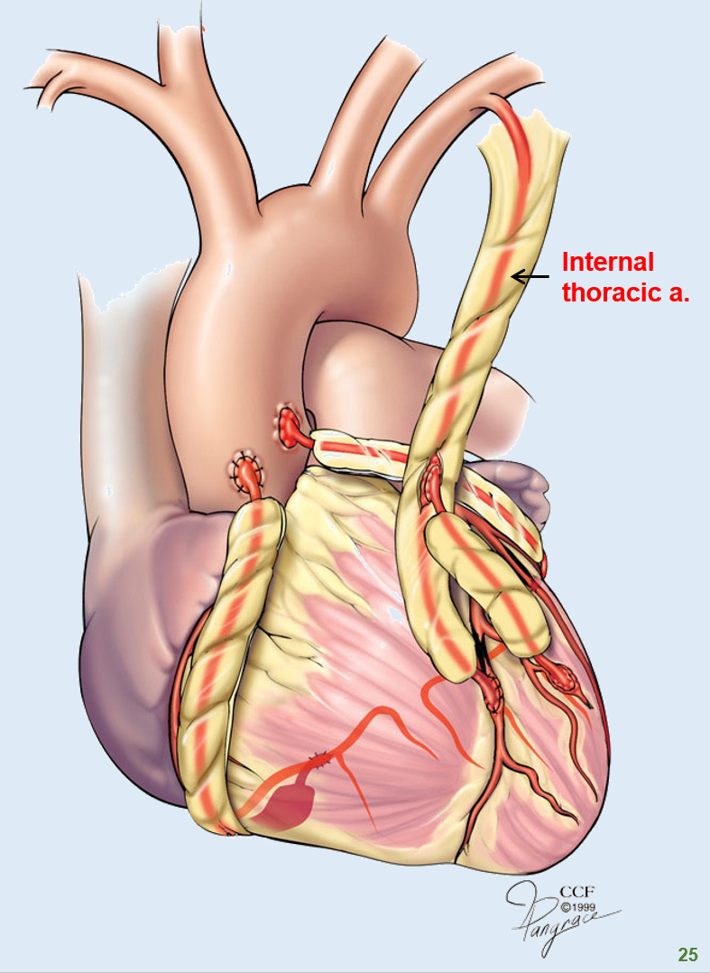
A surgical procedure that bypasses blocked coronary arteries using a graft (usually from the internal thoracic artery or a vein)
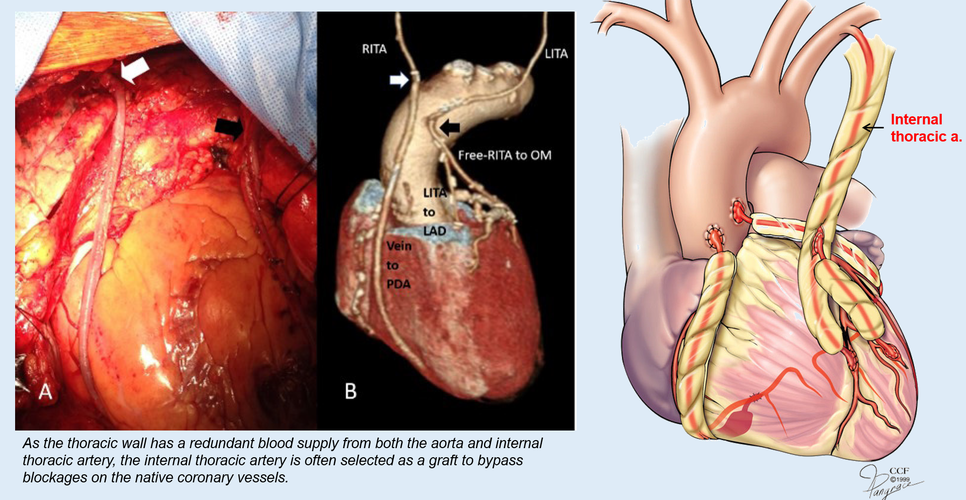
Why is the internal thoracic artery often used in coronary artery bypass grafting?
It has a good blood supply and remains open longer than vein grafts, making it a preferred choice for bypassing blocked coronary arteries.
What are the three main spaces of the thoracic cavity?
Mediastinum – The central region containing the heart, major vessels, and other structures.
Right pleural cavity – Surrounds the right lung.
Left pleural cavity – Surrounds the left lung.
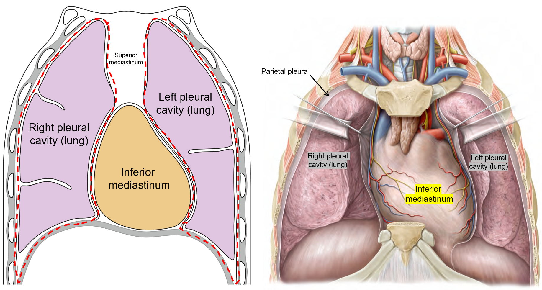
What is the function of the pleural cavities?
Protect and encase the lungs, allowing smooth movement during breathing with the help of the parietal pleura lining.

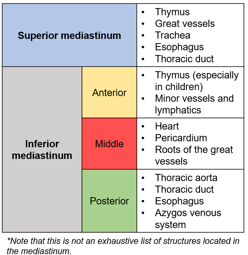
What is the mediastinum?
The space between the lungs, containing the heart, vessels, and other structures. It is divided into:
Superior mediastinum (above the sternal angle).
Inferior mediastinum (below the sternal angle at rib 2 and T4/T5 level).
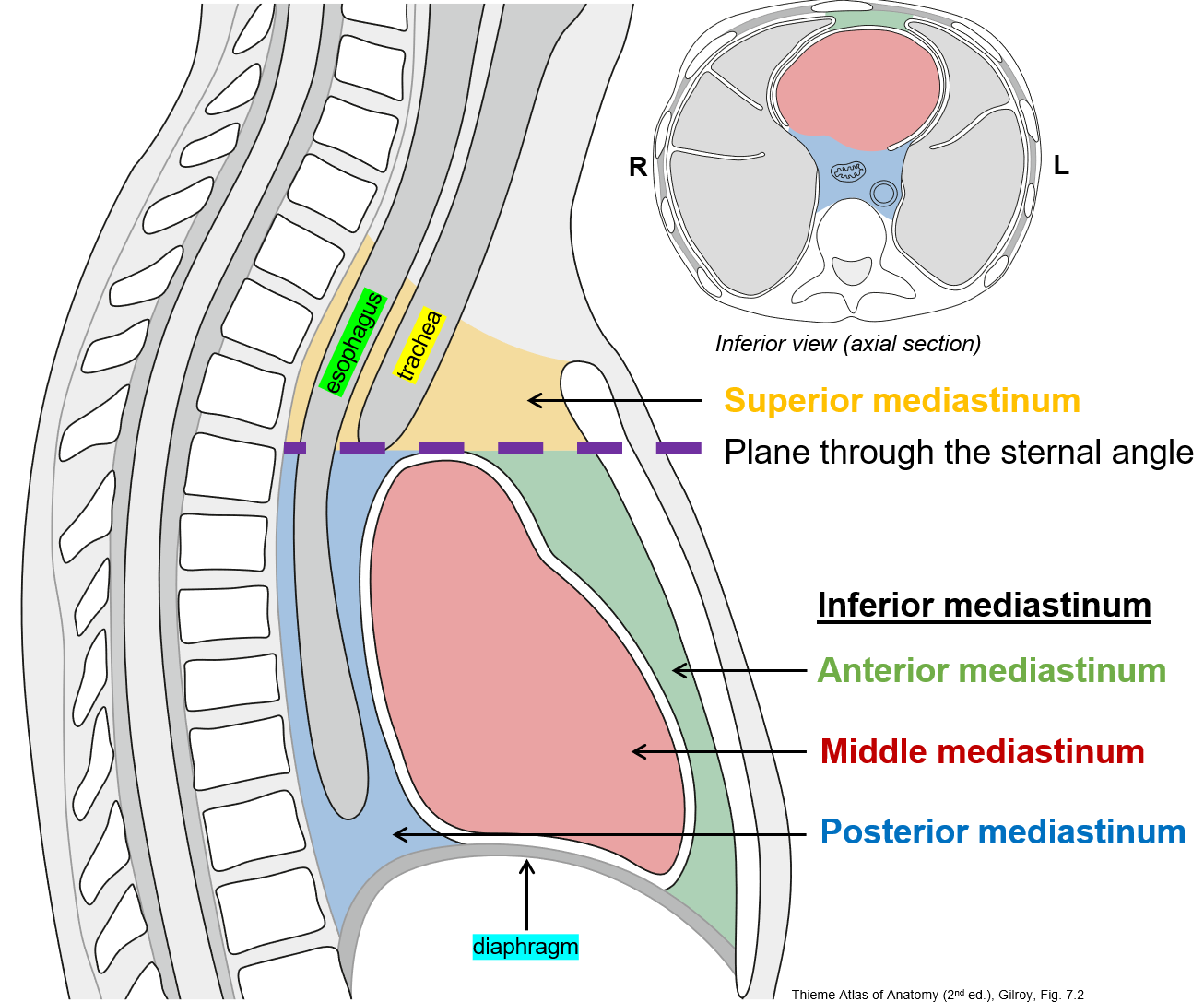
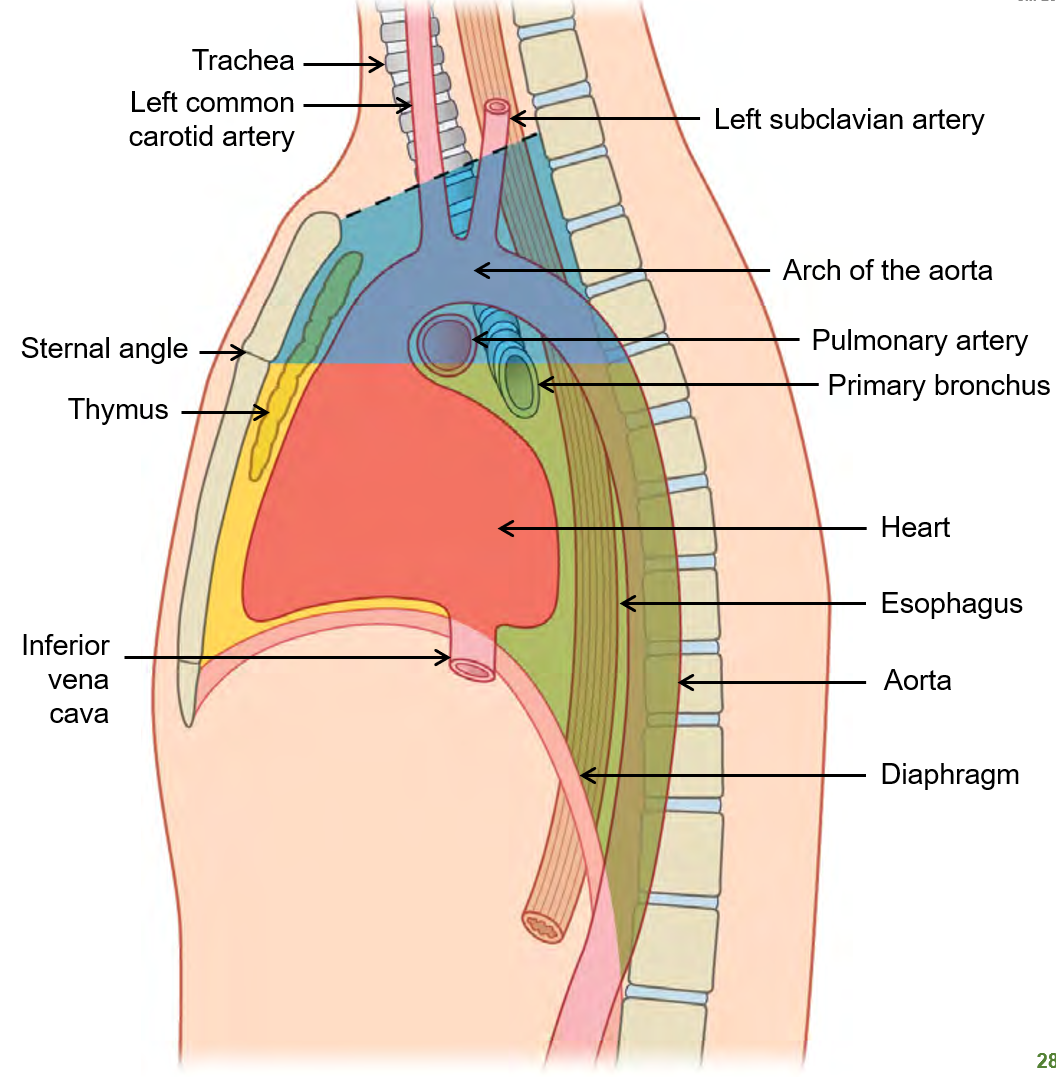
What are the main structures found in the superior and inferior mediastinum?
Superior Mediastinum:
Thymus
Great vessels
Trachea
Esophagus
Thoracic duct
Inferior Mediastinum:
Anterior: Thymus (in children), minor vessels, and lymphatics
Middle: Heart, pericardium, and roots of great vessels
Posterior: Thoracic aorta, esophagus, thoracic duct, and azygos venous system