PHS3341 Midterm
1/123
There's no tags or description
Looks like no tags are added yet.
Name | Mastery | Learn | Test | Matching | Spaced | Call with Kai |
|---|
No analytics yet
Send a link to your students to track their progress
124 Terms
Homeostasis
Process by which organisms maintain a relatively stable internal environment (Walter Cannon, 1930). Required to sustain Cell Function. Variables maintained within a predictable range.
internal milieu
Internal environment (ECF i.e. Extracellular Fluid).
Regulated Variables
Sensor that detects changes and makes adjustments to keep physiological system within limited range (Homeostatic regulation). Initiating effector responses that restore variables to their optimal range (narrow or wide). Ex. Baroreceptor or Thermoreceptor.
Controlled Variables
Remain within a normal range over time but are not homeostatically regulated. Ex. Blood hematocrit, Testosterone, Heart rate.
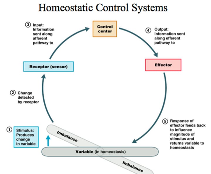
Homeostatic Control Systems (5 Parts)
1. Set point: Normal range for regulated variable.
2. Sensor: Detects value of regulated variable and transduce stimulus into a physiological signal (Input).
3. Error detector (Control): Compares actual value to set point.
4. Integrator (Control): Interprets error signal and determines the output of effectors (Output).
5. Effectors: Change value of regulated variable.
(Ex. Regulation of Core Temperature)
Homeostatic Imbalance Flaws
Flaws of System:
1. Depend on external resources
2. Sensors respond within a limited range stimulus values.
Caused by certain diseases (ex. diabetes). Possible to keep one property relatively constant by moving others away from their usual setpoint. Variables are connected (may cause changes in other variables as well).
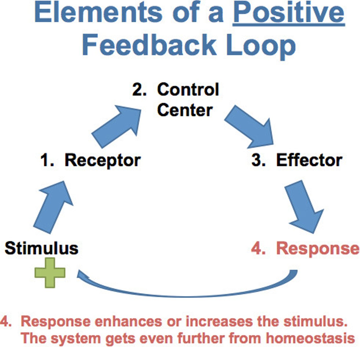
Positive Feedback
Pushes variable away from set point.
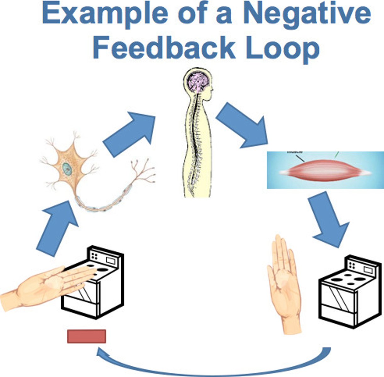
Negative Feedback
Returns variable to set point.
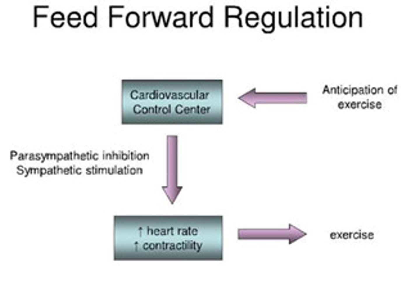
Feed-forward control
Anticipatory, minimizes changes to variable, activated before the change occurs.
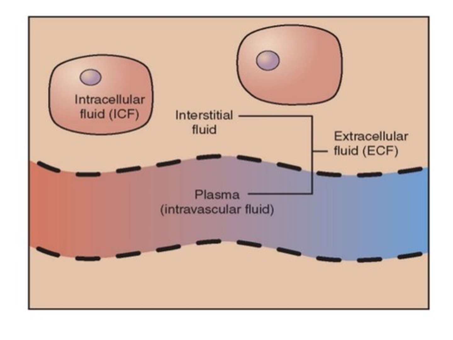
ECF (extracellular fluid)
Fluid in the blood (plasma) and between cells (interstitial fluid, ISF). ECF = 20-25% plasma, 75-80% ISF. Homogeneous in composition.

ICF (intracellular fluid)
Fluid within cells. Fluids enclosed in compartments, defined and maintained by barriers.
Passive transport
No energy input is required [high] to [low]
A. Simple Diffusion
B. Facilitated diffusion
C. Osmosis
Active Transport
Energy (ATP) is required to move substances against their concentration gradient (uphill).
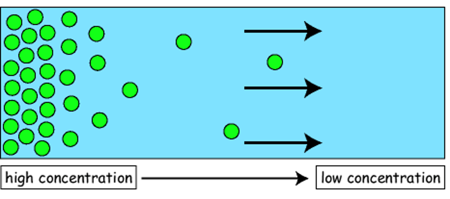
Diffusion
Movement of molecules from an area of higher concentration to an area of lower concentration due to intrinsic kinetic energy.
Diffusion Rate Factors (5 Factors)
1. Molecular Size: Smaller molecules diffuse faster.
2. Temperature: Higher temps increase kinetic energy which results in faster diffusion.
3. Concentration: The greater the difference of concentration between two areas, the faster diffusion occurs.
4. Surface area between compartments.
5. Medium: Less viscosity, faster the diffusion.
(Equilibrium is reached when there is no net movement of molecules in either direction).
Flux
The amount of substance diffusing across a surface in a unit of time. Net Flux determines the gradient direction.
Simple Diffusion Through Membranes
Nonpolar, lipid-soluble(hydrophobic) substances diffuse directly through phospholipid bilayer. Small amounts of very small polar substances, such as water, can even pass.
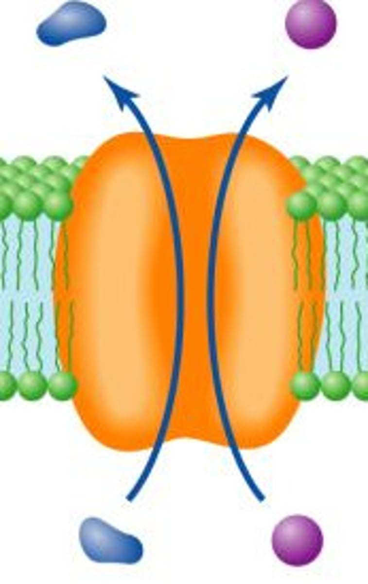
Facilitated Diffusion
Larger, non-lipid soluble, or polar molecules can cross membrane but only with assistance of carrier molecules via integral proteins in the membrane.
1. Carrier-mediated
2. Channel-mediated
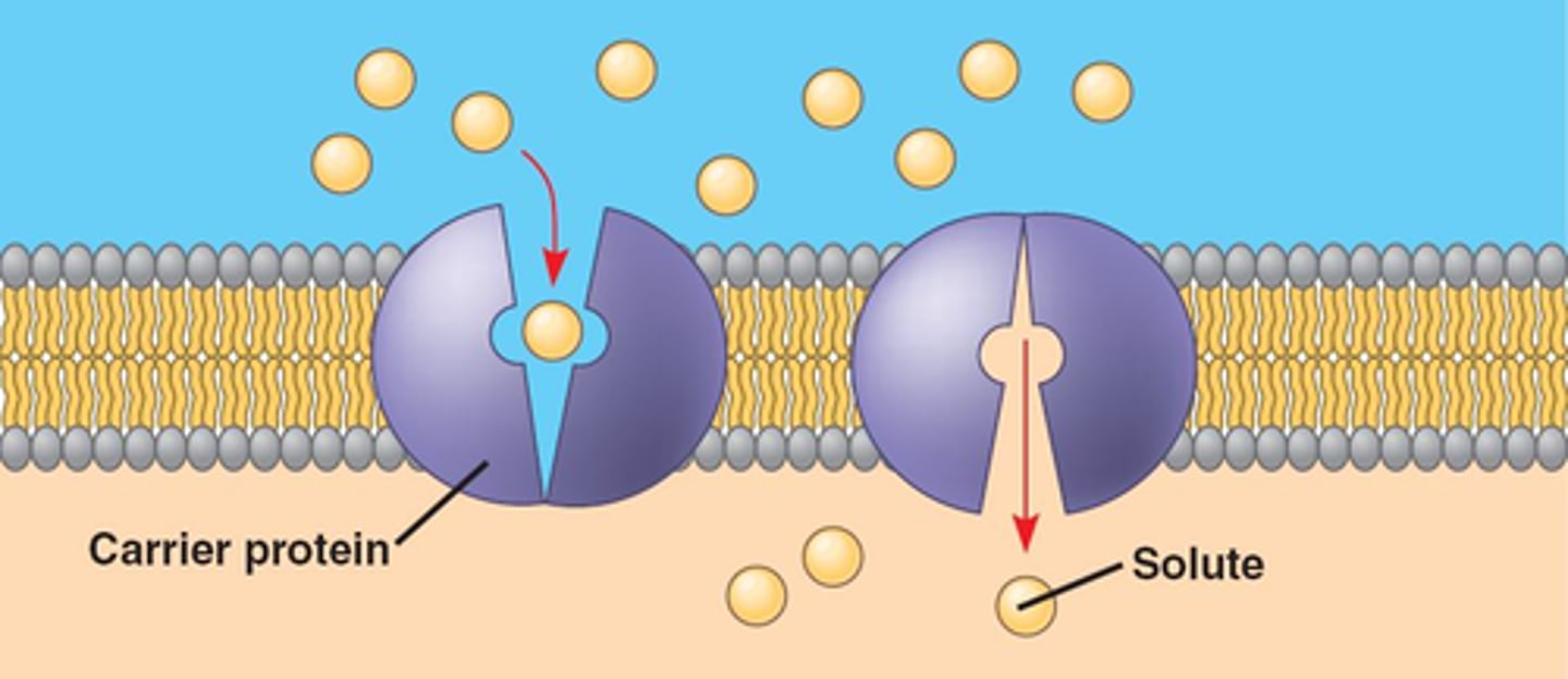
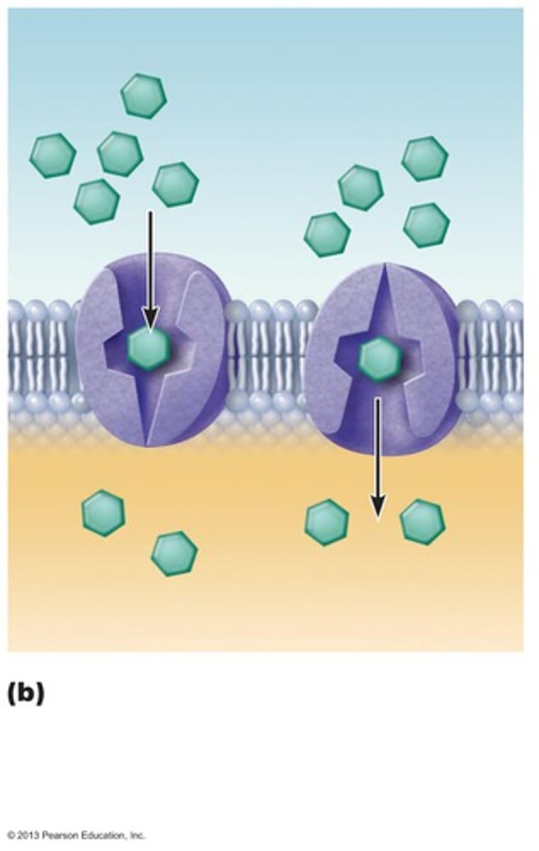
Carrier-mediated Facilitated Diffusion
Certain hydrophilic molecules transported passively down their concentration gradient by carriers (transmembrane proteins). Binding of molecule causes carrier to change shape and results in molecule being moved across membrane.
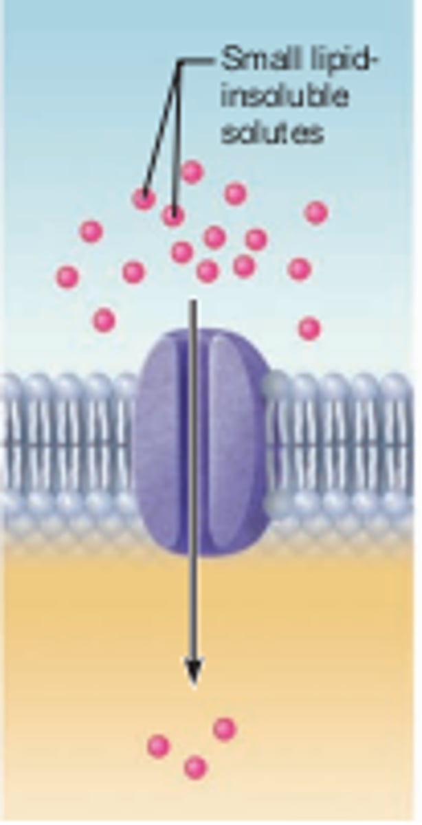
Channel-mediated Facilitated Diffusion
Ion channels show selectivity for a particular type(s) of ion based on channel diameter, charged residues lining pore, water of hydration.
Regulation of Diffusion Through Ion Channels (Gating)
Gating: Opening and Closing of the Ion Channel
Gated channels:
1. Ligand-gated: Based on if ligand is attached.
2. Voltage-gated: Based on Membrane potential.
3. Mechanically-gated: Based on Mechanical Pressure.
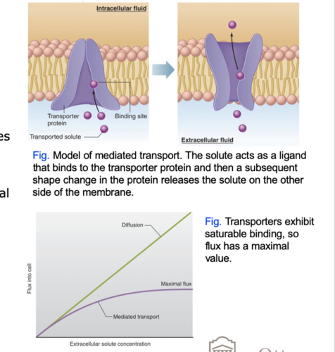
Carrier-mediated transport
Transmembrane proteins called transporters move solutes via conformational changes.
Types:
1. Facilitated diffusion
2. Active transport
Flux through Carrier-mediated transport (3 Factors)
1. Binding Site Saturation
2. Number of Transporters in Membrane
3. Rate of Conformational Change
Primary Active Transport
Active transport that relies directly on the hydrolysis of ATP. Transporters are called ATPases.
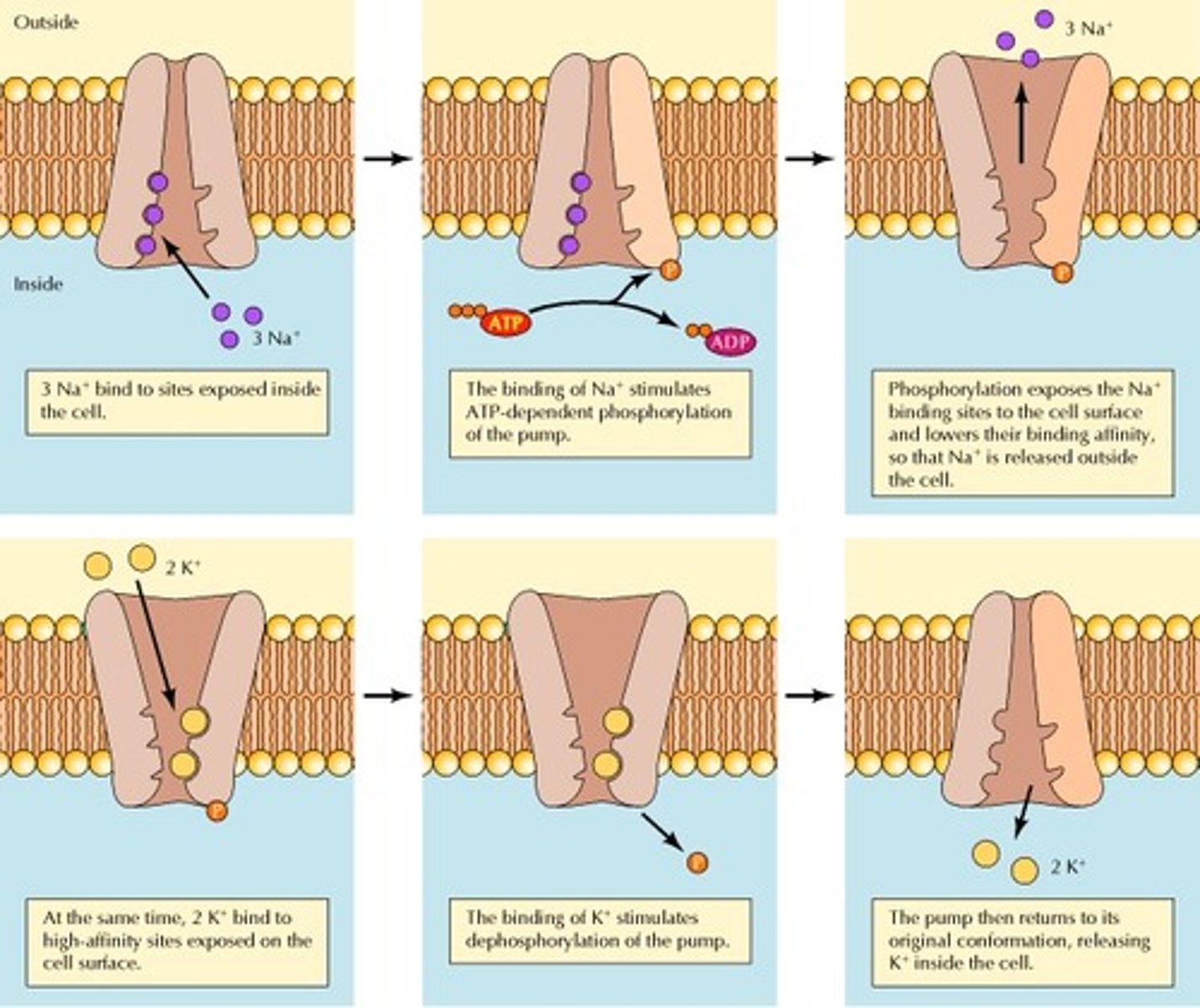
Na+/K+-ATPase pump (5 Steps)
Primary active-transport protein that hydrolyzes ATP and releases energy used to transport 3 sodium ions out of cell and 2 potassium ions in. Results in electrochemical gradient: high K+ and low Na+ inside (vice versa).
1. The transporter (with bound ATP) binds 3 Na+ on inside of cell (low affinity for K+ ).
2. ATPase activated. Auto-phosphorylation.
3. Conformational change and release of Na+ to outside.
4. Increased affinity for K+ allows 2 K+ to bind.
5. Dephosphorylation and return to original conformation. Release of K+ to inside.
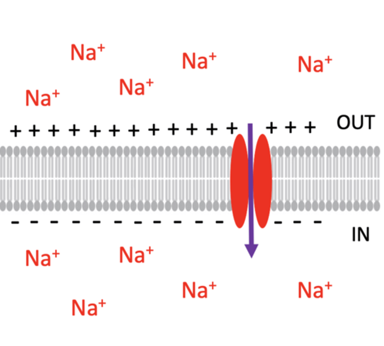
Electrical Forces in Membrane
Membrane potential (separation of charges across membrane). Electrochemical gradient caused by membrane potential and ion concentration. The inside of the cell has a net negative charge.
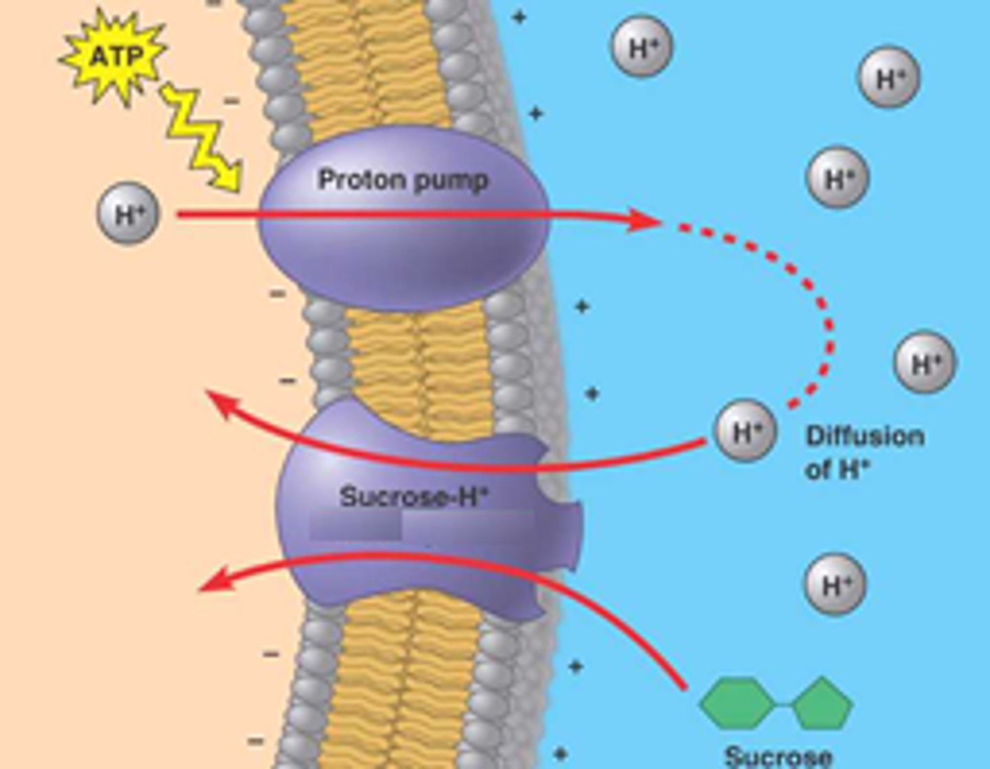
Secondary Active Transport
Form of active transport which does not use ATP as an energy source; rather, transport is coupled to ion diffusion down a concentration gradient established by primary active transport. These transporters have binding sites for an ion (usually Na+) and the co-transported molecule.
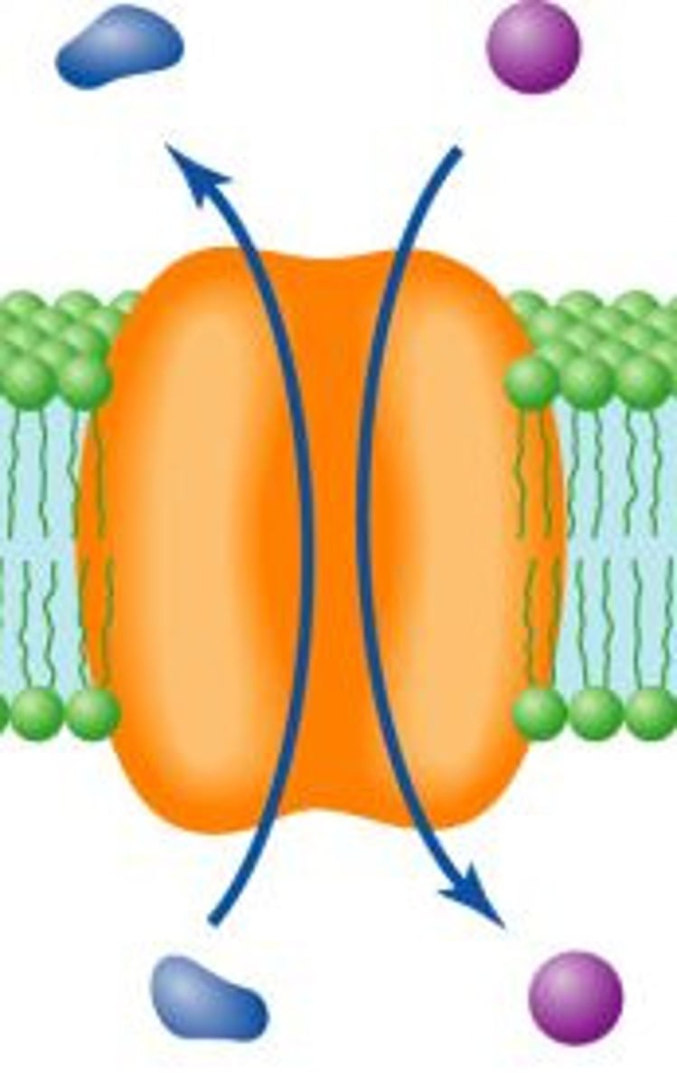
Antiport
Two substances move in opposite directions (Na+ down concentration gradient).
Symport
Two molecules travel in the same direction (Na+ down concentration gradient).
Osmosis
Diffusion of water/solvent through a selectively permeable membrane from [high] to [low]. Mostly through aquaporins(AQPs) and some through lipid bilayer.
Osmolarity
Total number of solute particles in a solution. Water moves by osmosis from areas of low solute (high water) concentration to high areas of solute (low water) concentration.
Ex. 1 M NaCl ionizes to Na+ and Cl- (2particles) is 2 Osm (osmolar).
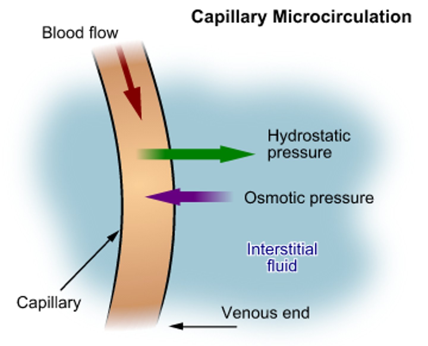
Hydrostatic pressure
Outward pressure exerted on cell side of membrane caused by increases in volume of cell due to osmosis.
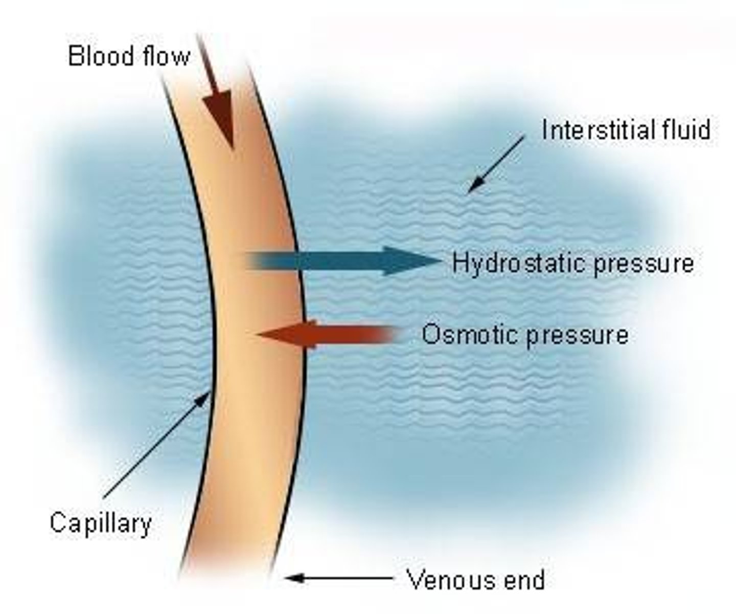
Osmotic pressure
Inward pressure exerted on cell side of membrane caused by osmosis (The more solutes inside a cell, the bigger the pull on water to enter).
Osmotic vs. Tonic
Hyper-osmotic, iso-osmotic and hypo-osmotic refers to the concentration of ALL solutes.
Hypertonic, isotonic and hypotonic refer to the concentration of non-penetrating solutes.
note:
- Na+, Cl-, K+ = non-penetrating solutes.
- Hyper = more on outside
- Iso = same on outside
- Hypo = less on outside
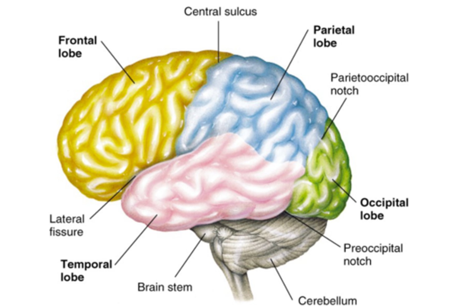
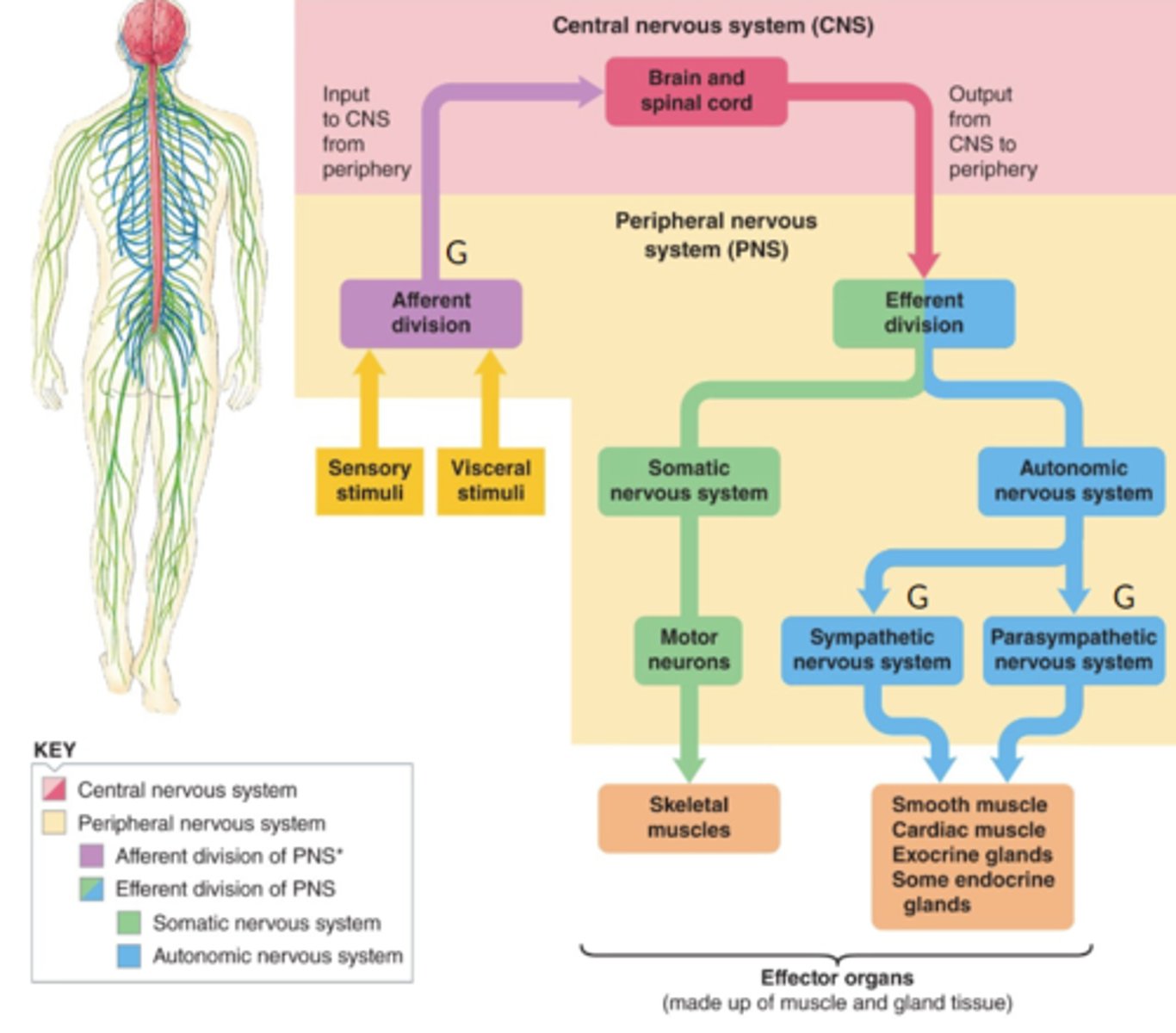
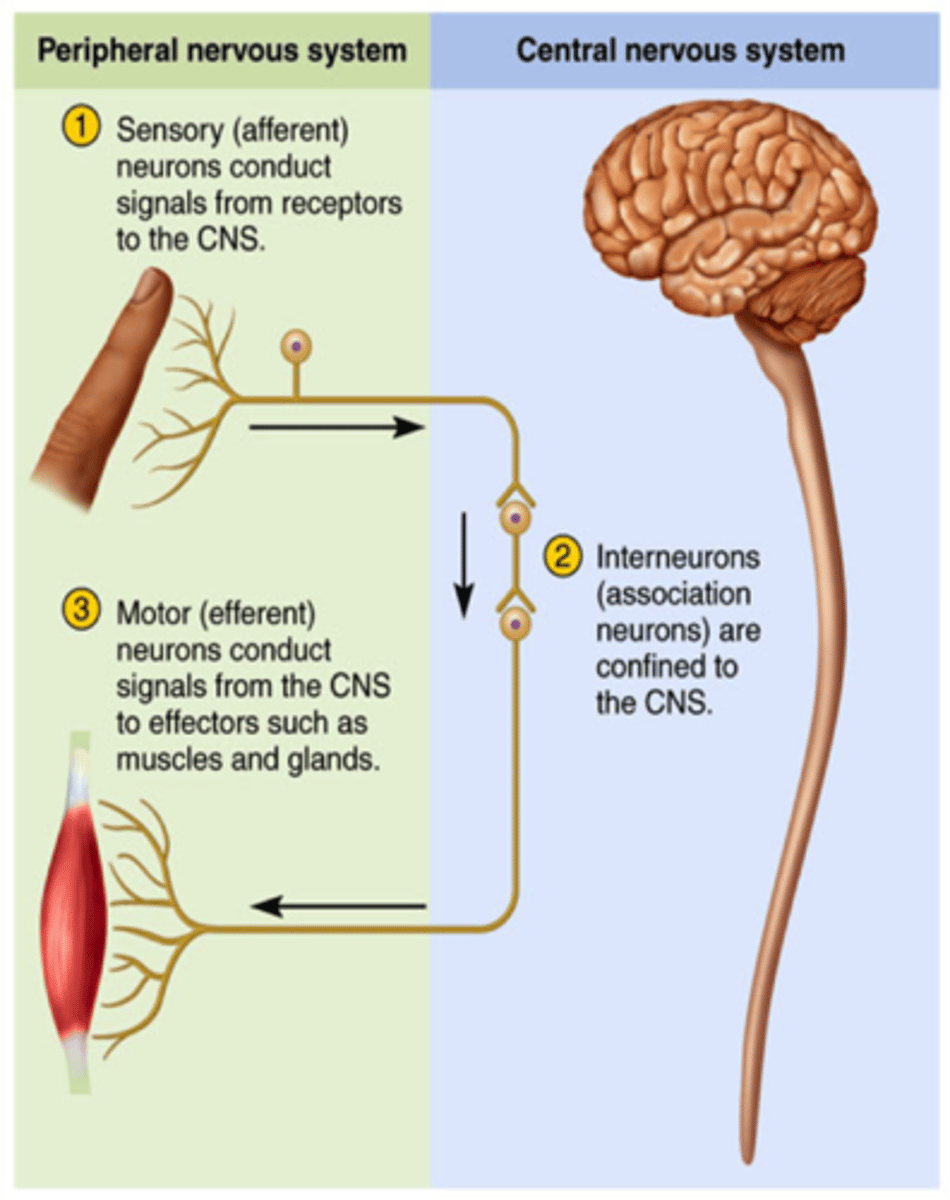
Major Divisions of the Nervous System
Central Nervous System (CNS)
- Parts: Brain + Spinal Cord
Peripheral Nervous System (PNS)
- Parts: Spinal and Cranial nerves + Ganglia
- Somatic
- Autonomic (Sympathetic and Parasympathetic)
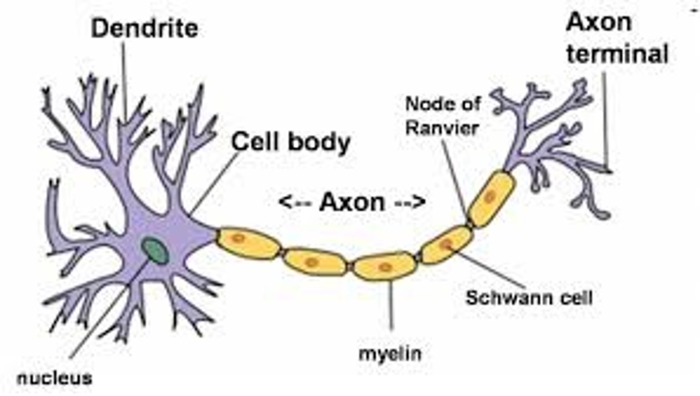
The Neuron
Basic functional unit of the nervous system.
- Dendrites: receive information via neurotransmitters from synapse.
- Axons: conduct action potentials, send signals towards axon terminals.
- Initial segment (axon hillock): AP trigger zone (between cell body and axon).
- Axon Terminal (bouton): Make synaptic connections with another nerve cell.
Glial Cells (Microglia and Macroglia)
Supporting cells of CNS and PNS (~90% of cells in CNS)
- Micro ganglia
- Astrocytes
- Oligodendrocytes (CNS)/Schwan Cells (PNS)
- Ependymal Cells
- Radial Glia
- Satellite Cells (PNS)
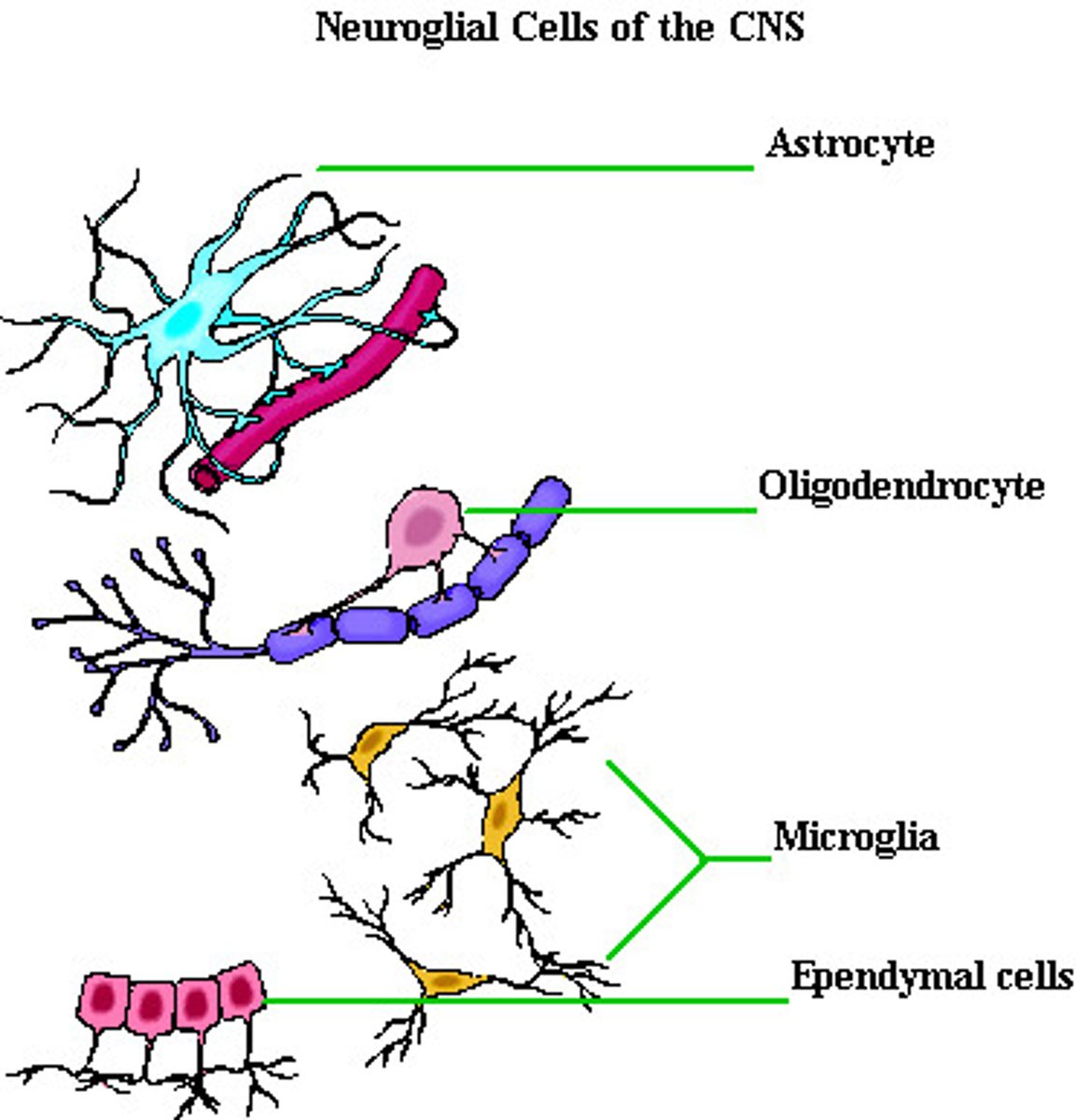
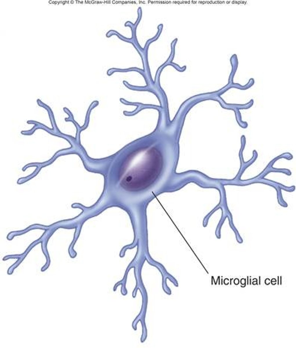
Microglia
Primary Immune defense of CNS. Similar to macrophages, phagocytize neuronal debris and neuromodulate functions.
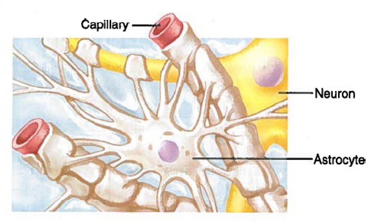
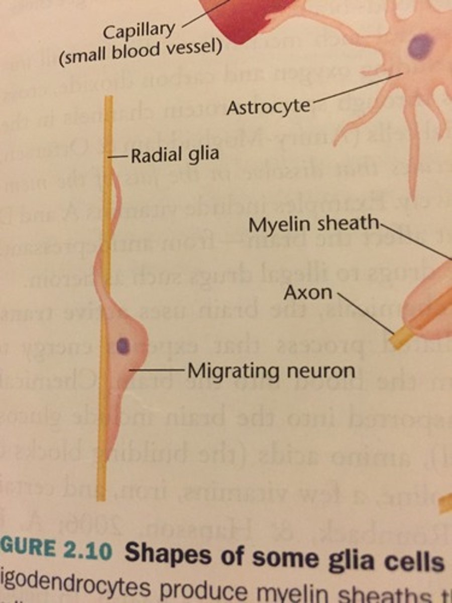
Astrocytes (CNS)
- Support, Repair, Communication, BB Barrier.
- Spatial buffering of extracellular K+.
- Neurotransmitter uptake and release.
- Guide neuronal migration in developing brain.
- Can respond to nerve impulses and neurotransmitters.
- Mediate neurovascular coupling.
- Modulation of synaptic activity.
- Promote Myelination of OD.
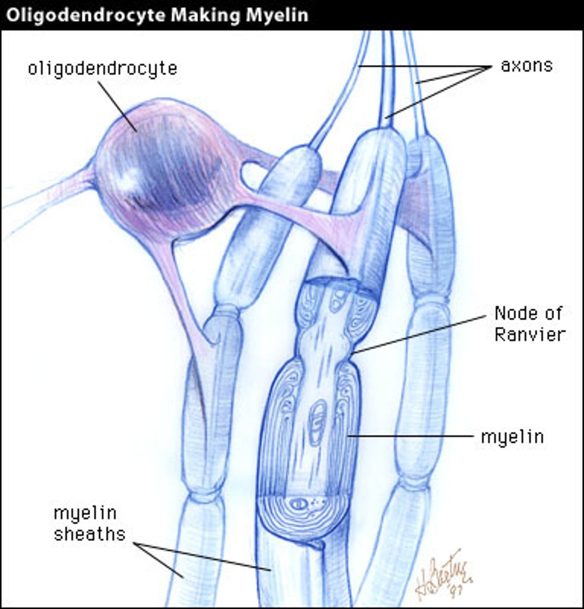
Oligodendrocytes (CNS)/Schwan Cells (PNS)
- Myelination (Insulation), speeds up conduction.
- Nodes of Ranvier: Spaces between adjacent sections of myelin = saltatory conduction.
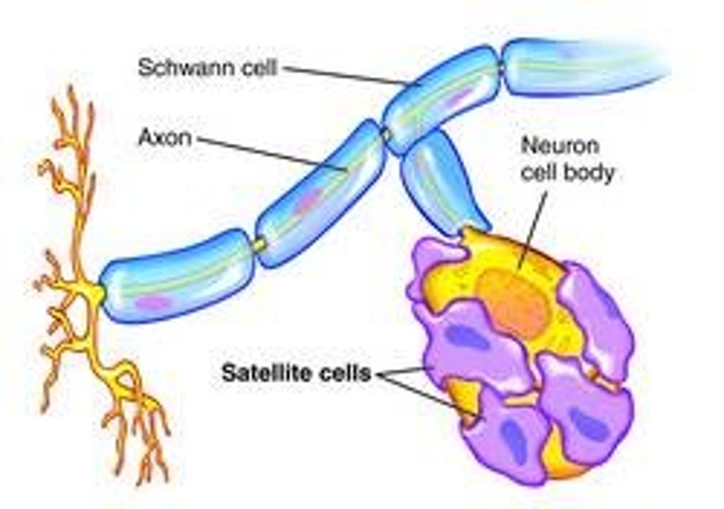
Schwan Cells: Only myelinate 1 axon, Pain and temperature fibers are unmyelinated (Still can be triggered).
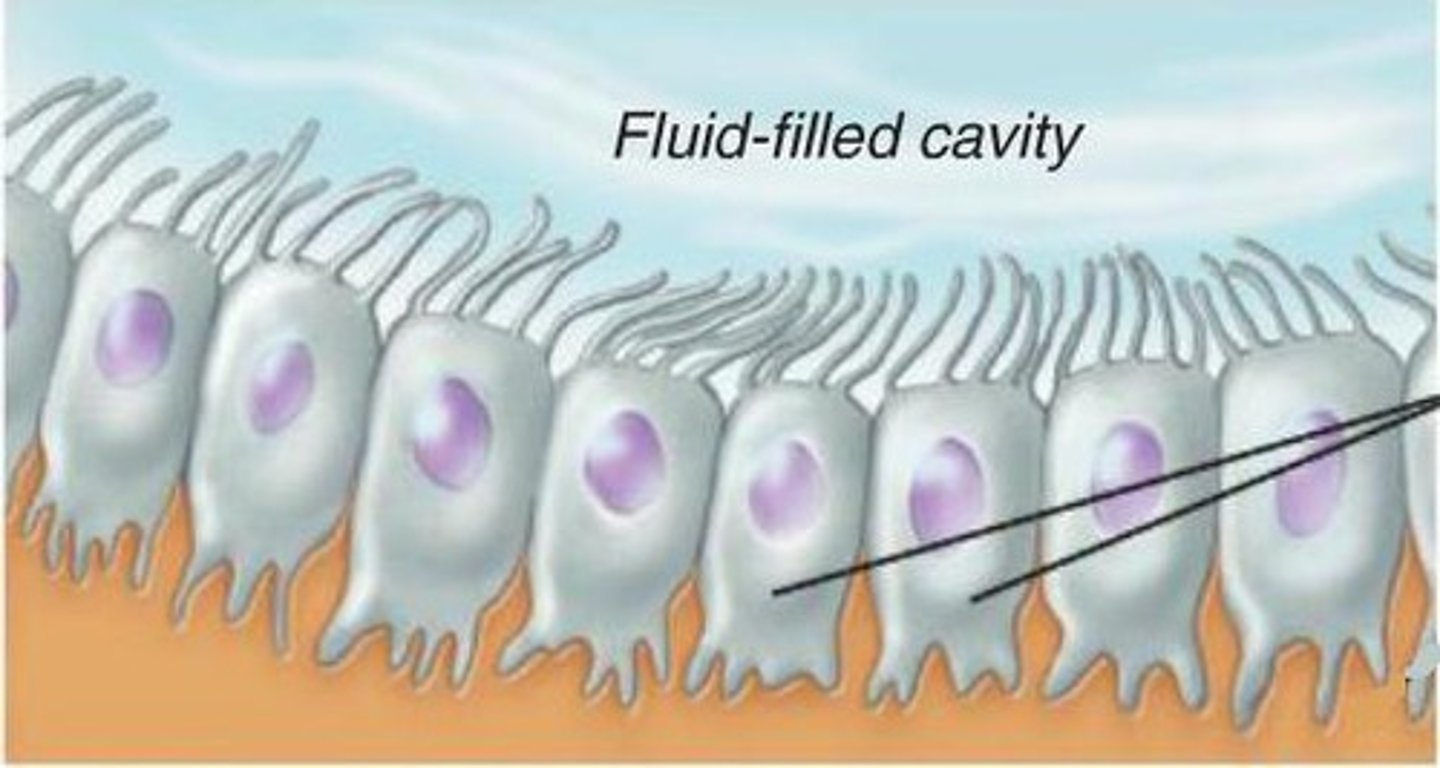
Ependymal Cells (CNS)
Found in Walls of Ventricles producing cerebrospinal fluid (many function for protection).
Radial Glia
Guide the migration of neurons and neurogenesis.
Satellite Cells (PNS)
Regulate neural environment, similar to astrocytes but for PNS.
Axonal Transport (2 Speeds)
1. Fast
- Bidirectional (because of Kinesin)
- ATP Dependent
2. Slow
- Unidirectional
- ATP Independent
Axonal Transport (2 Directions)
1. Anterograde (toward axon terminal)
- Synaptic Protein
- Neurotransmitters
- mRNA
2. Retrograde (toward cell body)
- Growth Factors
Axonal Transport (Microtubules)
- Kinesin (toward +) (w/Secretary vesicle)
- Dynein (toward -) (w/Recycling vesicle)
More Regulation
- Modification of Microtubules
- Kinesin Regulation via autoinhibition (undone via motor protein or attachment of cargo)
(actin side directions can be connected via myosin)
Resting membrane potential
ECF = High Na+, (Cl- varies)
ICF = High K+
Resting Membrane Potential = -70mV (Since K+ leak channels more likely to be open). Na+/K+ pump maintain resting membrane potential.
Na+ electrical and [Na+] gradient toward inside of the cell. K+ electrical but not [K+] gradient toward inside of the cell.
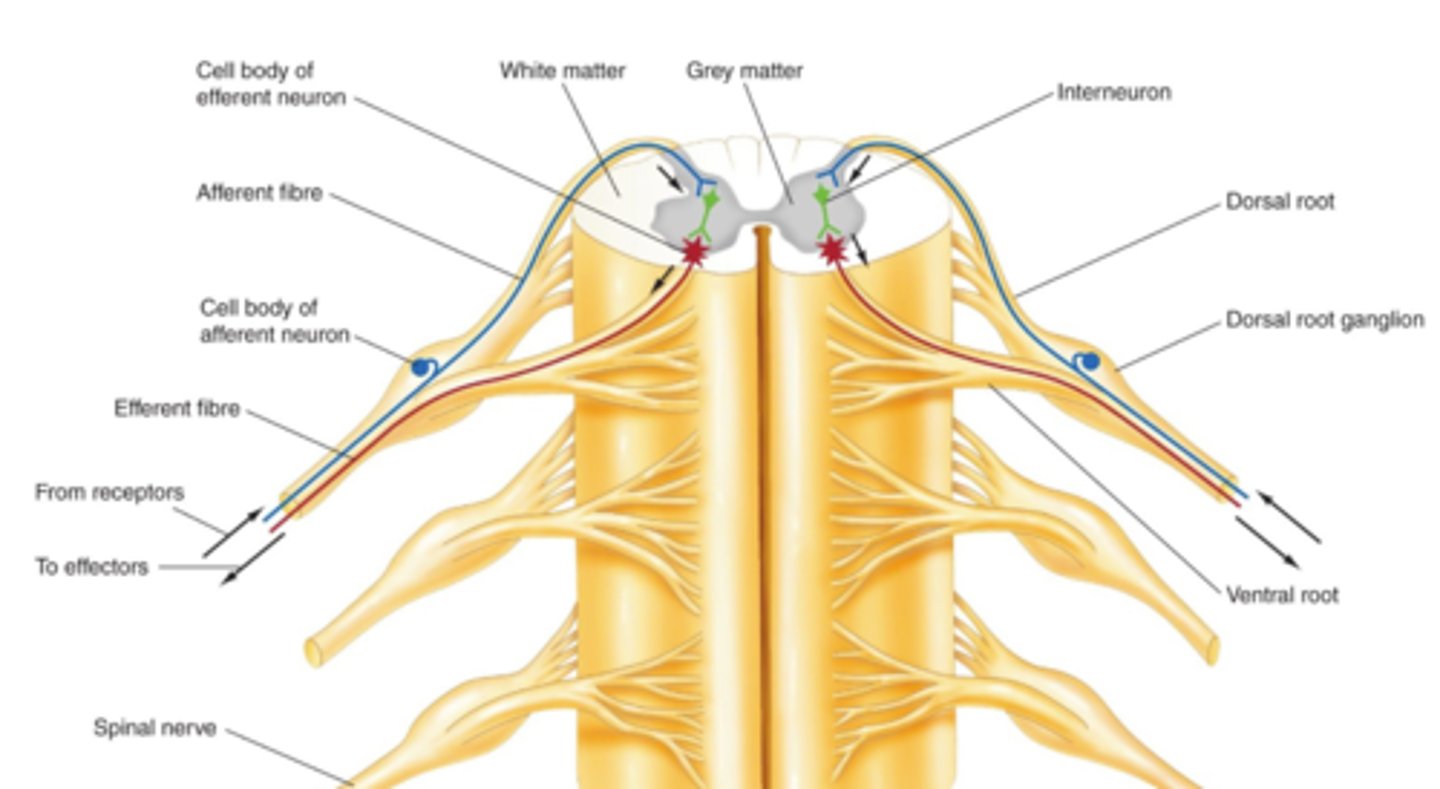
Classes of Neurons (3 Types)
STEPS:
(AFFERENT) Sensory receptor > Axon (PNS) > (After cell body) Axon (CNS) > (INTERNEURONS) > (EFFERENT) Axon (CNS) > Axon (PNS) > Axon Terminal.
Groups of afferent and efferent neurons form the nerves (bundles) of the PNS. Note that a nerve fiber is a single axon, and a nerve is a bundle of axons.
Synaptic Inputs
A neuron that conducts a signal towards a synapse is presynaptic. One that conducts signal away from the synapse is postsynaptic. A single Neuron can do both.
Connections:
- Axo-dendritic
- Axo-somatic
- Axo-axonic: No AP Trigger, affects neurotransmitter release i.e. presynaptic facilitation or inhibition.
Ohm's Law
Voltage (V) = Current (I) x Resistance (R)
- Insulator: substance with high electrical resistance
- Conductor: substance with low electrical resistance
Equilibrium Potential
The membrane potential at which chemical and electrical forces are balanced for a single ion (K+ = +60 mV or Na+ = -90 mV).
Nernst Equation: Eion = 61/Z log (Co/Ci)
- Z = valence
- Co = [outside]
- Ci = [inside]
![<p>The membrane potential at which chemical and electrical forces are balanced for a single ion (K+ = +60 mV or Na+ = -90 mV).</p><p>Nernst Equation: Eion = 61/Z log (Co/Ci)</p><p>- Z = valence</p><p>- Co = [outside]</p><p>- Ci = [inside]</p>](https://knowt-user-attachments.s3.amazonaws.com/f0624c79-f1ca-4b4c-bf5c-68a2198e0922.image/jpeg)
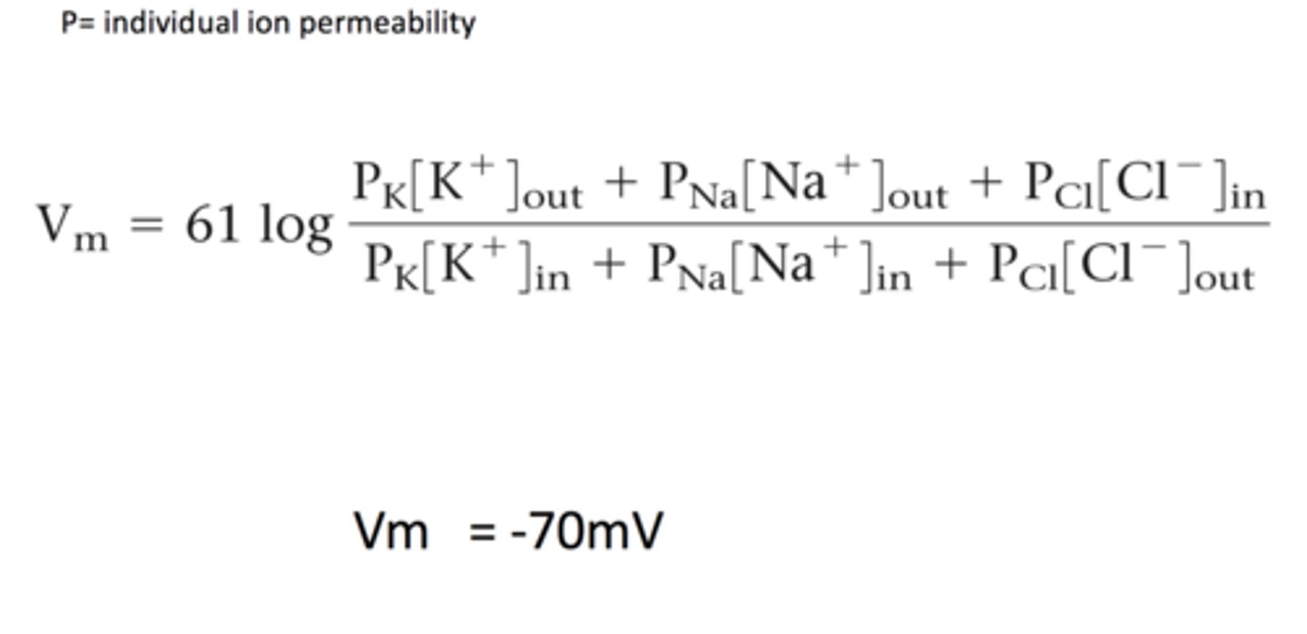
The Goldman-Hodgkin-Katz Equation
The membrane potential that results from the contribution of all ions that can cross the membrane. We need to consider the permeability (P).
Changes in Membrane Potential
Polarization: Change from RMP
- Depolarization: Decreased membrane potential (more +ve).
- Hyperpolarization: Increased membrane potential (more -ve).
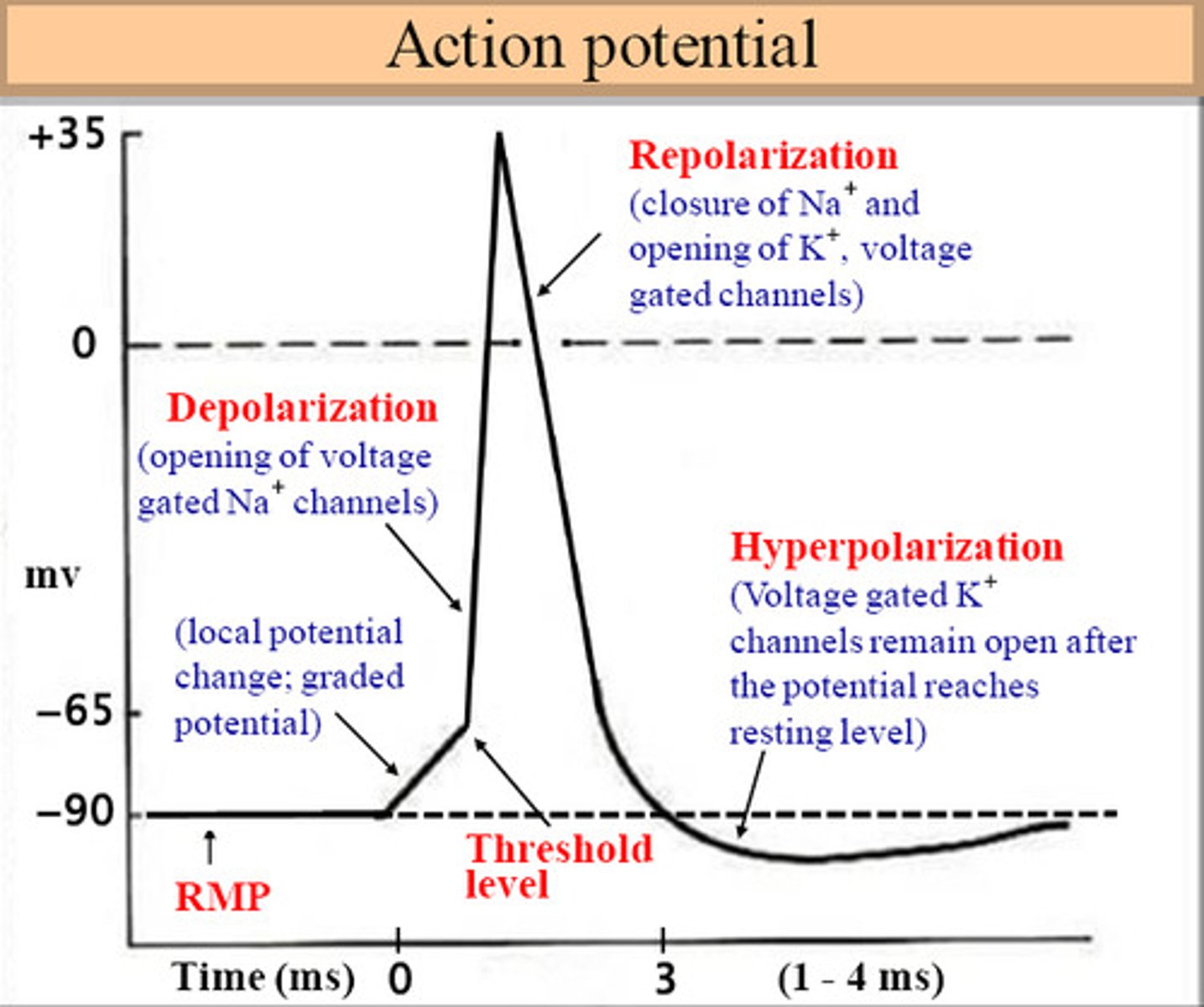
Action Potential
Long-distance neural communication (muscle cells and axons of neurons). Do not decay with distance.
Depolarizing stimulus trigger MP to -55mV (threshold) at axon hillock, further depolarization due to Na+ flowing inside cell through voltage gated channel (+ Feedback), until reaches +mV.
Absolute Refractory: K+ flows outside cell through voltage gated channels that took longer to open and Na+ channels become inactive (Repolarization) (+ Feedback)
Relative Refractory: Results of K+ channel causes MP of -80mV (below -70mV = Hyperpolarization) where Na+ channels become closed, then regulated back to RMP (-70mV) once K+ channels close.
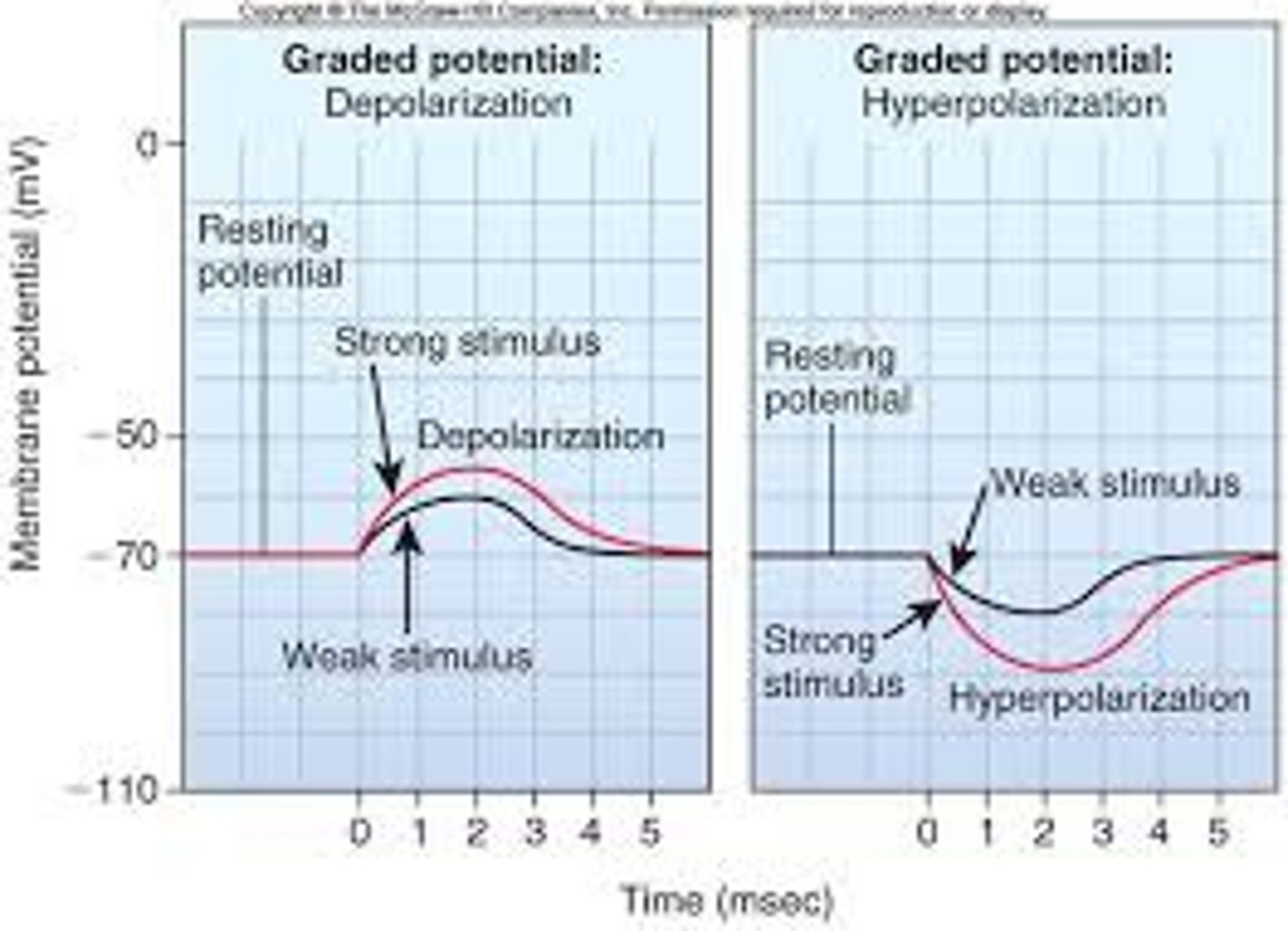
Graded Potential
A shift in the electrical charge in a tiny area of a neuron. Spreads but current decays with distance, time.
Examples:
- Receptor potential: receptors of sensory neurons.
- Postsynaptic potential: dendrite/soma of neuron or muscle cell.
Nervous System Encoding
Frequency of AP result in encoding. All or None Potential. The value of threshold can vary according to numerous factors (available channels and ion relative frequencies) (not always -55mV).
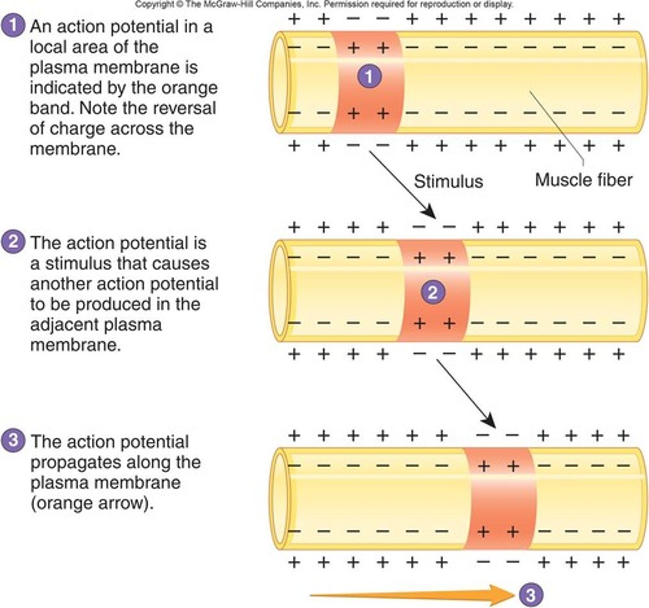
Action Potential Propagation
Propagation of AP from the initial segment to the axon terminal is typically one-way because the absolute refractory period follows along in the "wake" of the moving AP. The speed of propagation depends on fiber diameter (larger) and whether the fiber is myelinated.
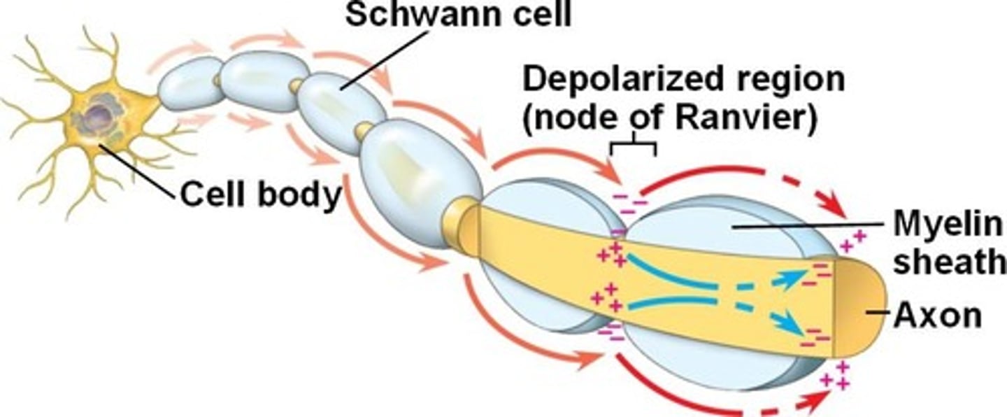
Saltatory Conduction
Rapid transmission of a nerve impulse along an axon, resulting from the action potential jumping from one node of Ranvier to another, skipping the myelin-sheathed regions of membrane.
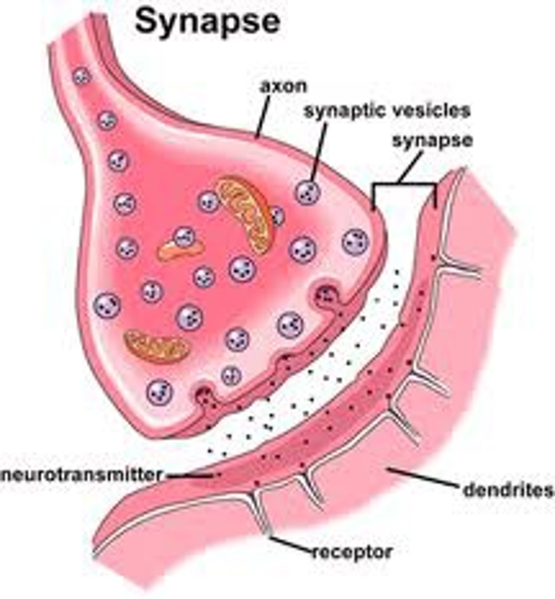
Synapse (Chemical vs. Electrical)
Synapse: Point of communication between two neurons (or neuron and muscle cell). All synapses are "tripartite", but more complex arrangements are frequently observed
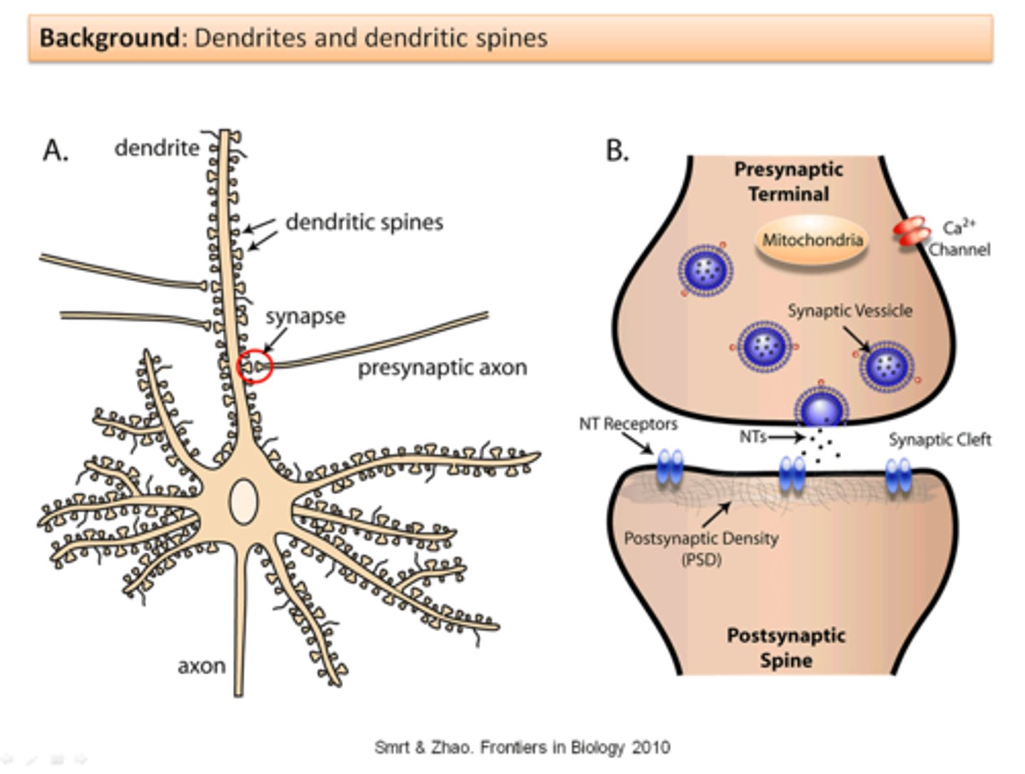
- Chemical synapse: neurotransmitters relay information from pre- to postsynaptic cell across synaptic cleft. Postsynaptic density area of postsynaptic neuron is where the neurotransmitter goes.
- Electrical synapse: pre- and post-synaptic cells joined by gap junctions. Allow local current to flow from arriving APs from one neuron to the next.
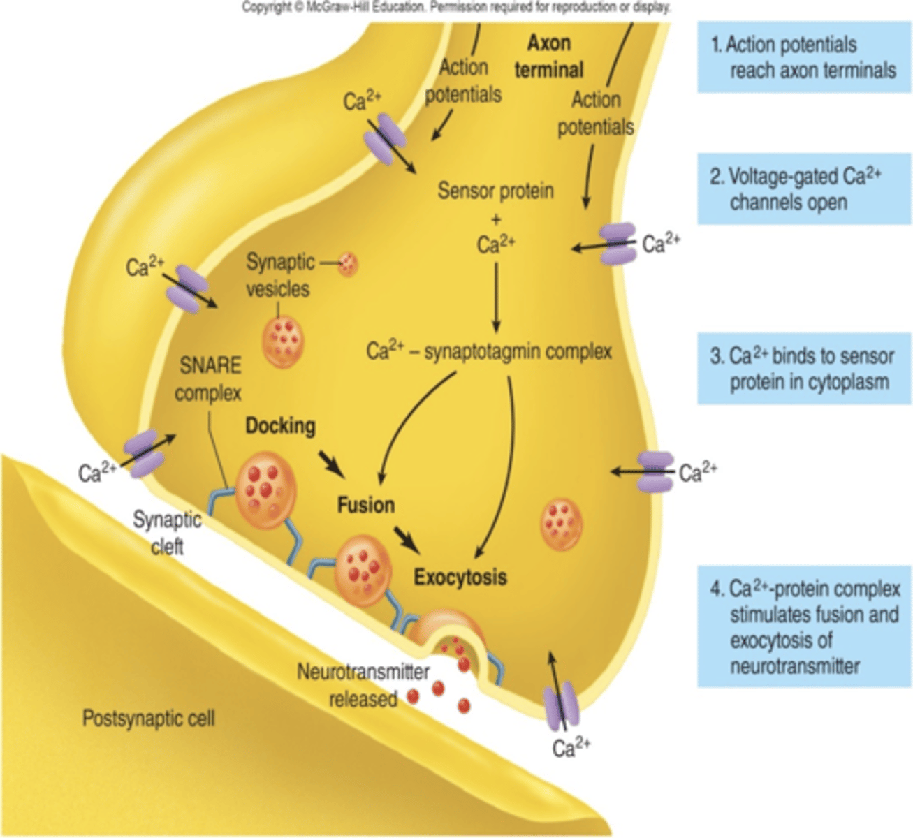
Mechanism of Neurotransmitter Release
1. Action potential, moving along axon's voltage-gated Na+ and K + channels, arrives at terminal.
2. Action potential opens voltage-gated Ca2+ channels (through depolarization).
3. Ca2+ influx triggers neurotransmitter release into the synaptic cleft (from the active zones) when AP reaches nerve terminal (bouton) via exocytosis mediated by SNARE pin.
4. The binding of neurotransmitters to receptor proteins in the postsynaptic membrane is linked to an alteration in its ion permeability. Example:
- Ionotropic receptors: ligand-gated ion channels
- Metabotropic receptors: mediate slower actions through G-protein second messengers.
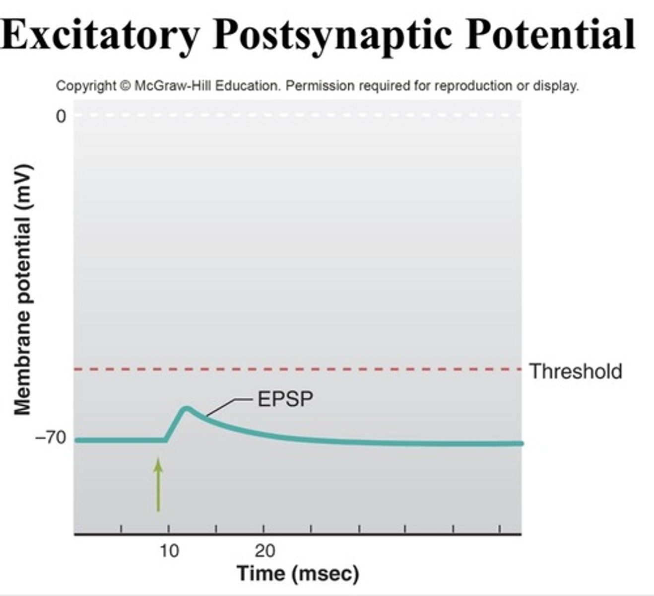
Excitatory Chemical Synapses
Generate an excitatory postsynaptic potential (EPSP) that serve to bring the membrane potential closer to threshold for generating an action potential. Main Example: Glutamate. Depolarization.
Glutamate Ligand-Gated Receptor Channels
The most common neurotransmitter in the brain (learning and memory). Excitatory.
iGluRs: non-selective cation channels (ionotropic)
(Fast Exitatory)
mGluRs: G Protein Coupling (metabotropic)
(Slow Excitatory G1)
(Slow Inhibitory G2 + G3)
Dendritic Spines
Protrude from the main shaft of dendrite, single synapse at head. Modified by activity and experience (via actin). Morphological basis for synaptic plasticity
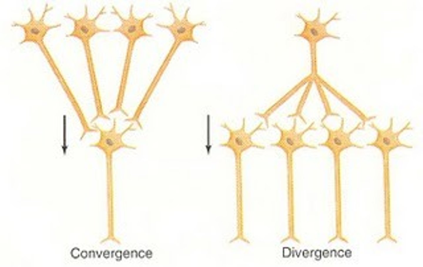
Neuron (Convergence and Divergence)
Convergence: many neurons connected to a single neuron.
Divergence: one neuron connected to many neurons.
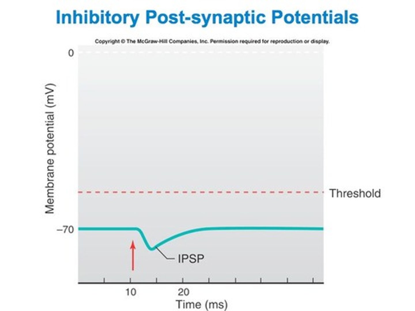
Inhibitory Chemical Synapses
Generate an inhibitory postsynaptic potential (IPSP) and make the cell's membrane potential more negative, making it harder to generate an action potential. Main Example: GABA, glycine. Cl- channel may keep Er even with Na+ open compete to equilibrate or increased K+ permeability for hyperpolarization.
Synaptic Integration (Summation)
Neurons undergo many EPSPs and IPSPs.
Temporal Summation: Rapid series of weak pulses from a single source into one large signal
Spatial Summation: Several weak signals from different locations are converted into a single larger one.
Axon Initial Segment and Action Potential
Lower threshold than axon for AP generation due to high VGSC concentration (Sensitive). Location of individual synapses on the postsynaptic cell is important (further from axon hillock less chance of firing AP).
Modulation of Synaptic Strength
Presynaptic
- Increase [Ca2+i] during high frequency stimulation increases NT release.
- Axo-axonic input (facilitation or inhibition).
- Auto-receptors: -ve feedback decreases NT release.
Postsynaptic
- Paired-pulse facilitation: Short-term, activity-dependent synaptic plasticity.
- Desensitization: Receptor responds then fails despite continued presence of NT (receptor internalization).
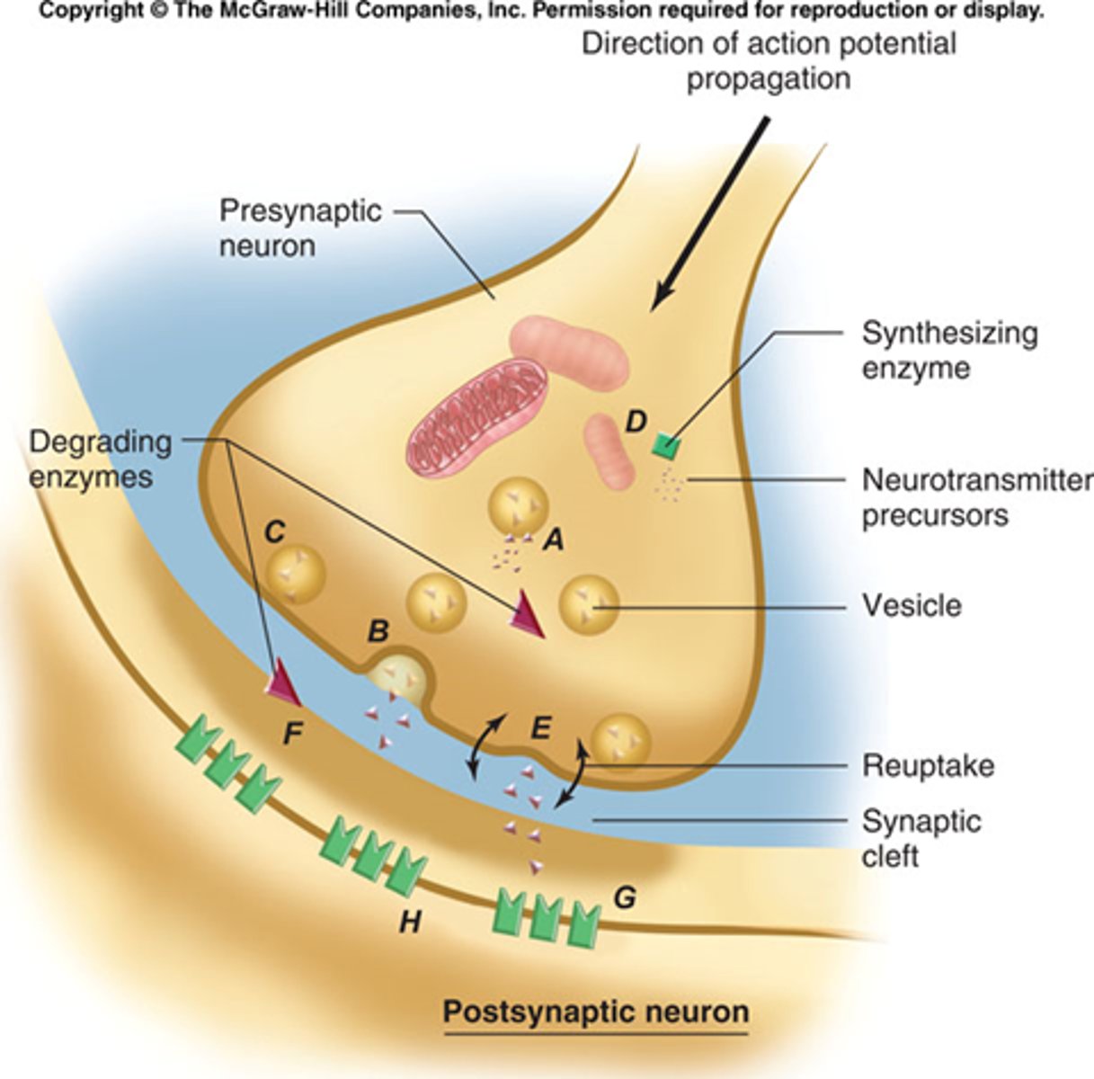
Drug effects on synaptic effectiveness
A. Release and degradation of the neurotransmitter inside the axon terminal.
B. Increased neurotransmitter release into the synapse.
C. Prevention of neurotransmitter release into the synapse.
D. Inhibition of synthesis of the neurotransmitter.
E. Reduced reuptake of the neurotransmitter from the synapse (e.g. Selective serotonin reuptake inhibitors(SSRIs) are antidepressants that affect serotonin levels in the brain)
F. Reduced degradation of the neurotransmitter in the synapse.
G. Agonists (evoke same response as neurotransmitter)or antagonists (block response to neurotransmitter)can occupy the receptors.
H. Reduced biochemical response inside the dendrite.
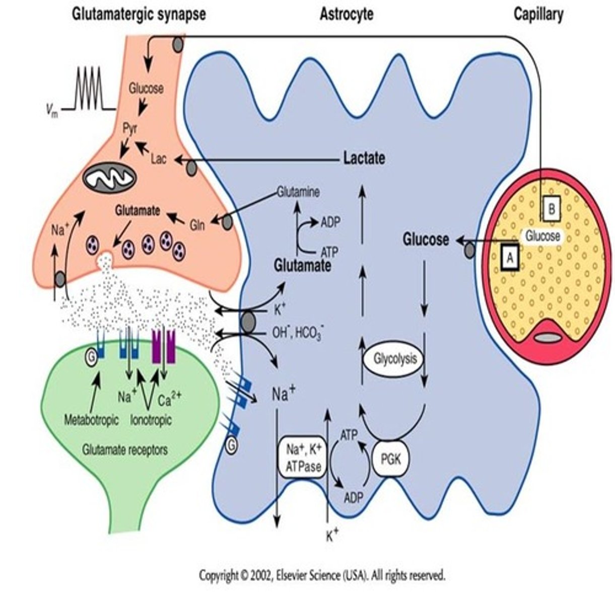
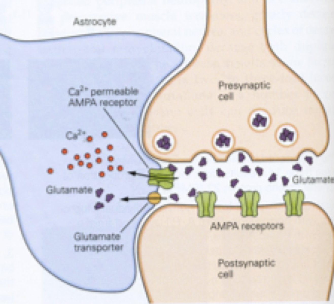
Neurotransmitter recycling
The process of astrocytes taking up certain neurotransmitters, metabolizing them, then transport the precursor back to the neuron.
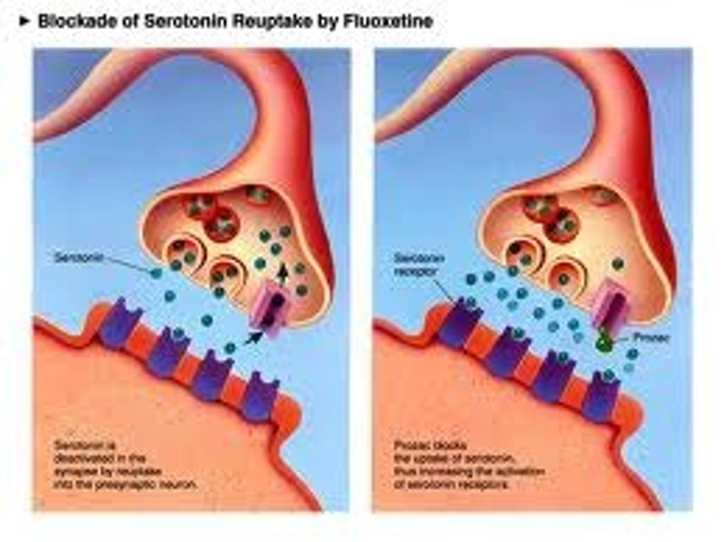
SSRI
Selective serotonin reuptake inhibitor. SSRIs block the SERT transporter on the presynaptic terminal. Serotonin neurotransmitter levels are elevated and prolonged in the synaptic cleft. Elevated serotonin reduces depression in many cases.
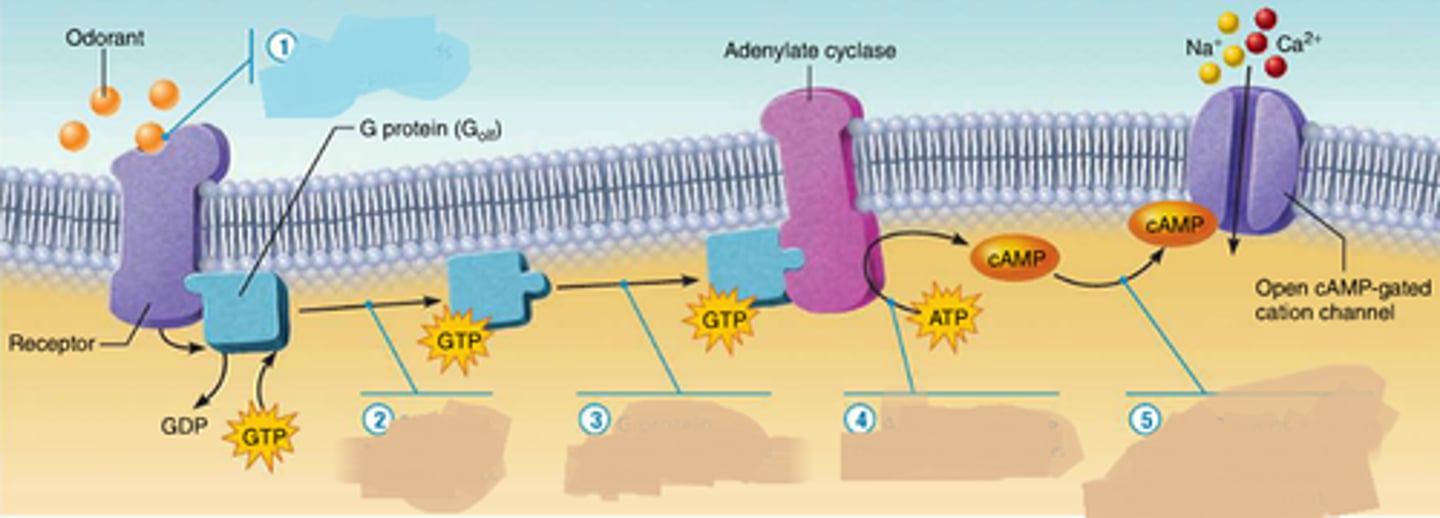
G protein coupled receptors
A signal receptor protein in the plasma membrane that responds to the binding of a signaling molecule by activating a G protein. Also called a G protein-linked receptor.
1. Ligand binds to GCPR inducing conformational change.
2. GDP phosphorylates to GTP resulting in alpha subunits dissociating from beta/gamma subunits.
3. Alpha subunit regulates Adenylate cyclase (ATP to cAMP) resulting in relay of signal via second messenger (cAMP).
4. Also, cAMP gated ion channel lets ions inside cell.
5. Undo steps via hydrolysis of GTP to GDP resulting in ligand dissociating.
Desensitization (3 Types)
Modulation of Synaptic Strength
1. Beta-arrestin does not allow G protein to dissociate
2. Regulator of G-protein hydrolysis GTP to GDP on alpha subunit.
3. Receptor is internalized and forms endosome which fuses to lysosome (degraded) or recycled.
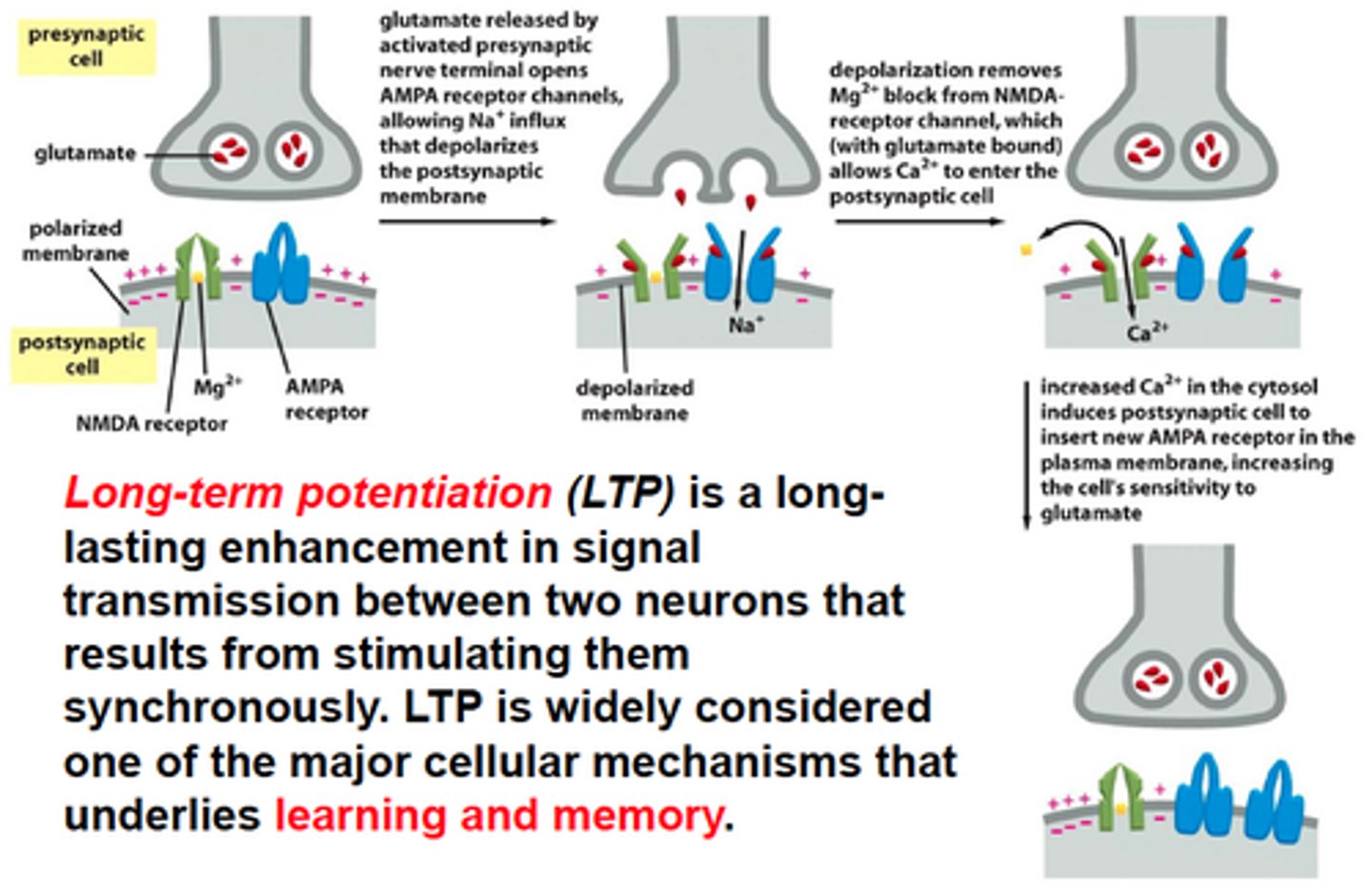
Long Term Potentiation (LTP)
Modulation of Synaptic Strength
Stimulation of AMPA receptor via Glutamate causes membrane to depolarize (Na+ influx). This removes the Mg2+ found in the NMDA receptor allowing Ca2+ to flow inside. This results in more AMPA receptors being added to the membrane resulting in greater responsiveness to glutamate (making the synapse stronger)
Closed at -70mV
Blocked by Mg2+ at -70mV (despite Glu ligand)
Open at 0mV and Ca2+ enters (Glu ligand)
Long-term potentiation is thought to underlie learning and memory. In the short term, insertion of AMPA receptors increases synaptic strength. Over longer time scales, calcium influx drives changes in gene expression that are necessary for sustaining LTP.
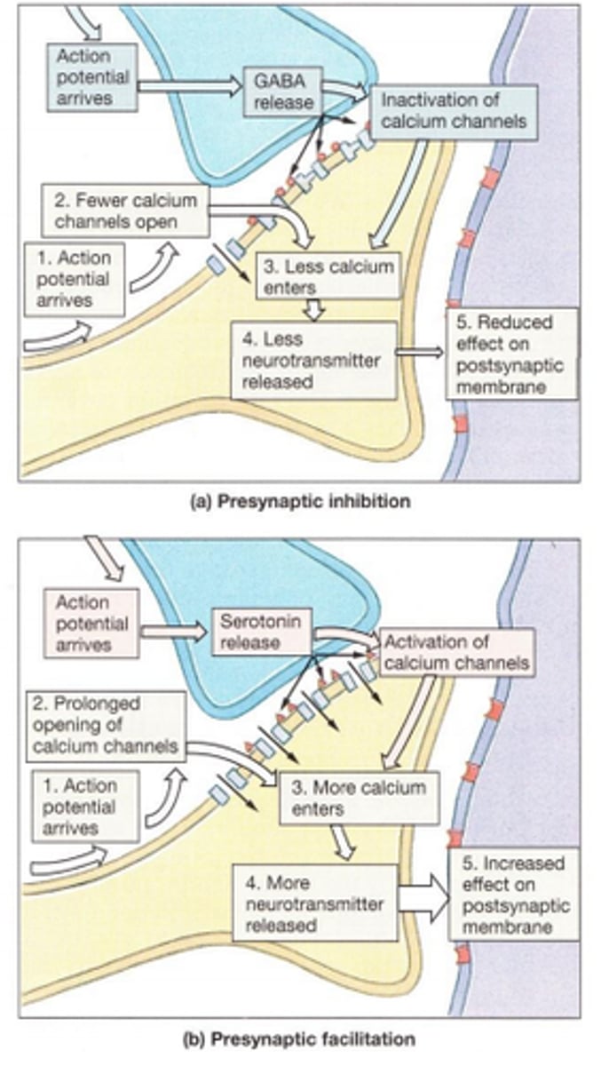
Presynaptic inhibition and facilitation
Inhibitory and Excitatory responses may occur not only at synapses involving the cell body and dendrites but also at synapses along the axon (axoaxonic synapse)
This can either lead to more or less neurotransmitter release at the presynaptic neuron thus causing facilitation inhibition, respectively.
Ex.
Inhibition: Presynaptic GABA receptor depolarization decreases Ca2+ influx and therefore inhibits synaptic transmission by lowering release probability.
Facilitation: Closure of K+ channels by second-messenger systems in the presynaptic terminal, which increases the duration of the presynaptic AP, thereby enhancing Ca+2 influx resulting in greater NT release.
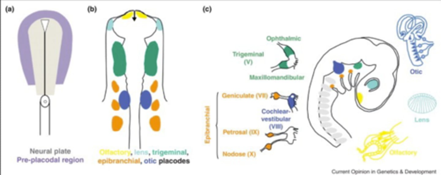
CNS vs. PNS development
Ectoderm forms neural plate folding into neural groove. This results in neural folds which fuse together. Full fusion results in formation of neural tube which form CNS.
The neural plate also forms the neural crest, cells of which will later migrate to different parts of the body and become most of the cells in the PNS.
The Brain Metabolic Demands
Why the Brain is so demanding:
- Constant blood supply
- ~20% of energy consumption
- Neurons cannot produce ATP in absence of oxygen
- The brain does not store glucose very well
- The brain has its own dedicated circulatory system
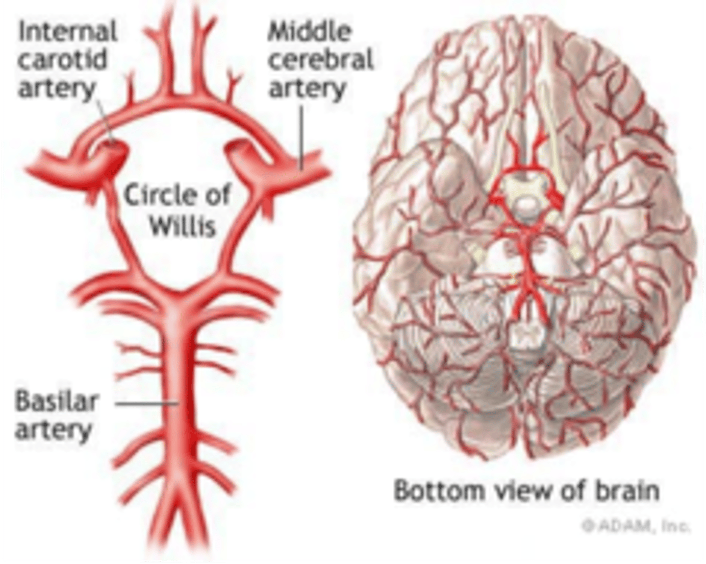
CNS Blood Supply
The anterior parts of the cortex and deep structures are supplied by two branches of the internal carotid artery - the anterior and middle cerebral arteries.
The posterior part of the cortex and deep structures are supplied a branch of the vertebral artery - the posterior (basilar) cerebral artery. The cerebellum and brainstem are supplied by various branches of the vertebral artery.
The anterior and posterior circulations are united by the circle of Willis.
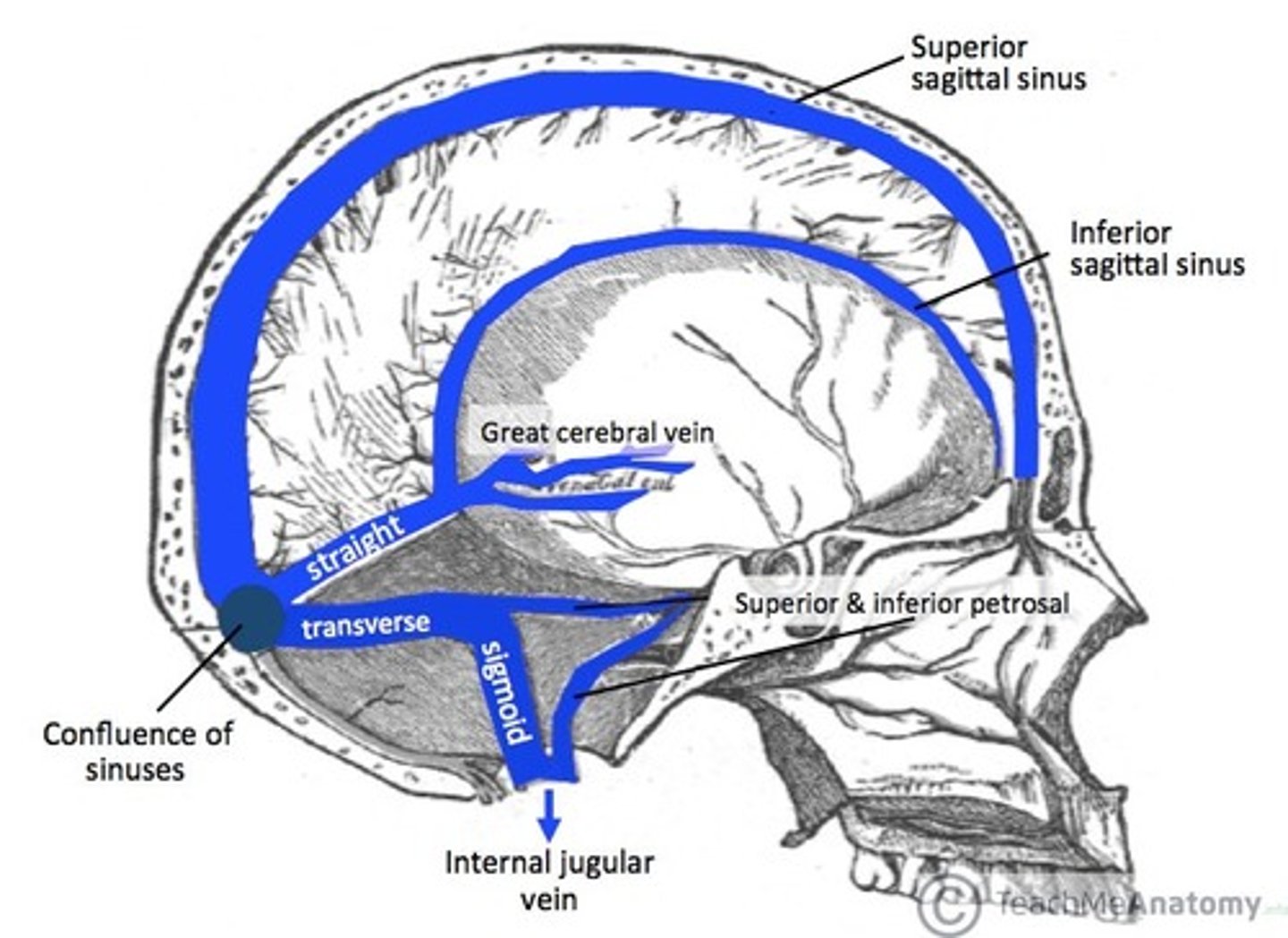
CNS venous drainage
The cerebral veins empty into the dural venous sinuses situated within the subarachnoid space. The superficial system drains into the superior sagittal sinus, while the deep system drains into transverse, straight and sigmoid sinuses.
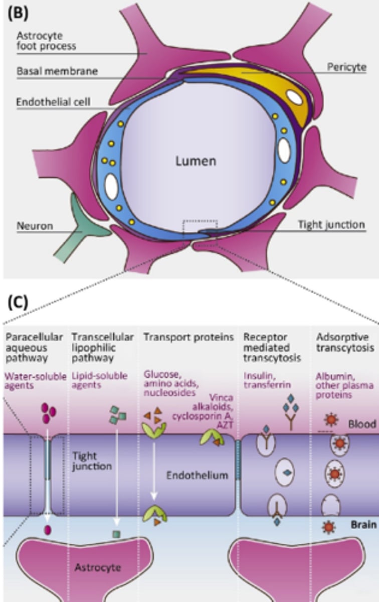
Blood brain barrier
Shields brain from harmful changes in the blood and toxins. Consists of endothelial cells wrapped by glial endfeet. Tight junctions prevent exchange across the capillary wall. Most substances only passage through specific pathways. Lipid soluble substances such as oxygen, alcohol, and certain drugs can penetrate the barrier.
CNS Anatomy
Brain is enclosed in hard, bony structures. Meninges lie between the bone and brain proper. Bathed in Cerebrospinal fluid. Blood-brain barrier.
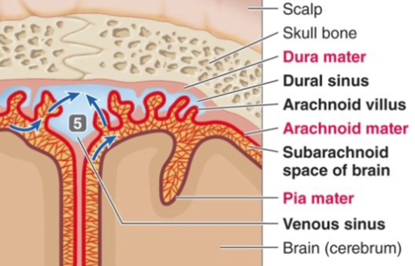
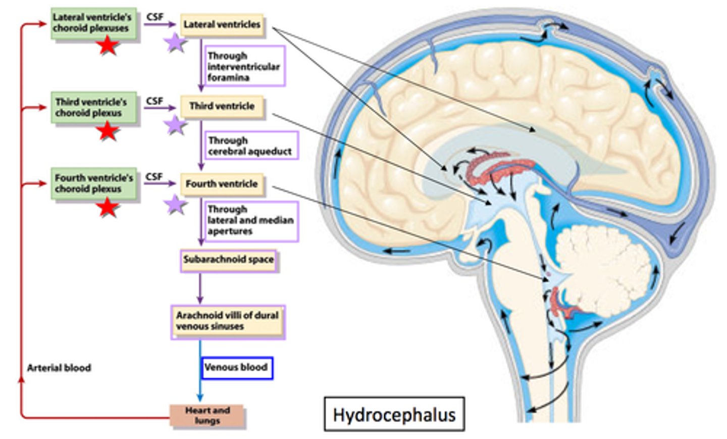
Cerebrospinal fluid
Fluid in the space between the meninges that acts as a shock absorber that protects the central nervous system as well as maintains a proper chemical environment for neural signaling.
CSF is produced in the lateral ventricles and is resorbed into the venous superior sagittal sinus.
Choroid plexus
- Secretes growth factors helping neurogenesis.
- Gateway for immune system to enter brain.
- Form blood CSF barrier.
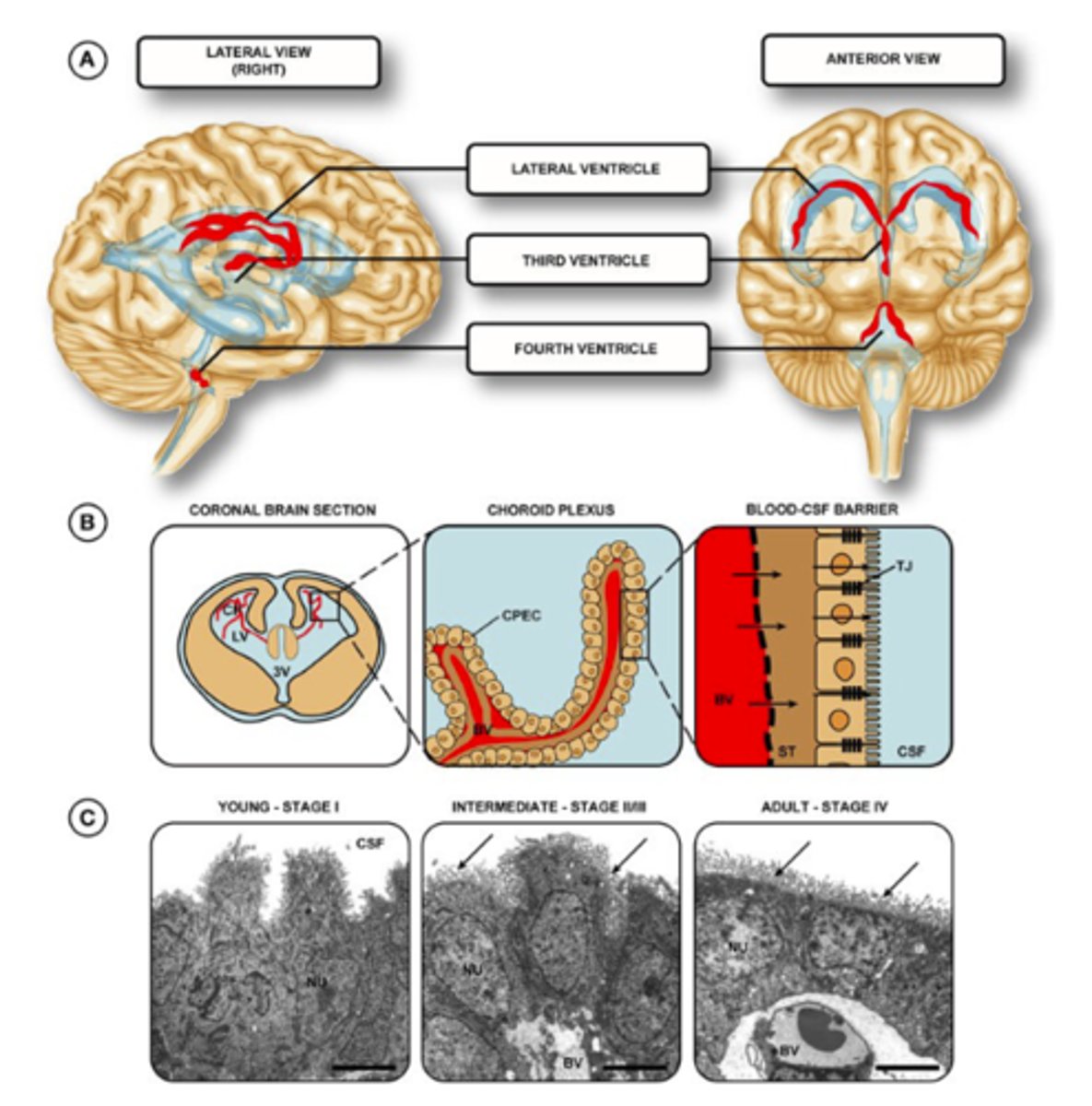
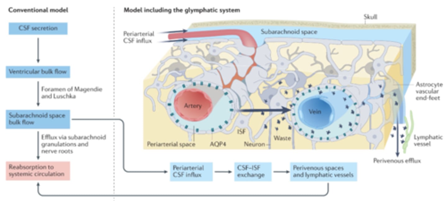
Glymphatic drainage
Fluid system that accesses all regions of the brain.
1. Influx of CSF
2. Exchange of CSF and ISF
3. Efflux of CSF
Tripartite synapse
The idea that a synapse includes not only the pre- and postsynaptic neurons involved but also encompasses many connections with astrocytes.
Decussate
To cross over to the other side of the brain.
The nervous system is crossed. Sensory information from the left side of the body will end up in the sensory cortex on the right-hand side and motor information from the right motor cortex we'll end up stimulating muscles on the left side of the body. This is because sensory and motor tracts decussate: cross over the midline.
Spinal Nerves (5 Types)
31 pairs of nerves arising from the spinal cord
8 cervical
12 thoracic
5 lumbar
5 sacral
1 coccygeal
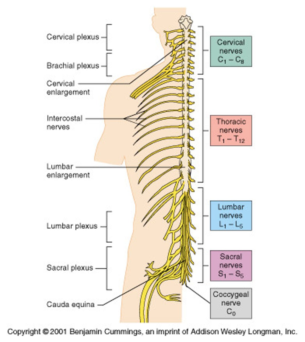
Spinal Nerve Anatomy
Spinal nerve begins at the junction of the dorsal root and ventral root. Each spinal nerve then branches into a dorsal ramus and a ventral ramus. The intervertebral foramen serves as the doorway between the spinal canal and periphery.
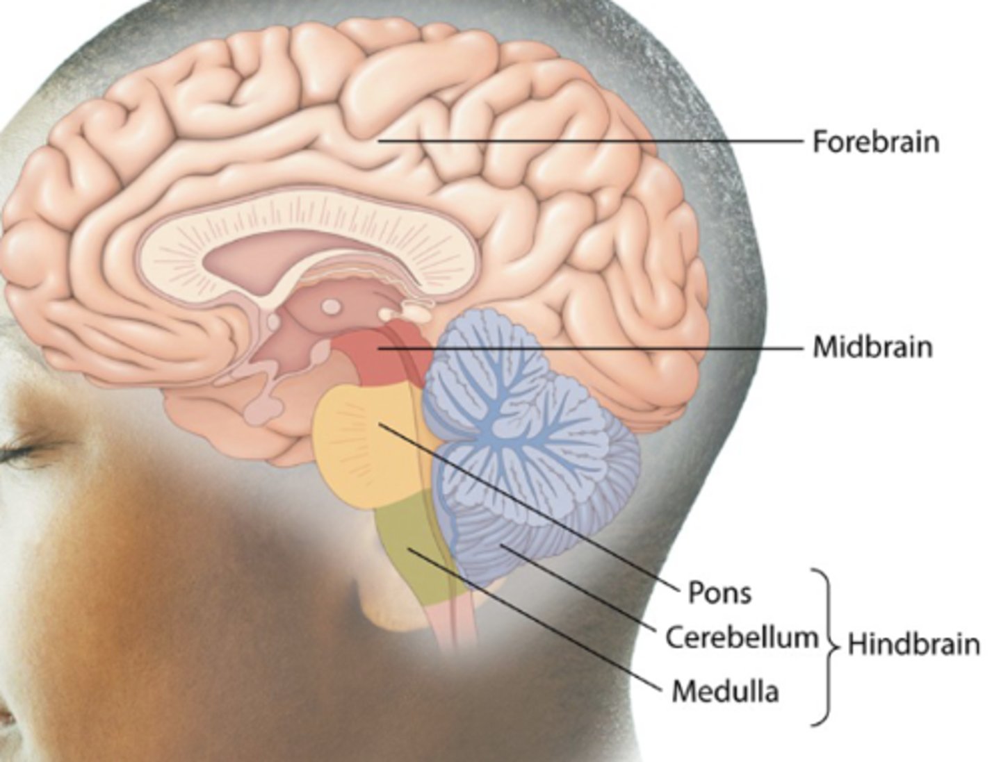
Brain (3 Parts)
Forebrain, midbrain, hindbrain.
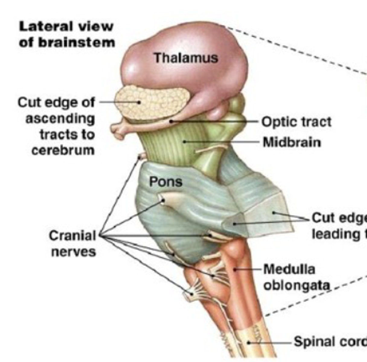
Brainstem
Interfaces between higher brain regions, cerebellum, and spinal cord. Regulates/controls some critical bodily functions (breathing, heart rate). Contains the efferent somatic motor neurons for the cranial nerves. Receives input from 10/12 cranial nerves (reflexes, sensorimotor functions, taste, autonomic functions, vision).
Parts:
- Thalamus (forebrain)
- Midbrain
- Pons (hindbrain)
- Medulla oblongata (hindbrain)
- Spinal cord
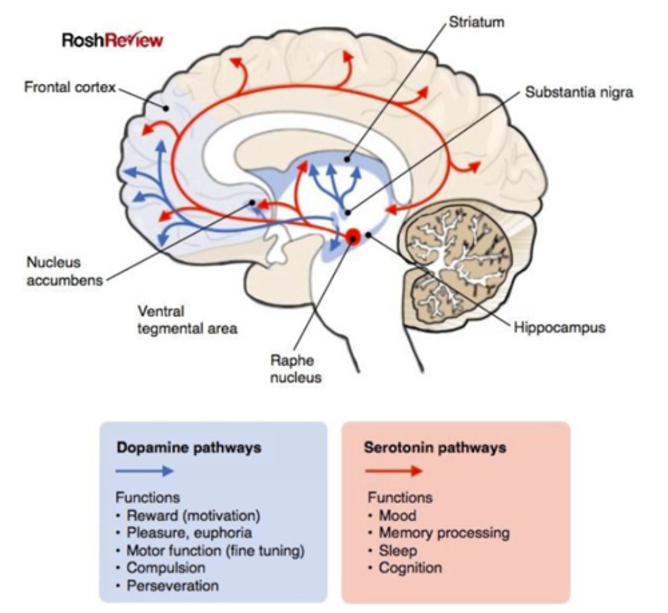
Midbrain
Critical for voluntary motor function (substantia nigra). Superior colliculus is a major relay station for the optic nerve. Regulates eye movements. Dopaminergic and serotonergic reward system.
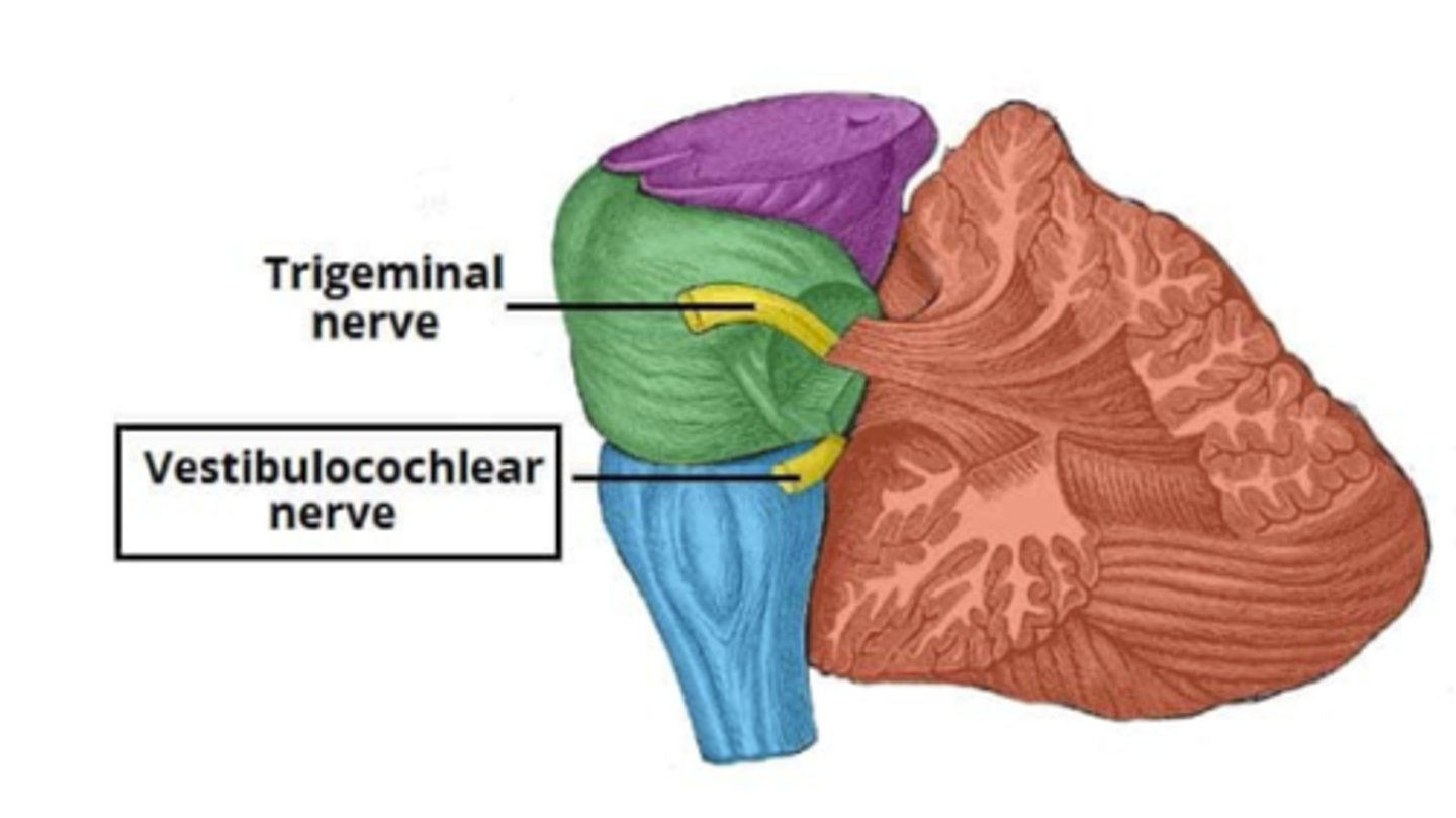
Pons
A brain structure that relays information from the cerebellum to the rest of the brain. Coordinates facial sensorimotor functions. Hearing and balance.
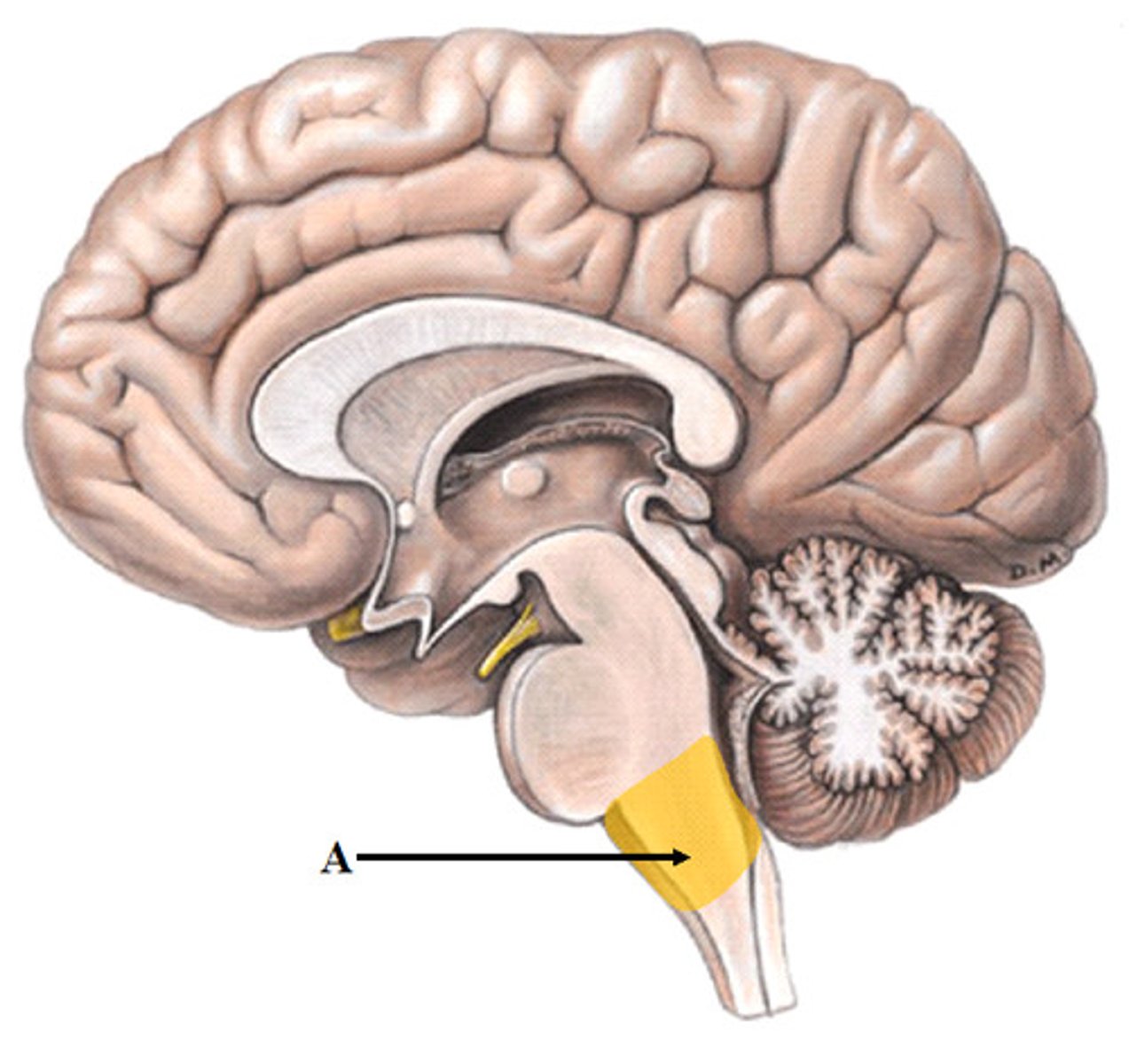
Medulla oblongata
An extension of the spinal cord into the skull that coordinates heart rate, circulation, swallowing, and respiration. Contains the reticular activating system (RAS) (sleep cycle and alertness).
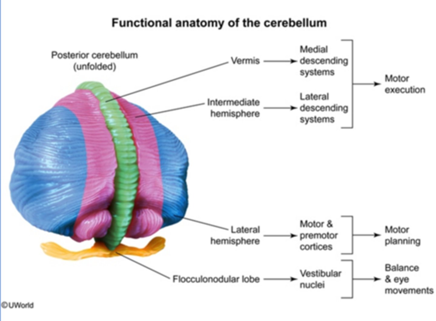
Cerebellum (3 Types)
The "little brain" at the rear of the brainstem; functions include processing sensory input and coordinating movement output and balance.
Spinocerebellum descending - spinal cord: execution
Cerebrocerebellum Ascending - motor cortex: planning
Vestibulocerebellum - vestibular nuclei (pons/medulla): balance and eye
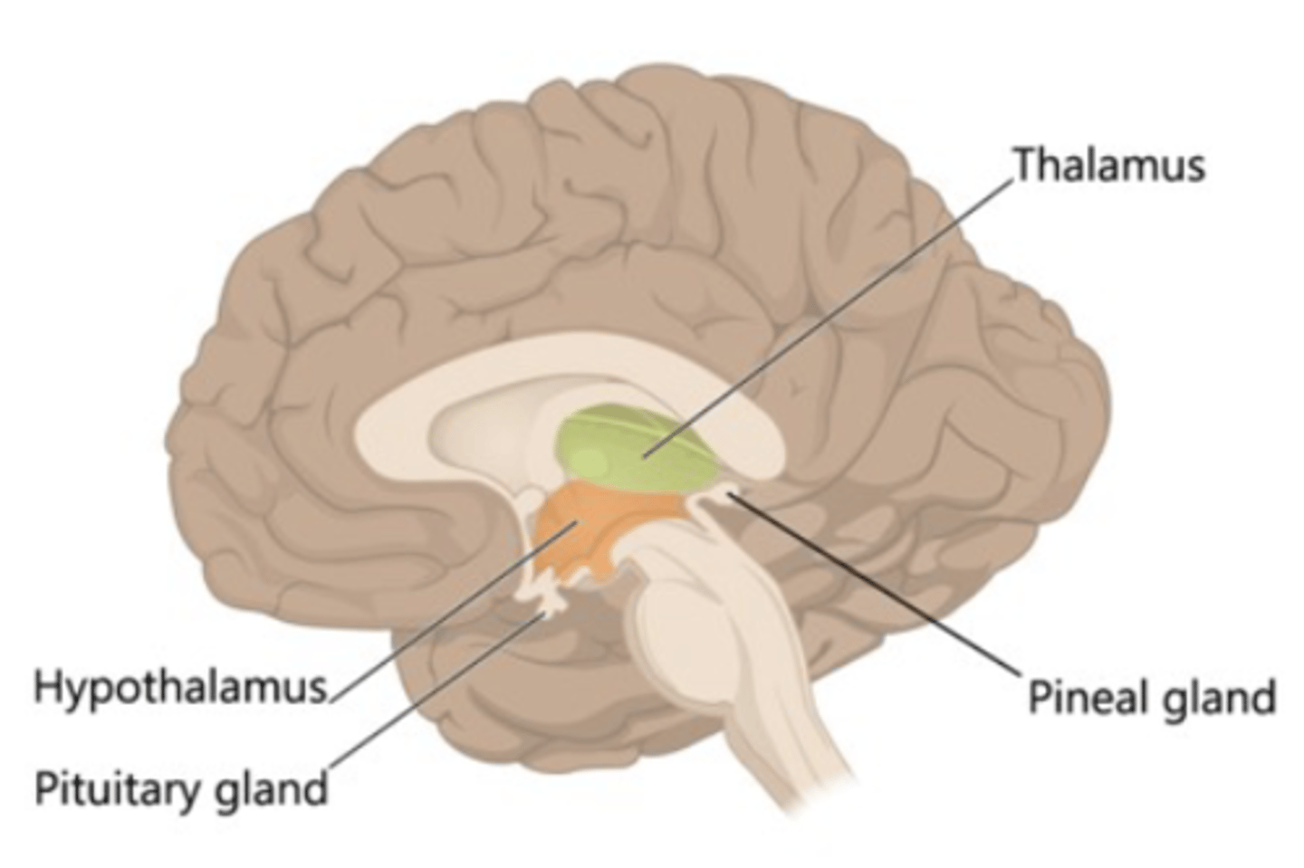
Diencephalon (Forebrain)
1. Thalamus: Major sensory relay Centre. Receives input from sensory nerves and projects to the primary sensory areas of the cerebral cortex. All sensory information is relayed through the thalamus except olfaction. Also participates in motor and memory circuits.
2. Hypothalamus: Critical homeostatic functions. Regulates autonomic and endocrine systems.
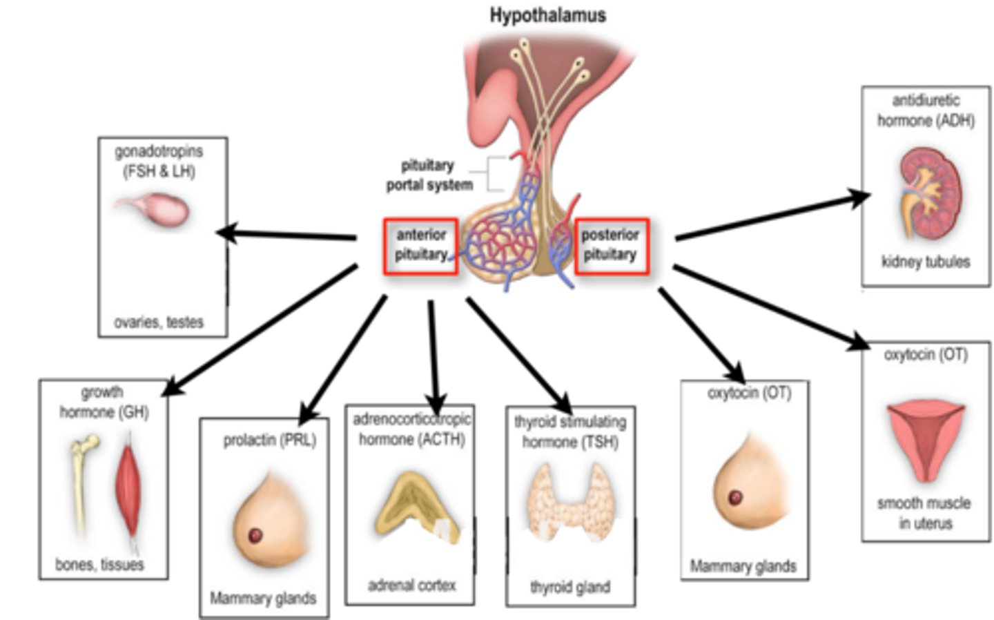
Anterior vs Posterior pituitary
Anterior pituitary is controlled by hypothalamic releasing and inhibiting factors.
Posterior pituitary is composed of axons which descend from hypothalamus. Neurons secrete hormones into bloodstream.
Telencephalon (Forebrain)
Cerebrum is the largest part of your brain, and it handles a wide range of responsibilities.
- Cerebral cortex
- Basal ganglia
- Limbic system
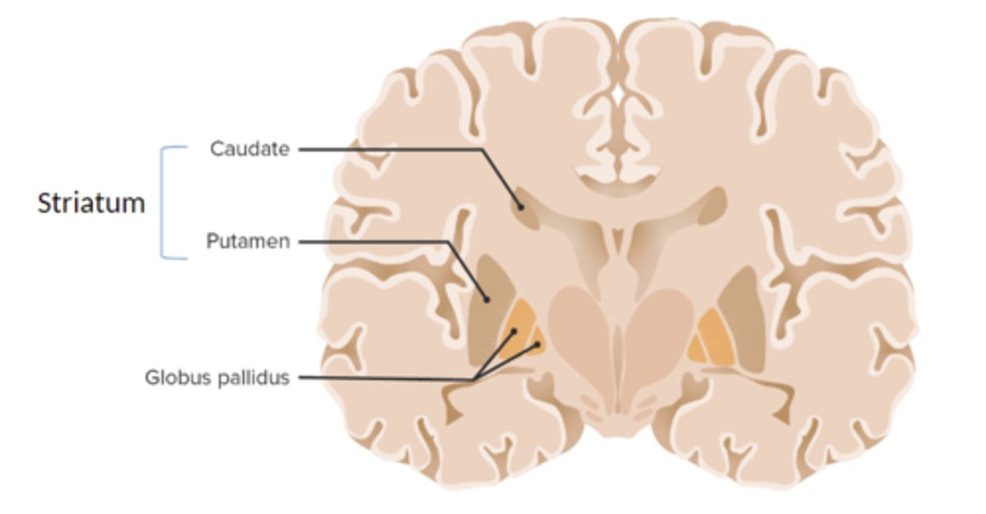
Basal ganglia
A set of subcortical structures that directs intentional movements (heavily connected with the motor cortex and the substantia nigra of the midbrain). Important for voluntary motor control. Also involved in the reward system.
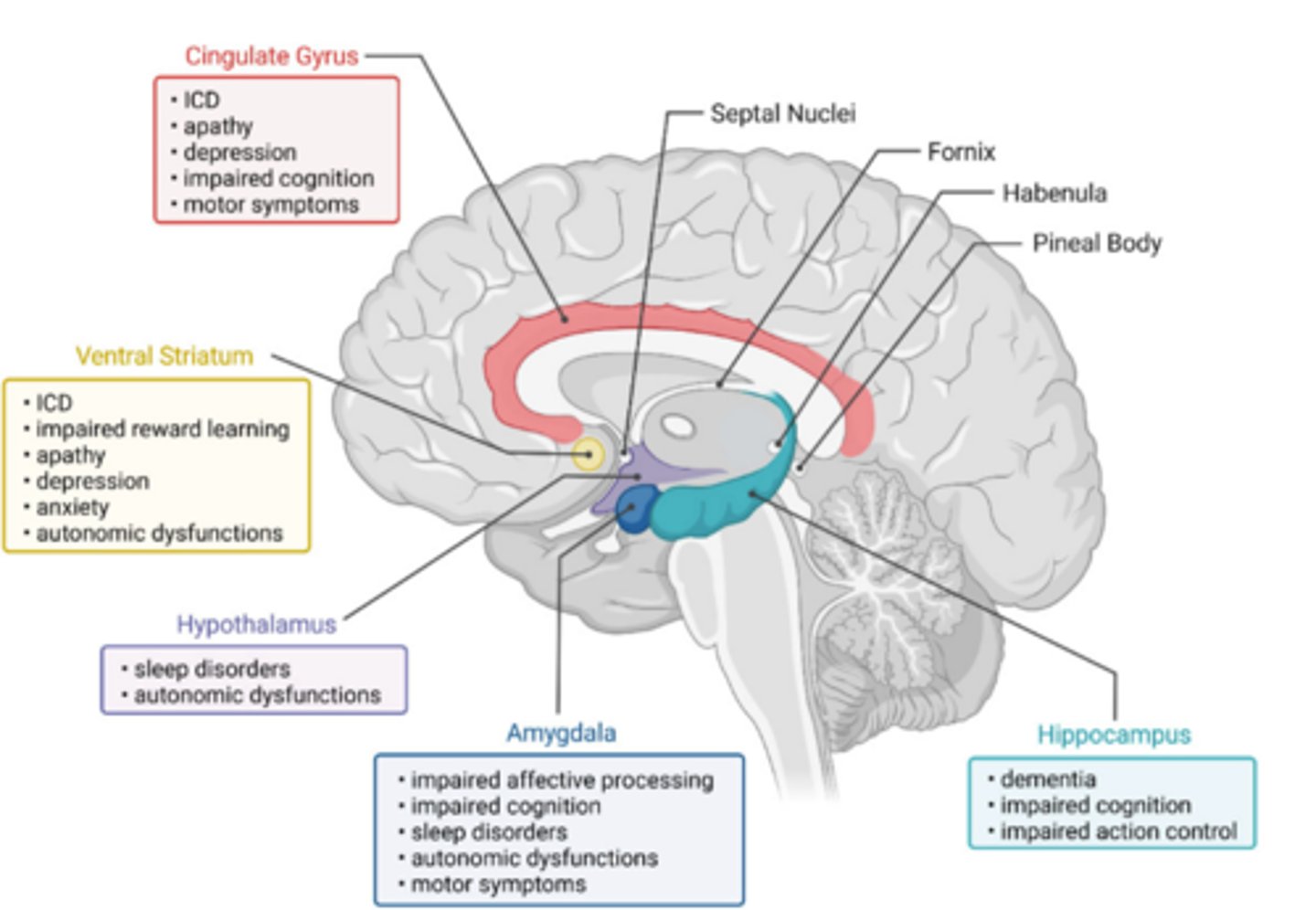
Limbic system
Neural system (including the hippocampus, amygdala, and hypothalamus).
- "Lizard brain"
- Emotion, anxiety
- Long-term memory
- Motivation
Cerebral cortex
The intricate fabric of interconnected neural cells that covers the cerebral hemispheres; the body's ultimate control and information-processing center.