Clinical Workup of the Optic Nerve
 Call Kai
 Learn
Learn Practice Test
 Spaced Repetition
 Match
Match Flashcards
 Knowt Play
1/9

There's no tags or description
Looks like no tags are added yet.
Name | Mastery | Learn | Test | Matching | Spaced | Call with Kai | Chat |
|---|
No analytics yet
Send a link to your students to track their progress
10 Terms
Signs & Symptoms of Optic Nerve Dysfunction
Reduced VA: non-specific sign, VA may be normal
Pupil dysfunction: esp in unilateral cases
Dyschromatopsia: mainly difficulty with red-green discrimination and acquired disease
Diminished light brightness sensitivity: ex. reduced red saturation
Reduced contrast sensitivity
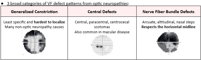
VF defects: may vary depending on underlying etiology
Pain is NOT common with many types of optic nerve diseases, but can be a very helpful symptom when present
Diagnosis of Optic Neuropathy
Characteristic (PATTERN) combo of findings = VA loss + color deficiency + VF defect + pupil dysfunction + (possible) abnormal ONH appearance
Workup: Key Tests
Main testing: case history, VA, pupils, EOMs, color vision, VF, ocular health eval
Ancillary testing: potential acuity testing, red saturation test, brightness comparison test, contrast sensitivity testing, exophthalmometry, OCT, OCT-A, FA, B-scan ultrasound, visual evoked potential (VEP)
Case History
Most common complaint = vision loss
Acute/rapid: sudden, over a few hours or days
Demyelinating (MS), ischemic, traumatic
Subacute: few days to 1-2 weeks
Inflammatory, infiltrative, nutritional/toxic, glaucomatous (some)
Gradual: over a few weeks or years
Hereditary, compressive, nutritional/toxic, glaucomatous
Transient vision loss? Diplopia? Proptosis? Other? → Pulfrich phenomenon
Pulfrich phenomenon = difference in quality of the 2 eyes; disorder of perception where 2D objects appear 3D
Due to delay in visual input of the affected eye
Occurs in unilateral/asymmetric optic neuropathy
Aggravating factors:
If transient vision loss w/ eye movement (gaze-evoked amaurosis) → suggests diseases that “deform” the optic nerve (ie. optic nerve sheath meningioma)
If vision worse w/ increased body temperature (Uhthoff phenomenon) → suggests demyelinating disease
If symptoms worsen w/ changes in posture → suggests papilledema and increased intracranial pressure
Visual Acuity
Non-specific for optic nerve disease
Is the least sensitive of all function tests
Pupil Testing
Afferent pathway = from the eye to the brain (CN II)
Efferent pathway = from the brain to the eye (CN III and oculosympathetic pathway)
Optic nerve disease can result in an afferent pupil defect (APD) IF the condition is asymmetric (“relative” to each other)
Abnormal red saturation and Brightness comparison test correlated well with the presence of an APD
Color Vision
Non-specific for optic nerve disease
Mismatch of good visual acuity and acquired color vision dysfunction → strongly suggestive of optic nerve dysfunction
Kollner’s rule:
Red/Green discrimination difficulty = optic nerve disease
Exceptions: glaucoma, dominant optic atrophy, chronic papilledema
Blue/Yellow discrimination difficulty = retinal disease
Exception: cone dystrophy
Visual Field
Contrast Sensitivity Testing
Contrast sensitivity usually reduced in optic neuropathy, even if good VA
Charts: Pelli-Robson, low-contrast Bailey-Lovie, Vistech, MARS
May be the most valuable in terms of long-term monitoring and in clinical trials
Optical Coherence Tomography (OCT)
Evaluates the peripapillary RNFL and macular Ganglion Cell Layer (GCL)
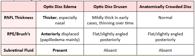
May be helpful in diagnosing early disc edema
Useful to monitor progression
Subretinal hyporeflective space (SHYPS)
Presence occurs in conditions where the optic disc is elevated
Hyporeflective space on OCT cross-section below the photoreceptors (neurosensory retina)
Smooth internal contour; lazy V pattern
Optic disc edema: smooth internal contour, “lazy V” pattern
Optic disc drusen: lumpy-bumpy internal contour, abrupt decline in SHYPS
OCT alone CANNOT definitely differentiate optic disc edema from pseudo disc edema