Lab Wk 1-2
 Call Kai
 Learn
Learn Practice Test
 Spaced Repetition
 Match
Match Flashcards
 Knowt Play
1/100

There's no tags or description
Looks like no tags are added yet.
Name | Mastery | Learn | Test | Matching | Spaced | Call with Kai | Chat |
|---|
No analytics yet
Send a link to your students to track their progress
101 Terms
What is a compound light microscope?
Has 2 lenses magnifying the object
What are the 2 lens of the compound light microscope?
Objective + Ocular Lens
Multiplying the magnifying power of the two compound lens will provide?
Total Magnification you will be seeing
Objective Lens Names:
Scanning (Red)
Low (Yellow)
High Dry (Blue)
Oil Immersion (White)
Objective Lens Magnification Red:
4X
Objective Lens Magnification Yellow:
10X
Objective Lens Magnification Blue:
40X
Objective Lens Magnification White:
100X
Ocular Lens Magnification:
10X for all Lens
Total Magnification (Objective + Ocular) Red:
40X
Total Magnification (Objective + Ocular) Yellow:
100X
Total Magnification (Objective + Ocular) Blue:
400X
Total Magnification (Objective + Ocular) White:
1000X
Light enters the microscope from?
A Source in the base
Light goes through a condenser-iris diaphragm unit which?
Converges the light beam —> beam passes through the slide + controls amount of light that passing through the slide to objective lens
Function of Objective Lens:
Magnifies the image before it passes ocular lens in the eyepiece
What is Ocular Lens?
Lens we look through
Mechanical Stage function:
Has adjustment knobs that can move the slide to optimum position
Coarse Adjustment Knob function:
Changes the distance between the objective lens + slides rapidly
Fine Adjustment Knob function:
Changes the distance between the objective lens + slides slowly
Where is the dimmer dial located on microscope? How should it be set?
Base of scope + should always remain set to white line on dial
Which lens should oil immersion be performed on?
ONLY White oil objective one
When grown on solid media, individual bacterial cells multiple to form?
Visible masses called colonies
In order to study bacteria accurately, microbiologists must work with isolate colonies which are?
Colonies that are well separated from others on an agar plate.
An isolated colony allows us to?
Observe a single type of organism without interference from nearby growth
What is the purpose of streaking?
To spread bacteria across the surface of the agar so that individual cells are separated enough to form isolated colonies
Which microscope part controls the amount of light passing through the specimen?
Iris diaphragm
The large knob that moves the stage a relatively large distance and is used for initial focusing is the __________ adjustment knob.
Coarse
Which microscope part should be used to carry the microscope safely?
The arm, while supporting the base
A microscope has a 10× ocular lens and a 40× objective lens. What is the total magnification?
400X
Which objective should normally be used first when locating and initially focusing on a specimen?
4× scanning objective
A student can see bacteria under the 40× objective but loses them after rotating to 100×. What was the most likely problem?
The specimen was not centered before changing objectives.
Which material should be used to remove immersion oil from an objective lens?
Lens paper
Why should ordinary paper towels not be used to clean microscope lenses?
They may scratch or damage the lenses.
A student finishes microscopy and notices oil on the 40× objective. What most likely happened?
The student rotated the 40× objective through the oil.
Before putting the microscope away, the __________ objective should be rotated into position.
4X
What is the main purpose of preparing a bacterial smear?
To attach a thin layer of cells to a slide for staining and observation
When preparing a smear from bacteria growing on solid media, what should normally be placed on the slide before adding the bacteria?
A drop of water
Why should only a small amount of bacterial growth be used to prepare a smear?
A thick smear may stain unevenly and make individual cells difficult to observe.
Before heat-fixing a bacterial smear, the smear must be completely __________.
dry
What is the main purpose of heat-fixing a bacterial smear?
To attach the cells to the slide and help preserve their shape.
What should the microincinerator look like before students leave the laboratory?
Unplugged and turned off.
How can you tell when the microincinerator is ready to use?
The inside of the barrel is glowing red hot.
Why should the inoculating loop be allowed to cool before transferring microorganisms?
To avoid killing the microorganisms with heat.
What is the primary safety reason you should never let go of an inoculating loop or needle while it is inside the microincinerator?
The entire instrument could become extremely hot and cause serious burns when picked up.
What is the primary purpose of performing a streak-for-isolation technique?
To produce isolated bacterial colonies
What is aseptic technique?
“Correct technique”
What is the main purpose of a simple stain?
To make bacterial cells easier to see and evaluate their morphology and arrangement
How many primary stains are used in a simple-staining procedure?
One
After applying the stain for the required amount of time, how should the slide normally be rinsed?
Gently, so the smear is not washed from the slide
After rinsing, excess water should be removed by gently __________ the slide with bibulous paper.
Blotting
Role of Crystal violet:
Primary Stain
Role of Acetone-Alcohol:
Removes the primary stain from cells that cannot retain the crystal violet-iodine complex
Role of Safranin:
Counterstain
Role of Gram's iodine:
Mordant
Gram-stain reagents in the correct order:
1) Crystal Violet
2) Gram’s Iodine
3) Decolorizer
4) Safranin
What color should Gram-positive cells appear after a correctly performed Gram stain?
Purple
What color should Gram-negative cells appear after a correctly performed Gram stain?
Pink or red
A student applies the decolorizer for too long. What is the most likely result?
Gram-positive cells may incorrectly appear pink.
Why is methylene blue added to the cheek cell preparation before viewing under the microscope?
To stain the human cells because they are mostly clear and difficult to see without dye.
Before collecting cheek cells, what should be placed on the microscope slide?
A single drop of water.
How should cheek cells be collected for the smear?
Gently rub the inside of the cheek with a sterile toothpick.
Steps in the correct order for preparing a cheek smear:
1) Add a drop of water to the microscope slide.
2) Collect cheek cells with a toothpick and mix them into the water.
3) Place the cover slip at 45 degree angle.
4) Add methylene blue next to the cover slip.
After collecting the cheek cells, the used toothpick should be discarded in the __________ biohazard bin.
Sharps
Not cooling the loop using the micro incinerator can?
Kill the bacteria + Prevent growth
Failing to sterilize the loop between sections will lead to?
Too many colonies
Streaking over the same area repeatedly when in section 2 or 3 will?
Prevent separation of bacteria
Selecting bacteria from crowded areas instead of isolated colonies will result in?
Mixed or unclear observations
Making a smear that is too thick will prevent?
Proper staining and make it difficult to distinguish individual cells.
Not allowing the smear to airy dry completely can?
Cause cells to wash off during staining.
Over-decolorizing will cause all cells to? How about Under-colorizing?
Cells to appear Gram negative; Cells appear Gram positive

What are these examples of?
Cocci

Coccus

Diplococci
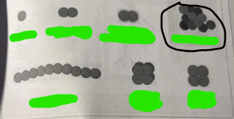
Staphylococci
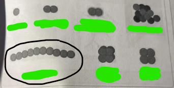
Streptococci
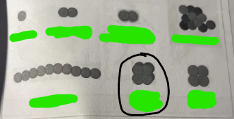
Sarcina coccus

Tetrad coccus

What are these examples of?
Bacilli

Coccobacillus

Bacillus

Diplobacilli

Streptobacilli

Palisades

Club rod
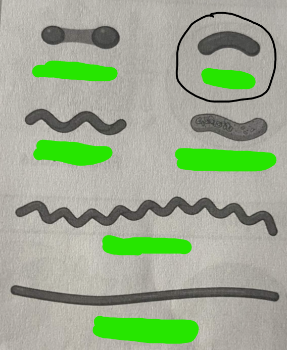
Vibrio

Spirillum

Helical form

Spirochete
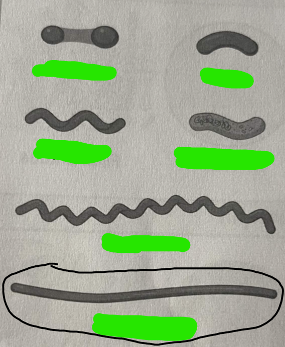
Filamentous

What is this category?
Shape

Circular

Filamentous

Rhizoid

Irregular

What is this category?
Margin
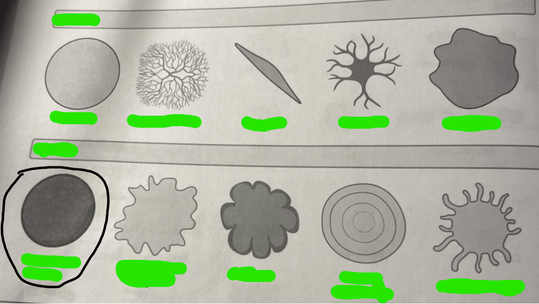
Smooth, entire

Undulate, irregular

Lobate
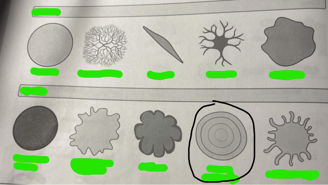
Curled, swarming