17.2 - Heart Anatomy + Blood Flow Pathways
 Call Kai
 Learn
Learn Practice Test
 Spaced Repetition
 Match
Match Flashcards
 Knowt Play
1/28

There's no tags or description
Looks like no tags are added yet.
Name | Mastery | Learn | Test | Matching | Spaced | Call with Kai | Chat |
|---|
No analytics yet
Send a link to your students to track their progress
29 Terms
What is the membranous structure surrounding the heart?
The pericardium; a sac
What are the 2 components of the pericardium?
Fibrous Pericardium: tough outer layer that attaches heart to surrounding structures
Serous Pericardium: thin inner serous membrane that produces serous fluid
What is the Fibrous Pericardium?
Composition
Function
Collagen bundles
Function:
Makes pericardium tough.
Anchors heart to diaphragm + great vessels.
Reduces distensibility = does not change shape upon being stretched.
What is the Serous Pericardium?
Composition
Function
Composition: Folds back on itself to form a sac around the heart; has 2 layers with space in between:
Parietal Pericardium (Outer)
Fused to inner surface of fibrous pericardium
Stratified Squamous ET
Pericardial Space
Between serous membrane
Filled with serious fluid = pericardial fluid
Visceral Pericardium; Epicardium (Inner)
Most superficial layer of the heart wall
Decreases friction from heart contraction.
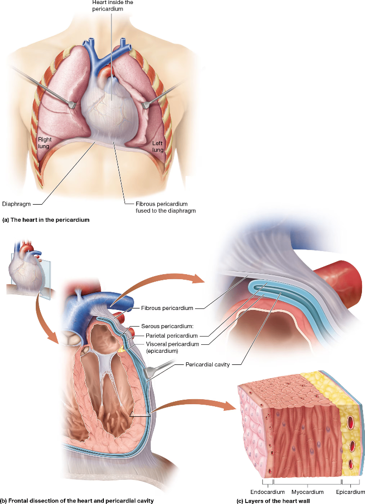
What are the 3 layers of the heart wall (most superficial --> deepest layer)
Epicardium aka Visceral Pericardium
Myocardium
Endocardium
What is the Myocardium layer of the heart wall?
Location
Composition and Function
Middle layer of heart wall.
Cardiac Muscle Tissue:
Cardiac myocytes, ECM, and Intercalated Disks
Arranged in spiral manner around heart chambers
Fibrous Skeleton:
DICT
Functions in structural support
Interventricular septum (wall)
Interatrial septum (wall)
around heart valves
Insulated heart for electrical activity
What is the Endocardium?
Deepest layer of heart wall.
Composed of endothelium = Simple Squamous ET
Lines the heart and blood vessels.
Composed of CT with elastic and collagen fibers.
Functions like the blood-brain barrier, but more of a blood-tissue barrier
What are great blood vessels?
Vessels that are directly attached to the heart.
Largest vessels in the body because:
they have yet to diverge into branches (arteries)
they have converged into one vessel (veins)
Arteries vs Veins
Arteries = vessels that transport blood away from the heart
Veins = vessels that transport blood away from capillaries; or towards the heart
What are names of the great vessels?
Superior + Inferior Vena Cava = Vein
Pulmonary Trunk = Artery
Branches into the pulmonary arteries, which branch into the pulmonary capillaries
Pulmonary Veins = Vein
Aorta = Artery
Function of Superior + Inferior Vena Cava?
Superior = Drains dO2 blood into the right atrium from the veins superior to the diaphragm.
Inferior = Drains dO2 blood into the right atrium from the veins inferior to the diaphragm.
Function of Pulmonary Trunk?
Receives dO2 blood from the right ventricle and pumps to the lungs.
Trunk --> Arteries --> Capillaries in the lungs
Largest and widest vessel of pulmonary circuit
Function of Pulmonary Veins?
Receives O2 blood from the pulmonary capillaries in the lungs and transports blood to the left atrium of the heart.
Function of Aorta?
Artery that transports blood from the left ventricle to the entire systemic circuit with O2 blood.
Thickest + Largest vessel in entire body.
Forms aortic arch; curving the the left.
Ventricles vs. Atria in structure
Ventricles are larger chambers than the atria.
This is because the ventricles are stronger pumps, which is needed to generate the pressure gradient for blood flow.
Right Atrium
Function and receiving vessels?
Structure
Receives dO2 from the body through the superior + inferior vena cava.
Large; thin walls, anteriorly positioned.
Left Atrium
Function and receiving vessels?
Structure
Receives oxygenated blood from pulmonary veins (from lungs).
Smaller than left atria; thick walled, posteriorly positioned.
Makes up base + posterior surface of heart
What are auricles?
External muscular pouch of the atria that expand to give the atria more space to hold blood.
Right atria has a larger auricle than the left.
How are the 2 atria separated?
Interatrial Septum; a thin wall.
Small indentation in the wall called fossa ovalis (depression), was once the foramen ovale (hole) in a fetal heart.
Right Ventricle:
Function and receiving vessels?
Structure
Recevies dO2 blood form the right atrium.
Wider and thinner-walled than left ventricle, crescent-shaped
Little resistance against pump
Left Ventricle:
Function and receiving vessels?
Structure
Receives oxygenated blood form the left atrium.
Smaller and thicker walled than right ventricle, circular-shaped.
Greater resistance against pump.
Works harder than right ventricle.
3x thicker wall than the right ventricle.
What are the Trabeculae Carneae?
Rigid surface along the walls of the ventricles caused by irregular protrusions of cardiac muscle tissue.
What are papillary muscles and chordae tendineae?
Papillary Muscles: Finger-like projections of the ventricles that hold the chordae tendineae.
Chordae Tendineae: Tendon-like chords on that protrude off the papillary muscles and attach to the valves.
Ensures valves can open and close.
What is the interventricular septum?
how does it differ from the interatrial septum?
Wall between the 2 ventricles.
Contracts with the rest of the ventricular muscle to help expel blood into the pulmonary trunk + aorta.
What are the functions in valves in the heart?
Ensures one directional flow
dO2 blood goes to pulmonary circuit
O2 blood goes to systemic circuit
Prevents backflow of blood
Contraction of ventricles can force blood to want to push back into the atria.
Blood flowing out of ventricles moves against gravity into high pressure arteries, which could cause blood to want to flow backward into the ventricles.
What are the valves of the heart?
Atrioventricular Valves (AV Valves)
Tricuspid Valve
Mitral (Bicuspid) Valve
Semilunar Valves (SL Valves)
Pulmonary Valve
Aortic Valve
What are the AV Valves?
Location
Type of Valve
Cusps?
What are the 2 AV Valves?
Function of chordae tendineae in the ventricles
Valves between the atria and the ventricles.
Inflow Valves: Blood flows through valves to enter the ventricles.
Cusps: Flaps composed out of endocardium and fibrous skeleton extensions.
Types:
Tricuspid Valve: Between right atrium and ventricle = 3 cusps (flaps)
Mitral (Bicuspid) Valve: Between left atrium and ventricle = 2 cusps
What are the function of the chordae tendineae and the cusps of the AV valves?
Chordae tendineae extend from the inferior end of each cusp (flap) and attach to the papillary muscles.
Papillary muscles contract before the ventricle eject blood to ensure the AV valves stay closed = prevents cusps from inverting backward into the atria and ventricular pressure increases.
What are the SL Valves?
Location
Function
Type of Valve
Cusps?
2 SL Valves?
Chordae Tendineae?
Between ventricles and the pulmonary trunk or aorta.
Prevents blood from backflowing back into the heart from the pulmonary trunk and aorta.
Outflow Valves: Blood passes through the valves to flow out of the heart.
Both valves have 3 cusps and are half-moon shaped; hence the name semilunar.
SL Valves:
Pulmonary Valves = Between right ventricle and pulmonary trunk.
Aortic Valve = Between left ventricle and aorta.
No chordae tendineae or papillary muscles.