1. Cell
1/83
There's no tags or description
Looks like no tags are added yet.
Name | Mastery | Learn | Test | Matching | Spaced | Call with Kai |
|---|
No analytics yet
Send a link to your students to track their progress
84 Terms
Histology
Microscopic anatomy or microanatomy, is the branch of biology which studies the microscopic anatomy of biological tissues
Gross anatomy
Looks at larger structures visible without a microscope
How histology usually performed?
Histology is performed by examining thin slices (sections) of tissue under a microscope. It should be used thin as light can pass through
What are the key capabilities of a light microscope in histological examination?
large areas (several cm2)
can be used with wide range of stains
up to ~ 1000 X mag
What are the key capabilities of an electron microscope in histological examination?
small areas (< 1mm2)
subcellular resolution (visualize structures smaller than individual cells)
up to ~ 100,000 X mag.
What is an electron micrograph, and how does an electron microscope differ from a light microscope in terms of its illumination source?
An electron micrograph is a micrograph prepared using an electron microscope
An electron microscope is a microscope that uses a beam of accelerated electrons as a source of illumination
What structures does Haematoxylin specifically stain in histological samples, what color does it produce, and how is it classified in terms of dye type?
Haematoxylin specifically stains the nucleus and nucleic acids in histological samples, producing a purple or blue color. It can be considered a basic dye.
What structures does Eosin stain in histological samples, what color does it produce, and what type of dye is it?
Eosin stains the cytoplasm, elastic fibers, and reticular fibers in histological samples, producing a red or pink color. It is an acidic dye and is used when the cell is intact.
What is Toluidine Blue used for in histology, and which cellular structures does it stain?
Toluidine Blue is a general cell stain used in histology that stains both the nucleus and cytoplasm blue.
What structures does silver stain in histological samples, and what color does it produce?
Silver stain targets nerve and reticular fibers in histological samples, producing a black or brown color.
What does the Periodic Acid-Schiff (PAS) stain specifically target in histological samples, and what color does it produce?
The Periodic Acid-Schiff (PAS) stain specifically targets carbohydrates in histological samples, producing a purple or blue color.
What structures does Alcian Blue stain in histological samples, and what color does it produce?
Alcian Blue stains the extracellular matrix of support cells and acidic epithelial mucins in histological samples, producing a blue color.
What does the Cyanin stain target in histological samples, and what color does it produce?
Cyanin specifically targets myelin in histological samples, producing a purple color.
What is the most commonly used staining system in histology, and what does it consist of
The most commonly used staining system in histology is called H&E (Haematoxylin and Eosin).
What colors do H&E produce for different cellular structures?
Haematoxylin stains the nucleus and RNA-containing parts of the cytoplasm purple, and Eosin stains the rest of the cytoplasm pink.
Define Morphology
Study of the form and shape of structures
Morphometry
Measurement of the shape of structures
Stereology
Examination of 2D images to gain information about 3D structures
What are the fundamental roles of cells in living organisms?
Cells are the building blocks of all animals and plants, and they are the smallest units that perform all vital physiological functions.
What is the origin of all cells?
All cells come from the division of pre-existing cells.
How do cells contribute to homeostasis (process by which the body maintains a stable internal environment despite changes in external condition)?
Each cell maintains homeostasis at the cellular level.
Define Cytology
Study of cellular structure and function
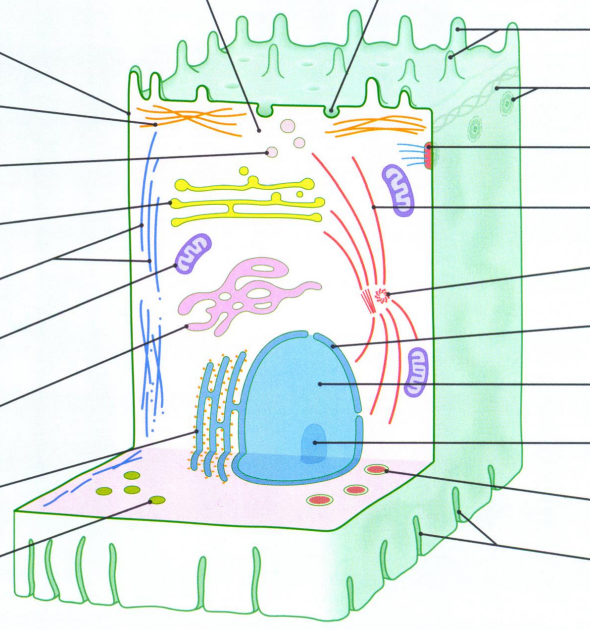
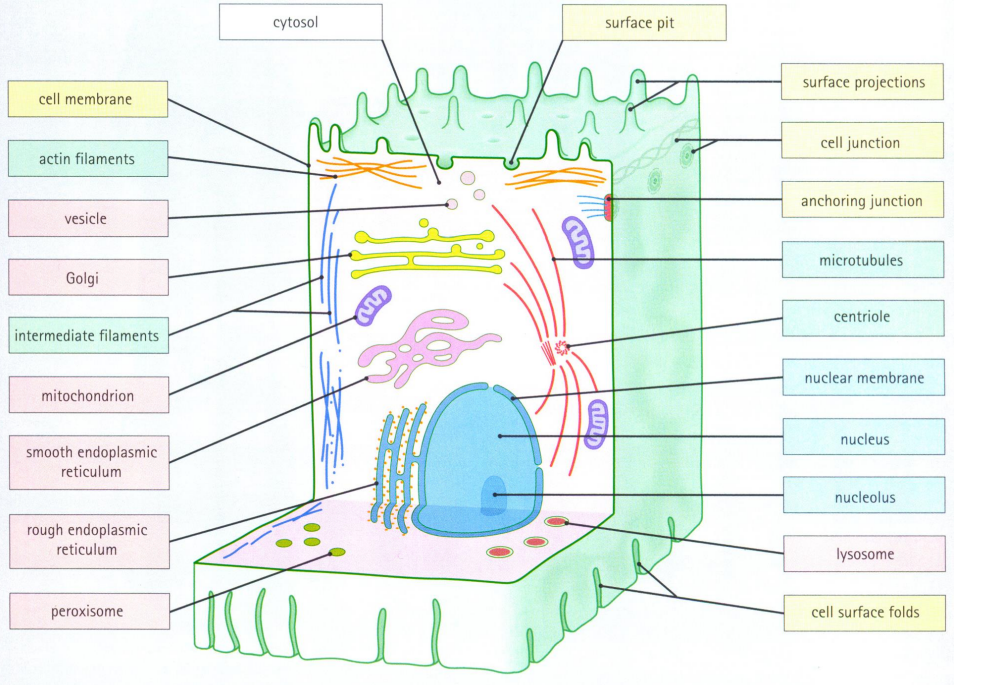
Name the main constituents of a cell
peroxisome
cell surface folds
centriole
anchoring junction
cell junction
How many different cell types are there in the human body?
Over 200 different cell types
How are different cell types and tissues organized in the human body, and why is each type of cell considered equally important?
Each type of cells is specialised to carry out a particular function, either solely, but usually by forming a particular tissue
Different tissues then combine and form specific organs, where the organ is like a factory where every type of cell has its own job
Since every tissue has its own function that contributes to the multifunctionality of an organ, every type of cell is equally important
What are the two major divisions of cells in the human body?
The two major divisions of cells are sex cells (gametes) and somatic cells.
What is the role of sex cells (gametes)?
Sex cells are involved in sexual reproduction and include sperm and oocytes.
What defines somatic cells, and what types of tissues do they make up in mammals?
Somatic cells are all other cells in the human body, excluding gametes, germ cells, gametocytes, or undifferentiated stem cells. In mammals, somatic cells make up all internal organs, skin, bones, blood, and connective tissue.
How many types of somatic cells are estimated to be in the human body?
There are approximately 220 types of somatic cells
What does the cell membrane separate?
Separates the cell contents (cytoplasm) from the extracellular fluid (interstitial fluid)
What are the main functions of the cell membrane?
Functions:
Physical isolation
Regulation of exchange with the environment
Sensitivity to the environment
Structural support
What does cytoplasm consists of?
cytosol + organelles
What does cell membrane consists of?
Membrane is formed by a lipid bilayer, with specialised proteins and surface carbohydrates
How lipid bilayer formed?
Forms a bilayer in water
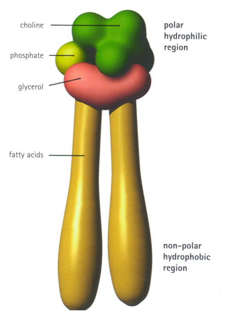
Describe the different parts of phospholipid
Each lipid molecule is amphipathic
hydrophilic end (phosphate)
hydrophobic end (lipid)
What are three different membrane lipids and their functions?
Phosphoglycerides (phospholipids)
~50% of membrane lipid
surround and anchor proteins
phosphatidylcholine, phosphatidylserine, phosphatidylethanolamine (only have to recognise name)
Cholesterol
stabilises membrane
Glycolipids
Intercellular communication
E.g. sphingolipids, gangliosides
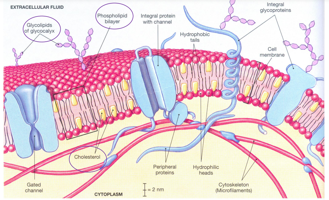
What are functions of membrane proteins?
Functions:
Attach cytoskeletal filaments to cell membrane
Attach cells to extracellular matrix
Transport molecules into and out of cells
Act as receptors for chemical signalling between cells
Possess specific enzymatic activity
What is an integral membrane protein (IMP), and how does it differ from peripheral membrane proteins?
An integral membrane protein (IMP) is permanently attached to the biological membrane, while peripheral membrane proteins adhere only temporarily and can attach to integral membrane proteins or penetrate the peripheral regions of the lipid bilayer.
What role do peripheral membrane proteins play, particularly in relation to ion channels and transmembrane receptors?
Peripheral membrane proteins can act as regulatory protein subunits for many ion channels and transmembrane receptors, influencing their function and activity.
What significance do integral membrane proteins (IMPs) have in an organism's genome?
Integral membrane proteins make up a substantial portion of the proteins encoded in an organism's genome.
What are some key functions of membrane carbohydrates?
Functions:
Lubrication and protection
Anchoring and locomotion
Specificity in binding
Recognition
Where are membrane carbohydrates primarily located and what is the name of the coating they form?
Mainly on extracellular surface, as a coating called the glycocalyx
Different types of transport across cell membranes
• Diffusion (gases, lipophilic or small molecules)
• Active transport (Na2+ ions)
• Bulk transport
- endocytosis
- pinocytosis
- phagocytosis
- exocytosis
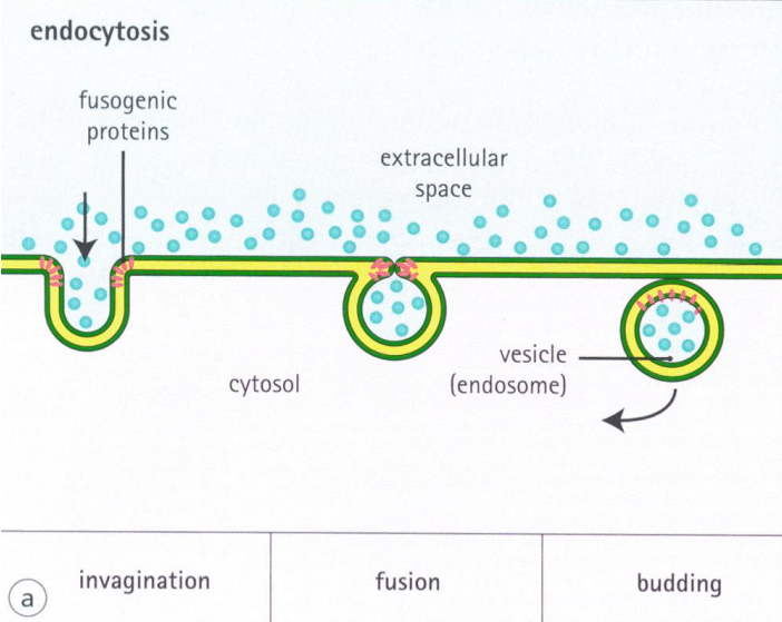
What is endocytosis and describe the process?
Endocytosis is a cellular process in which substances are brought into the cell. The material to be internalized is surrounded by an area of cell membrane, which then buds off inside the cell to form a vesicle containing the ingested material.
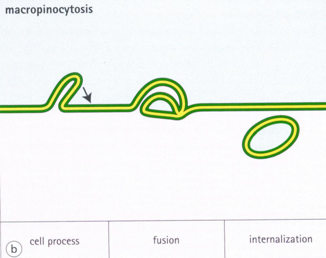
What is pinocytosis, and what are the steps involved in this process of endocytosis?
Pinocytosis, otherwise known as fluid endocytosis and bulk-phase pinocytosis, is a mode of endocytosis in which small particles suspended in extracellular fluid are brought into the cell through an invagination of the cell membrane, resulting in a suspension of the particles within a small vesicle inside the cell. These pinocytotic vesicles subsequently fuse with endosomes to hydrolyze (break down) the particles.
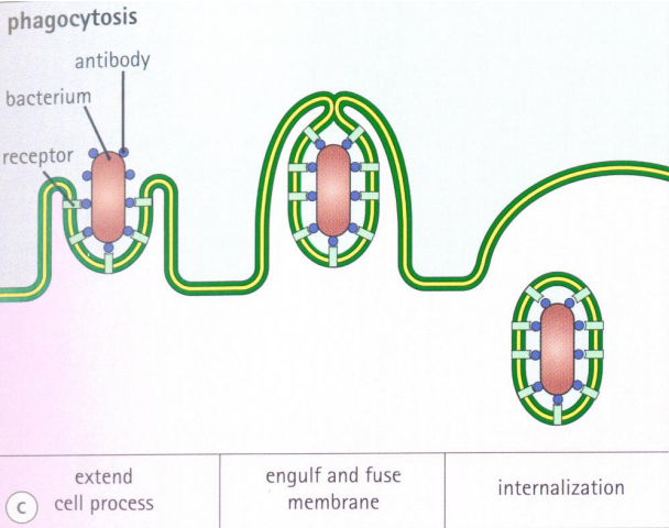
What is phagocytosis, and what role does it play in the immune system of multicellular organisms?
Phagocytosis is the process by which a cell uses its plasma membrane to engulf a large particle, giving rise to an internal compartment called the phagosome.
It is one type of endocytosis. In a multicellular organism's immune system, phagocytosis is a major mechanism used to remove pathogens and cell debris. It has to have receptor for antibody
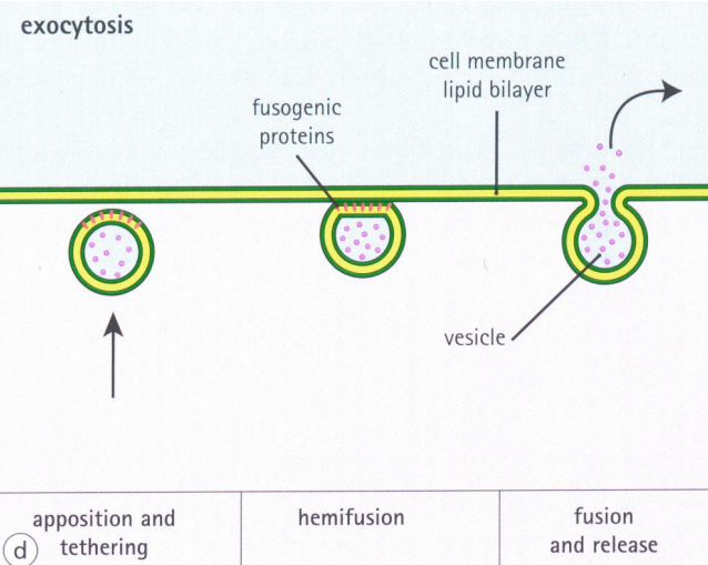
What is exocytosis?
Exocytosis is a form of active transport and bulk transport in which a cell transports molecules out of the cell by secreting them through an energy-dependent process. Opposite to endocytosis
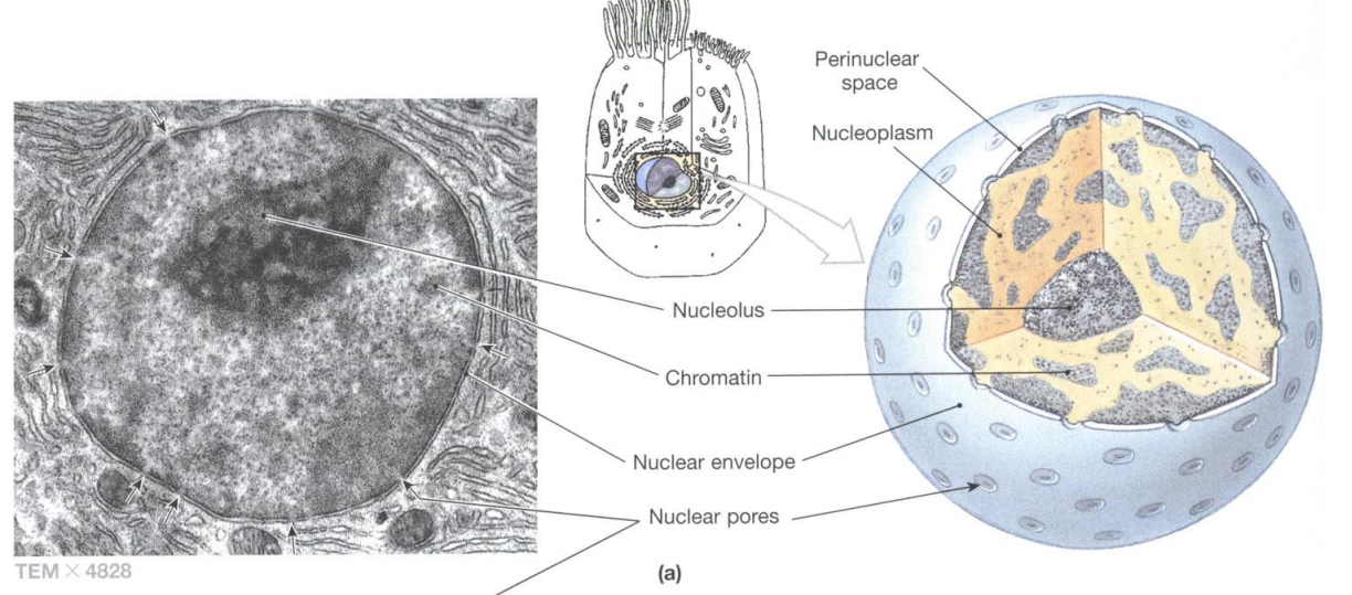
What are nucleus bounded by?
Nuclear envelope
How does the nucleus interact with the cytoplasm?
Communicates with cytoplasm through nuclear pores
What does nucleus contain?
- cellular DNA
- nucleoli (rRNA, mRNA, tRNA)
- nucleoproteins
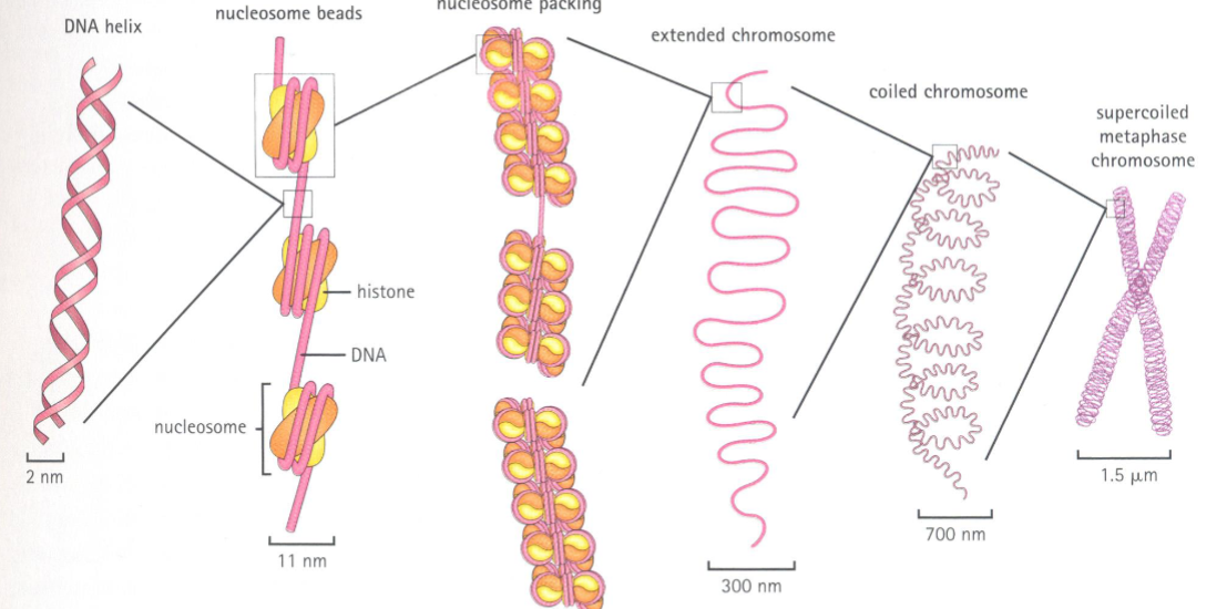
What is chromatin, and what is its primary function in eukaryotic cells?
Chromatin is a complex of DNA and protein found in eukaryotic cells. Its primary function is packaging long DNA molecules into more compact, denser structures
How does DNA interact with histone proteins to form euchromatin?
DNA wraps around histone proteins to form nucleosomes, creating a "beads on a string" structure known as euchromatin
What is heterochromatin, and how is it formed from nucleosomes?
Heterochromatin is the most compact form of chromatin, formed when multiple histones wrap into a 30-nanometer fiber consisting of nucleosome arrays.
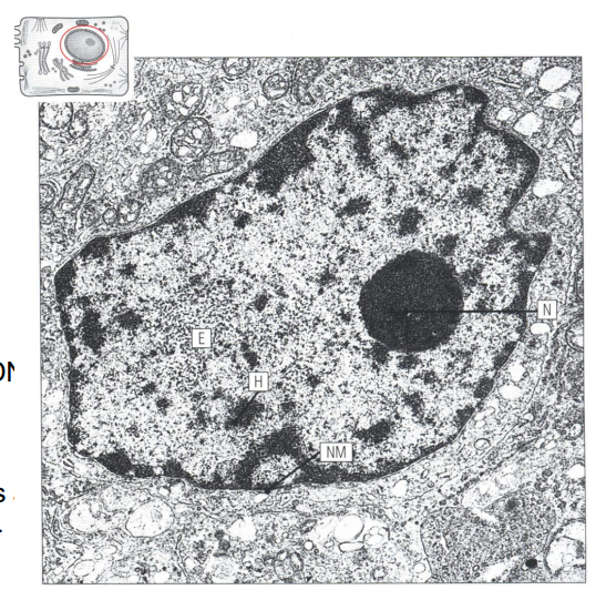
What are (N), (H) and (E) in this picture?
The nucleus (N) is clearly visible as an electrode-dense circular area. Nucleus chromatin is divided into two types: heterochromatin (H) is dense-staining, whereas euchromatin (E) is light-staining
How is DNA organized from nucleosomes to the supercoiled structure found in chromosomes?
DNA is organised around histones into nucleosomes. The nucleosomes are wound into a helix to form chromatin. In chromosomes this is then wound again into a supercoiled structure.
What is Endoplasmic Reticulum (ER)?
Network of membranous tubules, vesicles and cisternae
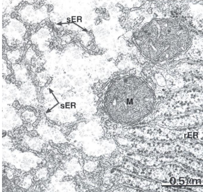
What are the two types of Endoplasmic Reticulum, and what are their respective functions?
The two types of Endoplasmic Reticulum are rough ER and smooth ER. Rough ER, with ribosomes on its surface, is involved in protein synthesis, while smooth ER functions in lipid synthesis, membrane synthesis, and repair.
What are the functions of the Golgi Apparatus in the cell, and what are its structural components?
The Golgi Apparatus is responsible for sorting, packaging, and transporting cell products.
It modifies macromolecules by adding sugars, performs proteolysis to activate peptides, sorts macromolecules into vesicles, transports lipids, and creates lysosomes. Its structural components include sacs or folds called cisternae.
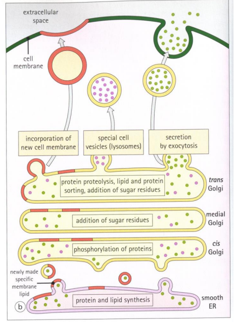
What are lysosomes, and what is their primary function in the cell?
Lysosomes are membrane-bound spherical organelles containing hydrolytic enzymes that break down various biomolecules engulfed by the cell. A lysosome has a specific composition, of its membrane proteins, and luminal proteins
What is the pH of the lysosomal lumen, and why is it important?
The pH of the lysosomal lumen is around 4.5–5.0, which is optimal for the enzymes involved in hydrolysis, similar to the activity of the stomach.
In which cellular processes are lysosomes involved, besides biomolecule breakdown?
Involved in various cell processes-plasma membrane repair, cell signalling, and energy
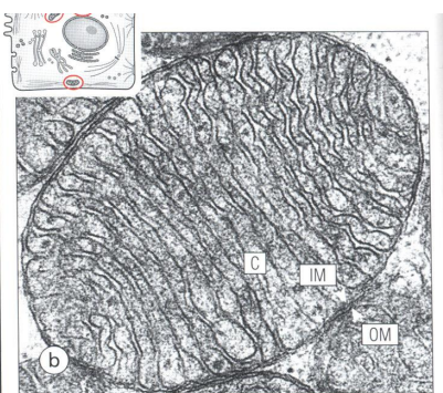
What is OM, IM and C in picture of mitochondrion?
Outer membrane (OM), inner (IM) membrane and cristae (C)
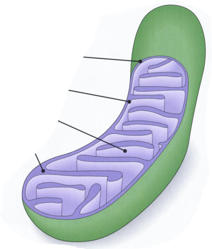
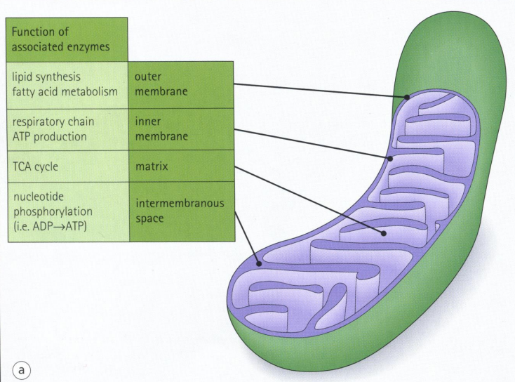
What is the structure of mitochondria and what are functions of different parts?
Intramembranous space
What are mitochondrias?
Mitochondria are membrane-bound organelles consisting of outer and inner membranes, separated by an intermembranous space.
What is the primary function of mitochondria within the cell?
Mitochondria are responsible for energy production in the form of ATP.
What components are found in the mitochondrial matrix?
Matrix contains many enzymes and small amounts of mitochondrial DNA
What are the three main components of the cytoskeleton in eukaryotic cells?
Microfilaments, intermediate filaments, and microtubules
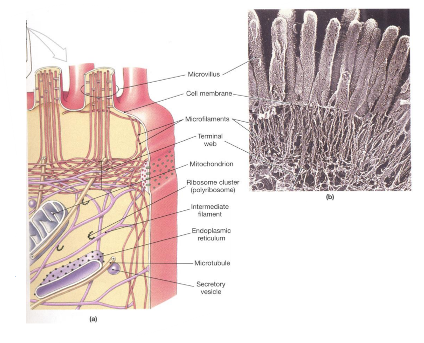
What are microfilaments composed of, and what are their key functions?
Microfilaments are long, thin filaments made of actin (5 nm in diameter). Their key functions include cytokinesis, amoeboid movement, cell motility, and changes in cell shape.
What are intermediate filaments, and how do they support durable structures like hair and fingernails?
Intermediate filaments, such as desmin, glial fibrillary acidic protein (GFAP), keratin, lamin, neurofilaments, and vimentin, are 10 nm cytoskeletal components. They provide stability and are found in durable structures like hair, fingernails, and scales
What role do microtubules play in the cytoskeleton, and what are they composed of?
Microtubules, composed of α and β tubulin (25 nm in diameter), provide platforms for intracellular transport and are involved in various cellular processes, including the movement of secretory vesicles and organelles.
What are the overall functions of the cytoskeleton in a cell?
The cytoskeleton provides strength and structural support for the cell and its organelles, facilitates movement of organelles, and enables changes in cell shape through interactions between its components.
How are nuclear pores formed, and what is the significance of the nuclear pore complex?
Nuclear pores are formed by concentric rings of subunits that create the nuclear pore complex, which facilitates transport between the nucleus and cytoplasm.
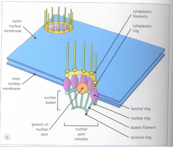
What is the function of nuclear pores regarding molecular transport?
Nuclear pores form channels for the transport of small molecules while restricting the passage of large molecules between the cytosol and the nucleus.
How are nuclear pores formed?
Pores are formed by concentric rings of subunits to form the nuclear pore complex.
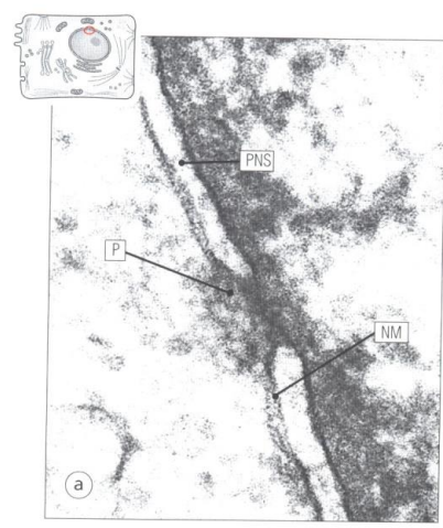
What is NM and P?
Double nuclear membrane (NM) is perforated by nuclear pores (P) –appear as gaps
What structures radiate from the rings above and below the large protein units in nuclear pores?
Filaments radiate from the rings above and below the large protein units in the nuclear pores, extending into nuclear and cytoplasmic spaces.

What structures radiate from the rings above and below the large protein units in nuclear pores?
Filaments radiate from the rings above and below the large protein units in the nuclear pores, extending into nuclear and cytoplasmic spaces.
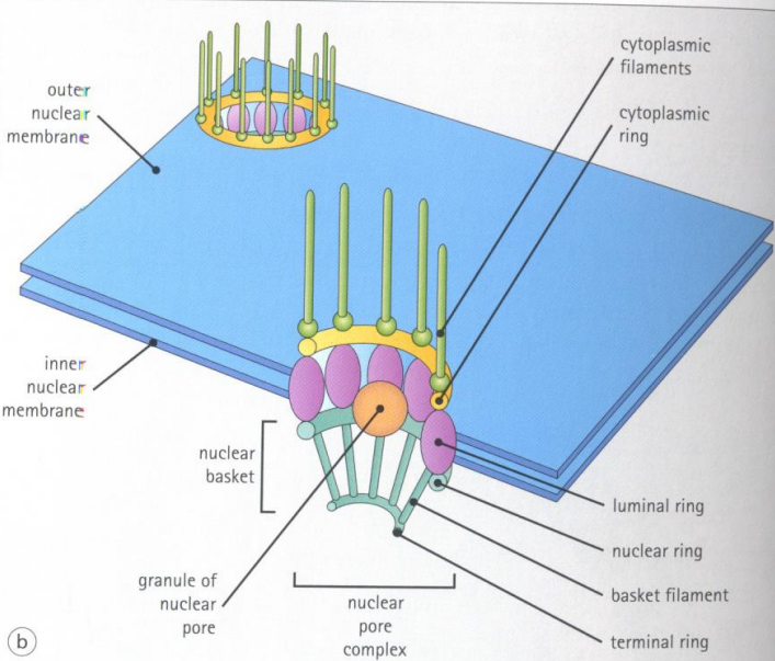
What is the nuclear basket?
The nuclear basket is the structure formed by the rings and filaments

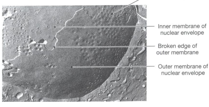
What technique is used to make the internal structures of a cell visible by breaking it apart?
The technique used is called freeze fracture or freeze etching
What specific structures are visible in the cell after applying the freeze fracture technique?
The nuclear envelope and nuclear pores are visible after applying the freeze fracture technique.
What effect does the freeze fracture process have on the outer membrane of the nuclear envelope?
The freeze fracture process breaks away part of the outer membrane of the nuclear envelope, allowing the cut edge of the nucleus to be seen.
How is DNA structured at its most basic level with histones?
At the most basic level, DNA wraps around histone proteins to form structures called nucleosomes, which resemble "beads on a string." This arrangement is known as euchromatin, a less compact form of chromatin.

What is the difference between euchromatin and heterochromatin in terms of DNA organization?
Euchromatin is a loosely packed form of DNA where nucleosomes form a "beads on a string" structure. Heterochromatin, on the other hand, is more compact, where nucleosomes are tightly packed into a 30-nanometer fiber.