Genetics Block Bev's Cards WEEK ONE
 Call Kai
 Learn
Learn Practice Test
 Spaced Repetition
 Match
Match Flashcards
 Knowt Play
1/338

Earn XP
Description and Tags
Week slide info with all the practice problems from workshop, quiz and practice problem worksheet
Name | Mastery | Learn | Test | Matching | Spaced | Call with Kai | Chat |
|---|
No analytics yet
Send a link to your students to track their progress
339 Terms
Central dogma
DNA to RNA to Protein
how many autosome pairs do humans have
22
Over half of the genome is
highly repetitive
How many of our genes code protein?
<2%
DNA sequence of any 2 individuals is
99.9% identical
tandem repeats
repeated DNA sequences are next to each other (adjacent).
Example: CAG-CAG-CAG-CAG
Dispersed repeats
Interspersed/dispersed repeats: repeated sequences are spread out in different locations in the genome.
Example: CAG-XXXXXX-CAG-XXXXXXXX-CAG
single copy sequence
• Protein coding sequence: ~1.5% genome
• Introns and noncoding RNAs: ~40%
• Gene Families
• Pseudogenes
Microsatellites
aka Short tandem repeats (STRs)
Clusters of very short tandem repeats, 2-6 bp
• (CA)n, where n= 5 to several hundred
Highly polymorphic (means that the number of repeat units can vary a lot between different people) – many possible numbers of repeats
• Analyze by PCR using primers
Dispersed repeats
• SINEs: Short repeated sequences scattered throughout genome
• LINE: Long repeated sequences scattered throughout genome
• Low copy repeats (LCR): Large DNA sequences repeated

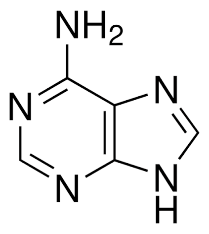
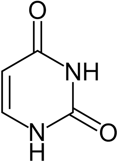
adenine
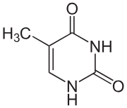
Thymine
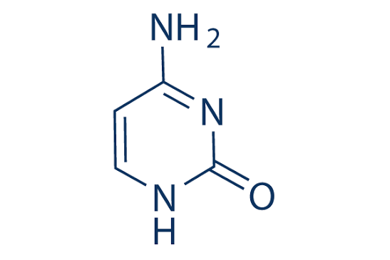
Cytosine
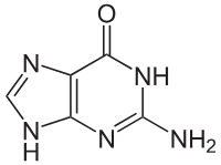
Guanine
Uracil
Chargaff’s Rules
%A ≈ %T and %G ≈ %C
The discovery of the double helix nature of DNA
Rosalind Franklin had been using x-ray crystallography to characterize the
structure of DNA
How does this DNA fit into such a small volume?
DNA is organized by histones & nucleosomes
These subunits can be modified post-translationally to affect chromatin accessibility and gene expression
• 8 histone subunits make up a nucleosome
RNA primase synthesizes
short priming sequences for DNA replication
DNA polymerase I
adds nucleotides in the 5’ -> 3’ direction
lagging strand requires formation of
Okazaki fragments and ligation activity to seal the nicks
RNA can be hydrolyzed due to
the 2’ OH
Regulation of transcription and production of mRNA (What determines whether a gene gets turned ON and an mRNA gets made?)
• Enhancers
• Core promoter
• TATA Box
• Transcription factors
• Transcript start site
Enhancers
An enhancer is a region of DNA that can increase transcription of a gene
Enhancers contain binding sites for transcription factors
an enhancer doesn't necessarily have to be right next to the gene
Core promoter
the region of DNA right around the beginning of a gene where the transcription machinery assembles.
TATA box
a specific DNA sequence found in the core promoter of some genes.
Proteins recognize this region and help position RNA Polymerase II correctly.
Transcription factors
Transcription factors are proteins that regulate transcription.
They bind to specific DNA sequences and can either:
Increase transcription → activators
Decrease transcription → repressors
mRNAs
make up ~5% of the total RNA in the cell and encode proteins
mRNAs are transcribed by
RNA polymerase II
mRNAs typically have (2 things)
• Almost all mRNAs in human undergo (blank)
mRNAs typically have a 5’ cap and a poly-adenine tail
• Almost all mRNAs in human undergo splicing
The 5’ cap protects the 5’ end of mRNA from degradation, and also recruits
translation initiation factors
Alternative splicing
Same DNA/gene → same initial pre-mRNA → different splicing → different mature mRNAs → different proteins
Nonsense-mediated decay
NMD detects certain mRNAs that contain a premature stop codon (>55 nucleotides away from the next splice junction) and destroys the mRNA
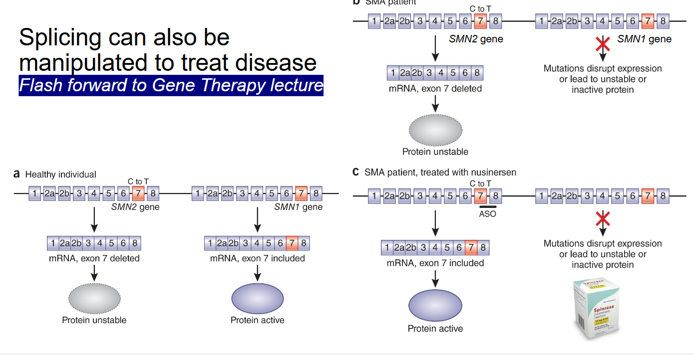
Splicing can be dysregulated in disease (disease example)
Hutchinson-Gilford progeria syndrome is caused by a genetic typo in the LMNA gene
Splicing can also be manipulated to treat diease
SMA patient treated with nusinersen (modifies the SMN2 gene)
RNA processing – cleavage& polyadenylation
mRNAs are cleaved and then polyadenylated by protein complexes
The polyA tail protects the 3’ end of the RNA from degradation
Ribosomal RNA makes of (% of RNA) and is transcribed by what?
Ribosomal RNA makes of >90% of the RNA in the cell
Ribosomal RNA is transcribed by RNA polymerase I
tRNA
• tRNAs are “charged” with amino acids
• tRNAs have an anti-codon loop that is complementary to mRNA
• tRNAs are transcribed by RNA polymerase III
• Some tRNAs must be spliced to be functional
Mutations in tRNA splicing machinery can cause
pontocerebellar hypoplasia and other diseases
Speed of translation can be influenced by
codon identity and tRNA abundance (which is why silent mutations can still be harmful)
Translation initiation
• Initiation factors recognize the 5’ cap
• The 40s subunit is recruited and begins scanning for AUG
• Upon reaching an AUG with a suitable Kozak sequence, the 60s subunit joins
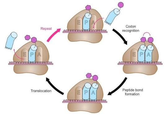
Translation Elongation
• The tRNA in the peptidyl (P) site is the one that is charged with the growing polypeptide chain
• An amino-acylated tRNA is delivered to the aminoacyl (A) site
• A new peptide bond is formed between the polypeptide chain and the charged tRNA in the A site
• tRNAs in the P and A site translocate into the exit (E) and P sites, respectively
Translation termination
• Eukaryotic release factor 1 (eRF1) recognizes the stop codon (UAG, UGA, UAA)
• Together with eRF3, it facilitates hydrolysis to release the nascent peptide chain
Proteins can be regulated post-translationally
• Ubiquitination
• Phosphorylation
• Sumoylation
• Dimerization
• Protein complex formation
MicroRNA
microRNAs are ~21 nucleotide long non-coding RNAs that exert regulatory activities on other RNA
Missense variant
different amino acid
Nonsense variant
stop codon
Frame-shift
change reading frame of message
DNA → RNA
transcription
RNA → protein
translation
“Variant” versus “Mutation”
avoid negative connotations of the word “mutation”
5 Classifiers are used to describe the effect of a variant
Pathogenic (P)
Likely
Pathogenic (LP)
Variant of Uncertain Significance (VUS)
Likely Benign (LB)
Benign (B)
SNVs
Single Nucleotide Variant
Genomic nomenclature
based on sequencing ALL DNA (intergenic, intragenic, exons, and introns
NC- Nomenclature Chromosome
LMNA NC_000001.11:g.156138613C>T
cDNA nomenclature
based on exons OR exons + some intronic (+/-100bp)(exome sequencing)
NM- Nomenclature mRNA
Example: LMNA NM_170707.4:c.1824C>T
Protein nomenclature
based on amino acid change resulting from DNA variant(s)
Example: LMNA NM_170707.4:c.1824C>T p.(G608G) OR p.(G608=)
cDNA – what is it?
DNA complementary to the mRNA sequence (rna degrades too quickly)
+ (plus) Nomenclature rule
indicate intronic variants;
c.123+45A>G = 45 nucleotide after the previous exon’s last nucleotide
(You are MOVING FORWARD)
- (minus) Nomenclature rule
used in nucleotide numbering, indicate intronic OR promoter/5’UTR variants;
c.124-56C>T = 56 nucleotides before the next exon’s first nucleotide
(YOU ARE MOVING BACK)
+1 or +2 in nomenclature (general area)
5′ splice donor site
-1 or -2 in nomenclature (general area)
3′ splice acceptor site
* (asterisk) nomenclature
to indicate a translation termination (stop)
examples:
c.*32G>A A G was changed to an A, 32 DNA letters after the stop codon
p.Trp41*
c.*32G>A
A G was changed to an A, 32 DNA letters after the stop codon
p.Trp41*
At position 41 of the protein, the amino acid tryptophan (Trp) was changed into a STOP signal
_ (underscore) nomenclature
is used to indicate a range; g.12345_12678del
fs nomenclature
indicates a frameshift
p.Arg456Glyfs*17
At amino acid 456, the sequence gets shifted, changing Arg to Gly, and the new incorrect sequence continues for 17 amino acids before hitting a STOP
Variant Allele Frequency (VAF)
% of cells in which a variant is found
VAF of 0.5 or 50%
means the variant is heterozygous in 100% of cells
VAF >50%
may indicate homozygosity and/or deletion of the other allele
VAF ~30-60%
may also be germline in a neoplastic assay
haploinsufficiency
happens when a single working copy of a gene is not enough to keep the body working normally
NONSENSE VARIANTS
Create premature stop codons
Abnormal mRNA may undergo (blank) IF the variant is >55nt upstream of the natural stop codon
nonsense-mediated decay (NMD) ,
this means no protein will be made
<55nt to natural STOP
truncated protein (abnormal, shortened protein) could lack function, may not be localized properly, or cause other problems
LMNA-related dilated cardiomyopathy (DCM) driven by
haploinsufficiency (AD) occurs when a single functional copy of the LMNA gene is insufficient to produce normal levels of nuclear lamin A/C proteins
nuclear lamina
which is involved in maintaining nuclear structure, chromatin organization, and gene regulation
INDELS
insertion/deletion
Can cause FRAMESHIFT variants
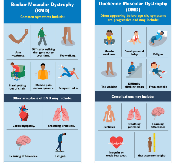
DMD/BMD (X-linked)
Most patients have large deletions of multiple exons
In-frame = Becker Muscular Dystrophy (milder symptoms)
Out-of-frame = Duchenne Muscular Dystrophy
CNVs
Copy Number Variants-the number of copies of a DNA segment varies between people.
Deletion or duplication
Copy Number Variants (CNVs): Deletion Disease example
Deletion of one or more copies of HBA or HBB can result in thalassemia traits or disease
Copy Number Variants (CNVs): Duplication Diseased Example
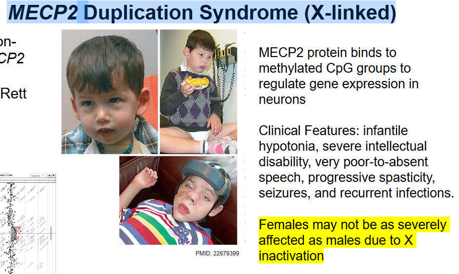
MECP2 Duplication Syndrome (X-linked)
How can synonymous variants cause phenotypic effects if the amino acid does not change?
Affect splicing & mRNA stability/regulation & Affect translation efficiency
SMA (Spinal Muscular Atrophy)
In people with SMA, both copies of the SMN1 gene are missing or faulty
SMA is an autosomal recessive condition
Our “backup” SMNA2, the more copies an SMA patient has, the better the outcomes
SNPs
Single Nucleotide Polymorphisms
To be polymorphic, at least 1% of the population has a variant allele (major vs minor) without obvious phenotypic effect
MISSENSE variants can have many effects
Loss of Function
Gain of Function
Dominant Negative
Stop-Loss
Loss-of-function
(Autosomal Recessive/Autosomal Dominant)
if one copy of a functional gene is sufficient, Heterozygotes (carriers) are NOT affected
Gain of function
Autosomal Dominant
results in protein with a new function OR excessive normal function (hypermorph)
Dominant negative
Autosomal Dominant
results in abnormal gene product affecting the function of the NORMAL gene product from the other allele
Stop-loss
Autosomal Dominant
results in loss of a stop codon, causing readthrough; may or may not result in an abnormal gene product
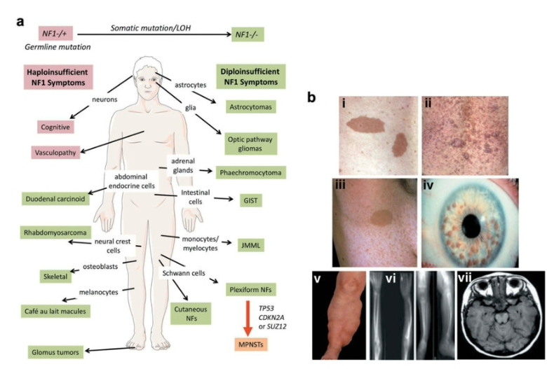
NF1
Neurofibromatosis type 1 (NF1)
MISSENSE AD Loss Of Function (LOF)
one of the most common AD genetic conditions
Familial Hypercholesterolemia
MISSENSE AD Loss Of Function (LOF)
Co-dominance – Both alleles in a trait are fully expressed but heterozygous and homozygous carriers may have different phenotypes
Sickle Cell Anemia
MISSENSE Autosomal Recessive LOF
(p.Glu7Val)
Achondroplasia
MISSENSE AD Gain-of-function (GOF)
FGFR3 gene
DOMINANT NEGATIVE Variants
4 Mechanisms:
1. Structural Disruption: Abnormal protein integrates into a multi-protein
complex and weakens the integrity of the complex
2. Competitive Inhibition: competes with the WT protein for binding, but does not
function properly, if at all
3. Sequestration: Abnormal protein prevents the normal protein (or another protein)
from functioning
4. Multimer interference: Abnormal protein subunit can still form multimer, but it
prevents the complex from functioning
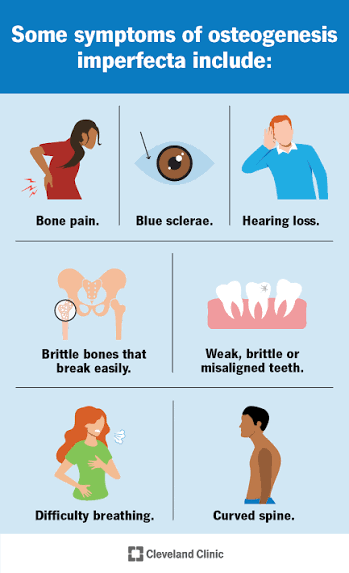
Osteogenesis Imperfecta
COL1A1
MISSENSE - Dominant Negative - Structural Disruption
Pathogenic missense SNVs alter the structural integrity of the collagen triple helix
Hutchison-Guilford Progeria syndrome
MISSENSE - Dominant Negative - Structural Disruption
C to T missense
Cryptic Splicing

von Willebrand Disease
MISSENSE - Dominant Negative- Competitive Inhibition
The most common inherited bleeding disorder
Abnormal vWF and wild type vWF both compete for platelet (PLT) binding
PCSK9-mediated familial hypercholesterolemia
MISSENSE - Dominant Negative- Sequestration
variants make abnormal pro-PCSK9 that is not cleaved and sequesters the normal PCSK9, preventing both from leaving
the Endoplasmic Reticulum
Abnormal p53 protein
MISSENSE - Dominant Negative - Multimer Interference
forms the tetramer with wild type and prevents p53 tumor suppressor activity
Can evolve to homozygous into neoplasia
Li-Fraumeni Syndrome