Week 4 Cellular Energetics + Cells, Environment, and Signaling
 Call Kai
 Learn
Learn Practice Test
 Spaced Repetition
 Match
Match Flashcards
 Knowt Play
1/94

There's no tags or description
Looks like no tags are added yet.
Name | Mastery | Learn | Test | Matching | Spaced | Call with Kai | Chat |
|---|
No analytics yet
Send a link to your students to track their progress
95 Terms
energy demand
what ATP production is driven by
glucose catabolism, lipolysis, protein catabolism
examples of metabolic pathways
oxidative phosphorylation
the primary energy-generating process in cells, taking place in the mitochondria, where the energy from the oxidation of nutrients is used to synthesize ATP (adenosine triphosphate).
UTP, GTP, ITP
energy equivalents to ATP
UTP
energy equivalent of ATP but used in RNA synthesis, glycogenolysis, nucleic acid synthesis mainly (pyramidine)
GTP
energy equivalent of ATP but used in ETC, protein synthesis, cell signaling, membrane dynamics (endo/exocytosis)
ITP
energy equivalent of ATP but used in nucleic acid synthesis mainly (purine)
translation
requires greater than or equal to 4 ATP equivalents to make a single bond in a protein (1 or 2 ATP to bind tRNA and amino acid bond, 1 GTP to deliver tRNA to the amino acid complex, 1 GTP to move ribosome, and +1 GTP for each initiation/termination, plus additional costs for editing!)
metabolic pathway purpose
function to synthesize ATP in response to decrease in ANC (adenine nucleotide charge), the response rate is related to enzyme concentration, enzyme activity, and availability of substrates
enzymes
typically the limiting factor in a reaction, since it can saturate and will no longer bind its substrate
end-product inhibition (in branched metabolic pathway)
regulatory mechanism for metabolic pathways, accumulation of the final product can inhibit enzymes in the reaction in order to favor different branches of the pathway (or directions of the pathway)
PKU deficiency
causes the metabolic pathway of Phenylalanine to not run correctly as one branch of the pathway doesn’t run, causing a buildup of phenylalanine and phenylpyruvic acid, which is bad for the system
about 4.4mM (relatively low)
concentration of ATP generally acroos tissue types
about 7.47 mM
concentration of ATP in cardiac muscle cells
about 5.86 mM
concentration of ATP in skeletal muscle
cardiac muscle, skeletal muscle, retina, brain
tissues that have higher ATP concentration compared the the rest of the body’s tissues
ATP demand
varies substantially across tissue types and even under differing conditions (ex: skeletal muscle needs 0.78 mmol/g/min at rest but 400 mM/min while sprinting)
constant
ATP levels are kept this way, even under greatly increased demand
adenine nucleotide charge
amount of adenosine in the form of ATP to all other forms of adenosine in the cell, marker of energetic state of tissue
([ATP] + 0.5[ADP]) / ([ATP] + [ADP] + [AMP])
ANC equation
0.80
resting ANC, can be lowered with increased ADP and AMP even if ATP is constant
glucose
primary fuel source during exercise and physical activity, typically delivered via blood from the liver, which stores much of the glycogen (muscles have intracellular stores)
glycogen
stored polymer of glucose, predominant in liver and skeletal muscle, is made in order to keep glucose concentrations down so that glucose uptake remains energetically favorable (low concentration)
epinephrine, ADP, AMP, drop in blood sugar
factors that stimulate glycogenolysis
increased blood sugar, insulin, ATP
factors that inhibit glycogenolysis
transporters on the sarcolemma
blood glucose entry happens in cells by way of these, works through facilitated diffusion that is regulated differently in different tissues
hexokinase
converts simple glucose to G6-P in an irreversible process so that glucose is retained for either glycolysis or glycogen synthesis
ATP cost of phase I
first 4 steps of glycolysis that required 2 ATP
ATP yield of phase II
last 5 steps of glycolysis that generates 4 ATP (so a net of 2 for all of glycolysis), 2 NADH, 2 pyruvate
2 ATP, 2 NADH, 2 ADP, 2 Pyruvate
Net products of glycolysis
outside of the mitochondria
where glycolysis occurs
lactate metabolism
occurs when there is an abundance of pyruvate, ie when there is low O2 and intense metabolic demand, “inadequate” TCA enzymes, Lactate dehydrogenase breaks down the excess pyruvate into lactate and regenerates NAD+, which allows glycolysis to continue
lactate
a marker for fatigue, is likely not a causative factor but more so just an indicator, but can also sustain activity as a fuel source by the cori cycle, ALSO directly works as a signal for increased oxidative phosphorylation
Cori Cyle
process in which liver takes up lactate and converts it into pyruvic acid where it can then go to the TCA cycle or continue on in the liver to be made into glucose!
Aerobic CHO metabolism
pyruvate not converted to lactate is taken up by the mitochondria, further catabolized to produce CO2 (in the Krebs) and eventually ATP (in the ETC)
matrix of the mitochondria
where the Krebs cycle is carried out
Acetyl-CoA
pyruvate is converted to this by PDH, which can then be used for many divergent metabolic pathways such as converted into cholesteral, fatty acids, ketone bodies, or to the citric acid cycle to create CO2
1 ATP (from 1 GTP), 4 NADH, FADH2, 2 CO2
Product of the TCA cycle
oxidative phosphorylation process
uses electrons and protons from FADH and NADH to create proton gradient, gradient is then used by ATP synthase to phosphorylate ADP, final electron acceptor is molecular oxygen
34 ATP
total yield from aerobic CHO metabolism of one glucose
beta oxidation
ATP regeneration via lipid metabolism is through this process, lipases break down the lipid storage forms into FFA for breakdown, occurs in mitochondria
carnitine shuttles
transfers FFA across the membrane to be broken down via beta oxidation (though may vary depending on tissue and FFA length)
MCFAs
(6-8) - (8-10) carbon chain
VLCFAs
22+ carbon chain
Beta oxidation products
though uses 1 ATP initially, produces more ATP than oxidative phosphorylation would (efficient!), a chain with n carbons gives n/2 - 1 cycles
protein catabolism
amino acids cleaved from protein strand, called deamination, generally undesirable because of buildup or urea and ammonia, is usually a last resort (cachexia is an example that happens to cancer patients whose bodies begin to break down their protein leading to wasting and fatigue and weight loss)
cell in situ
cells interface with other cells in an extracellular environment, mostly aqueous, temperature, pressure, pH, osmolarity, concentration, chemical composition may have an impact on the cell, due to variability, causes form and function changes potentially
solute
is dissolved into the solvent, examples are electrolytes (form ions), ions (charged species), non-electrolytes (don’t form ions)
solvent
the aquesous substance that contains solutes (H2O for biological systems)
molarity
number of moles of solute per liter of solution
molality
measure of amount of solute in a solution relative to the mass of solvent
equivalents
accounts for charge, is important in excitability, moles of an ion x |valence|
osmoles
non-diffusible in some part of system
osmolarity
function of number of solute particles in solution, ABSOLUTE measure, molecules/volumes (mass) H20
normal osmolarity inside the cell
280-294 mOsmoles/unit, slightly higher in plasma
tonicity
relative measurement within intracellular value as reference relates, describes the effect of a solution on cell volume by considering only the non-penetrating solutes across a cell membrane
hypotonic
tonicity that describes all solution with less solute than the cell, causes water to go into cell, leading to them bursting
isotonic
same concentration in and outside, no net increase or decrease of water in the cell
hypertonic
greater concentration of solute outside the cell, water moves out of the cell
equilibrium state
open systems tend to this due to brownian motion (random thermal movement), moves from high concentration to low concentration and leads to no net movement or flux
function of membrane in relation to equilibrium
membranes act as a barries to ions, polar molecules, and large molecules, which means they can separate aqueous compartments of dissimilar composition and maintain them, so may not be in a state of equilibrium
extracellular fluid high in:
Na+, Cl-, HCO3-, C6H12O6, fatty acids, amino acids, CO2
intracellular fluid high in:
K+, Mg2+, PO4-, proteins
diffusion
occurs based on concentration differences, from high concentration to low
things that affect diffusion
membrane barrier, changes in the difference in concentration across the membrane, thickness of the membrane (inhibits), area of the membrane (increases)
communication between cell and within cells often involves
changing barrier properties of membranes, increase or decrease permeability to some molecule
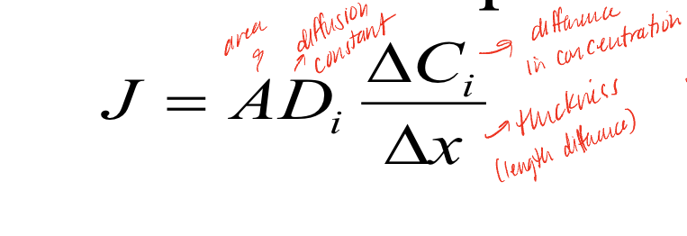
Fick’s diffusion Equation
J is the net flux, which is affected by the area, diffusion constant, change in concentration, and thickness of the membrane, additional factors not included in equation: temperature (higher increases J), size (lower - more diffusion), Ki (partition coefficient of solute i, higher causes more diffusion)
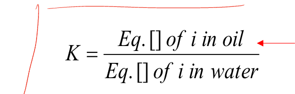
Ki
partition coefficient of solute i, how easily a solute can get across oil, which is similar to the lipid bilayer, the better then will diffuse into the cell
simple diffusion
type of passive transport, diffuses straight through the membrane
facilitated diffusion
type of passive transport, but goes through channels, gates, receptors, proteins due to concentration gradients (doesn’t require ATP)
active transport
goes against the concentration gradient, requires energy
co-transport
type of active transport in which the potential energy of a gradient for one molecule is used to transport another molecule against its gradient, can by symport (moving in the same direction), or anitport (moving in opposite directions)
osmosis
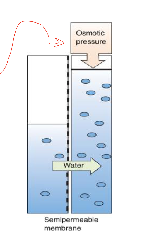
happens when solutes can’t move across the membrane to resolve concentration differences, net displacement/flow of of volume across a barrier due to concentration difference in matter, similar to diffusion but typically in biological systems is the movement of water
osmotic pressure
force that builds up as a counter pressure to the flow of water in osmosis, the build-up of fluid creates hydrostatic pressure to oppose further movement
reflection coefficient (sigma)
how good a sieve the membrane is, if = 1 is totally impermeable to solute, if 0 no discrimination between solute and water ( change in pi = osmotic pressure)

chemical signaling
signaling that occurs between cells and within cells or may come from the environment
mechanical signaling
example of this signaling is osmotic stress (creatine is an example?)
electrical signaling
signaling that occurs in excitable cells
endocrine signaling
hormone gland that doesn’t have ducts, example is thyroid, releases signaling hormone into the circulation for a distant target cell
autocrine signaling
signal is released from a cell and the target cell is that cell itself
paracrine signaling
one cell releases a signal, the target cell is a nearby cell
sensors
can be environmental, intracellular, intercellular, are needed to detect change, initiate signal and transmission, mostly at membranes (cell or organelles)
signaling components
sensors, transmission, response
receptors
membrane proteins, can be channels, gates, enzymes (examples are phophatases/kinases), lipases, ATPases, messengers (direct and indirect)
mechanical signal examples
piezo2 channels in merkel cells, as the surrounding keratinocytes move, the piezo channels are physically stretched open, allowing for the flux of neurotransmitters and a signal to occur, mechanical perturbation of collagen and laminin fibers in the extracellular matrix sends a mechanical signal to the integrins and focal adhesion complex, which sends another mechanical signal down the actin cytsokeleton and signals the nucleus to do proliferation, differentiation, protein synthesis, etc
2nd messenger examples
G-protein coupled receptors are examples, they first bind to a B-adrenergic receptor, activating the GDP into GTP, then they bind to adenylate cyclase(the effector), which leads to the conversion of ATP to cyclic AMP (the second messenger), which results in target reaction of activation of protein kinase A
insulin receptor signaling
another example of receptor mediated signaling, binding one receptor uses 2 different messenger systems to activate targeting and translocation of receptor vesicles
RAS pathway
example of growth factor signaling, binding of the growth factor activates the RAS complex, which then leads to the activation of the MAP kinase pathway, activating transcription. Normally, the pathway is inactivated by hydrolysis of GTP, but in cancer cases, the mutant RAS blocks this hydrolysis, and the “brakes” are taken off of the mechanism, leading to uncontrolled cell proliferation
cell communication: electrical signaling
the properties of biological membranes allow the development of electrical potential, due to selective permeability, and lipid is a good insulator/capacitor (can store charge), can use changes in potential to initiate and transmit signals
resting membrane potential
cells and extracellular fluid are electrically neutral, still, the characteristics of the membrane allow for a difference of charge on either side of the membrane, as the inside has a negative charge layer, and the outside of the cell has a net positive charge, this happens due to the sodium potassium pump, it pumps 3 Na+ out and pumps 2 K+ inside (only a change in 0.0000015% of the K+ is required to created it)
excitability
electrical phenomenon, critical to certain forms of information transmission, especially when rapid and localized, examples of cells with these properties are neurons, myocytes, beta cells, and mitochondria as well
diffusion of ions/electrolytes
influenced by two gradients, concentration and charge, still tends to equilibrium with equal distribution and no NET movement
impermeant
ion on one side of membrane, produce an equilibrium potential across the membrane with unequal distribution of ions, balance charge and concentration forces bc doesn’t allow that ion across the membrane at all
Gibbs-Donnan Equilibrium
systems with impermeant ions will tend to equilibrium of charges and concentration, this happens in our excitable cells, may lead to uneven distribution of concentrations of certain ions but has balanced charge, no energy is put into system
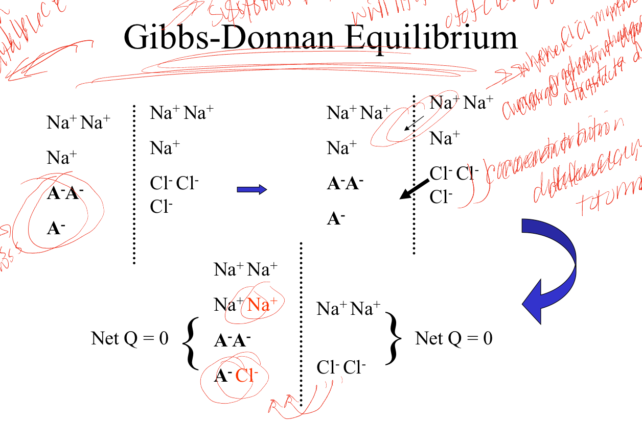
important distinction of Gibbs-Donnan Equilibrium
though compartments are neutral, for each diffusible ion there is an electrical and chemical gradient, also equilibrium is established by
