The Blood System
1/28
Earn XP
Description and Tags
Name | Mastery | Learn | Test | Matching | Spaced |
|---|
No study sessions yet.
29 Terms
Draw a diagram of the heart
Chambers
Two atria (singular = atrium) – smaller chambers near top of heart that collect blood from body and lungs
Two ventricles – larger chambers near bottom of heart that pump blood to body and lungs [Left ventricle has a thicker myocardium [=wall of heart]]
Heart Valves
Atrioventricular valves (between atria and ventricles) – bicuspid valve on left side ; tricuspid valve on right side
Semilunar valves (between ventricles and arteries) – aortic valve on left side ; pulmonary valve on right side
Blood Vessels
Vena cava (inferior and superior) feeds into the right atrium and returns deoxygenated blood from the body
Pulmonary artery connects to the right ventricle and sends deoxygenated blood to the lungs
Pulmonary vein feeds into the left atrium and returns oxygenated blood from the lungs
Aorta extends from the left ventricle and sends oxygenated blood around the body
![<p>Chambers</p><ul><li><p><strong>Two atria (</strong>singular = atrium) – smaller chambers near top of heart that collect blood from body and lungs</p></li><li><p><strong>Two ventricles</strong> – larger chambers near bottom of heart that pump blood to body and lungs [Left ventricle has a thicker <strong>myocardium</strong> [=wall of heart]]</p></li></ul><p>Heart Valves</p><ul><li><p>Atrioventricular valves (between atria and ventricles) – <strong>bicuspid</strong> valve on left side ; <strong>tricuspid</strong> valve on right side</p></li><li><p><strong>Semilunar valves</strong> (between ventricles and arteries) – aortic valve on left side ; pulmonary valve on right side</p></li></ul><p>Blood Vessels</p><ul><li><p><strong>Vena cava (inferior and superior)</strong> feeds into the right atrium and returns deoxygenated blood from the body</p></li><li><p><strong>Pulmonary artery</strong> connects to the right ventricle and sends deoxygenated blood to the lungs</p></li><li><p><strong>Pulmonary vein</strong> feeds into the left atrium and returns oxygenated blood from the lungs</p></li><li><p><strong>Aorta</strong> extends from the left ventricle and sends oxygenated blood around the body</p></li></ul>](https://knowt-user-attachments.s3.amazonaws.com/cf49954d-51d4-4000-9d2d-99ed134a2030.jpeg)
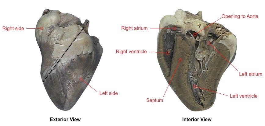
Identify components of a Dissected Sheep heart
Outline William Harvey’s discovery regarding the circulation of blood
Arteries and veins were part of a single connected blood network (he did not predict the existence of capillaries however)
Arteries pumped blood from the heart (to the lungs and body tissues)
Veins returned blood to the heart (from the lungs and body tissues)
Outline the double-circulatory system
The human heart is a four chambered organ, consisting of two atria and two ventricles
The atria act as reserviors, by which blood returning to the heart is collected via veins (and passed on to ventricles)
The ventricles act as pumps, expelling the blood from the heart at high pressure via arteries
The reason why there are two sets of atria and ventricles is because there are two distinct locations for blood transport
The left side of the heart pumps oxygenated blood around the body (systemic circulation)
The right side of the heart pumps deoxygenated blood to the lungs (pulmonary circulation)
There is therefore a separate circulation for the lungs (right side of heart) and for the rest of the body (left side of heart)
The left side of the heart will have a much thicker muscular wall (myocardium) as it must pump blood much further
Define the Systole
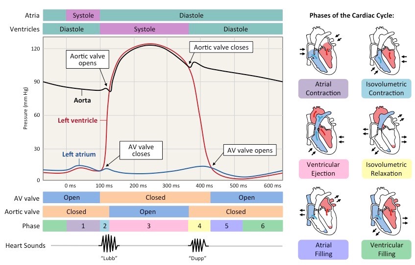
Blood returning to the heart will flow into the atria and ventricles as the pressure in them is lower (due to low volume of blood)
When ventricles are ~70% full, atria will contract (atrial systole) after signal from SA node, increasing pressure in the atria and forcing blood into ventricles
As ventricles contract, ventricular pressure exceeds atrial pressure and AV valves close to prevent back flow (first heart sound)
With both sets of heart valves closed, pressure rapidly builds in the contracting ventricles (isovolumetric contraction = No blood is ejected, and blood volume in ventricles remains constant)
When ventricular pressure exceeds blood pressure in the aorta and pulmonary artery, the aortic/pulmonary valve opens and blood is released into the aorta/pulmonary artery
Define the Diastole
As blood exits the ventricle and travels down the aorta/PA, ventricular pressure falls
When ventricular pressure drops below aortic/PA pressure, the aortic/PA valve closes to prevent back flow (second heart sound) = End of systole/Beginning of Diastole
When the ventricular pressure drops below the atrial pressure, the AV valve opens and blood can flow from atria to ventricle
Ventricles relax with all valves closed = Drop in pressure, no change in volume
Throughout the cycle, aortic pressure remains quite high as muscle and elastic fibres in the artery wall maintain blood pressure
Ventricular filling begins when Ventricular pressure drops below atrial pressure = Opens AV valve
Passive flow of blood from Atria to ventricles [as in first stage of systole]
Outline pressure changes during systole and diastole
Atrial Contraction (Ventricular filling)
AV valve closes
Isovolumetric Contraction of Ventricles
Semi-Lunar Valves open (Ventricular pressure exceeds aortic/PA pressure)
Ventricular ejection into Aorta/Pulmonary Artery
Semi-Lunar valves close
Isovolumetric Relaxation of the Ventricles (Atrial Filling)
AV valve opens (once Atrial pressure > Ventricular)
Passive filling of Ventricles
(repeat)
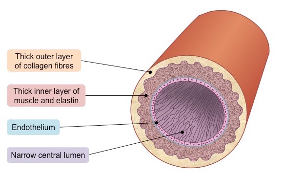
Outline the specialized structure of arteries to carry blood at high pressure away from the heart
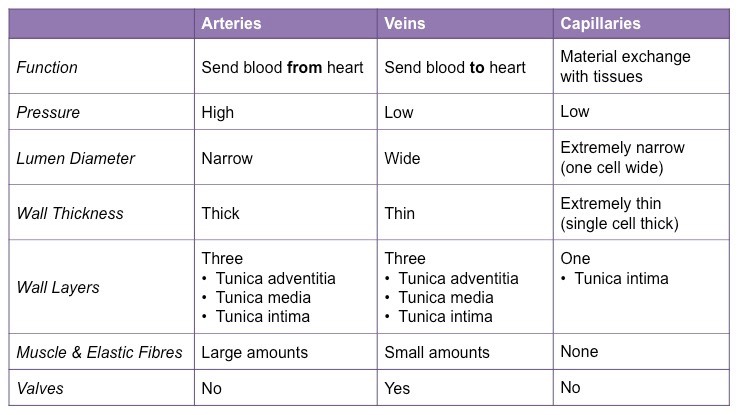
They have a narrow lumen (relative to wall thickness) to maintain a high blood pressure (~ 80 – 120 mmHg)
They have a thick wall containing an outer layer of collagen to prevent the artery from rupturing under the high pressure
The arterial wall also contains an inner layer of muscle and elastic fibres to help maintain pulse flow (it can contract and stretch)
Outline how muscle and elastin fibres aid in maintaining pressure between pumps
The muscle fibres help to form a rigid arterial wall that is capable of withstanding the high blood pressure without rupturing
Muscle fibres can also contract to narrow the lumen, which increases the pressure between pumps and helps to maintain blood pressure throughout the cardiac cycle
The elastic fibres allow the arterial wall to stretch and expand upon the flow of a pulse through the lumen
The pressure exerted on the arterial wall is returned to the blood when the artery returns to its normal size (elastic recoil)
The elastic recoil helps to push the blood forward through the artery as well as maintain arterial pressure between pump cycles

Outline the specialized structure of veins to carry blood at low pressure to the heart atria
They have a very wide lumen (relative to wall thickness) to maximise blood flow for more effective return
They have a thin wall containing less muscle and elastic fibres as blood is flowing at a very low pressure (~ 5 – 10 mmHg)
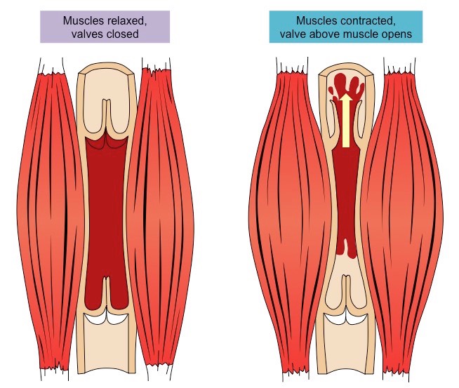
Because the pressure is low, veins possess one-way valves to prevent backflow and stop the blood from pooling at the lowest extremities
What’s the relationship between skeletal muscle and valves of veins
Veins typically pass between skeletal muscle groups, which facilitate venous blood flow via periodic contractions
When the skeletal muscles contract, they squeeze the vein and cause the blood to flow from the site of compression
Veins typically run parallel to arteries, and a similar effect can be caused by the rhythmic arterial bulge created by a pulse
Outline the function of capillaries
The function of capillaries is to exchange materials between the cells in tissues and blood travelling at low pressure (<10mmHg)
Arteries split into arterioles which in turn split into capillaries, decreasing arterial pressure as total vessel volume is increased
The branching of arteries into capillaries therefore ensures blood is moving slowly and all cells are located near a blood supply
After material exchange has occurred, capillaries will pool into venules which will in turn collate into larger veins
Outline the specialized structure of capillaries to perform material exchange
They have a very small diameter (~ 5 µm wide) which allows passage of only a single red blood cell at a time (optimal exchange)
The capillary wall is made of a single layer of cells to minimise the diffusion distance for permeable materials
They are surrounded by a basement membrane which is permeable to necessary materials
Explain how capillary structure may vary based on location in the body and its role
The capillary wall may be continuous with endothelial cells held together by tight junctions to limit permeability of large molecules
In tissues specialised for absorption (e.g. intestines, kidneys), the capillary wall may be fenestrated (contains pores)
Some capillaries are sinusoidal and have open spaces between cells and be permeable to large molecules and cells (e.g. in liver)
Explain material exchange in the capillaries
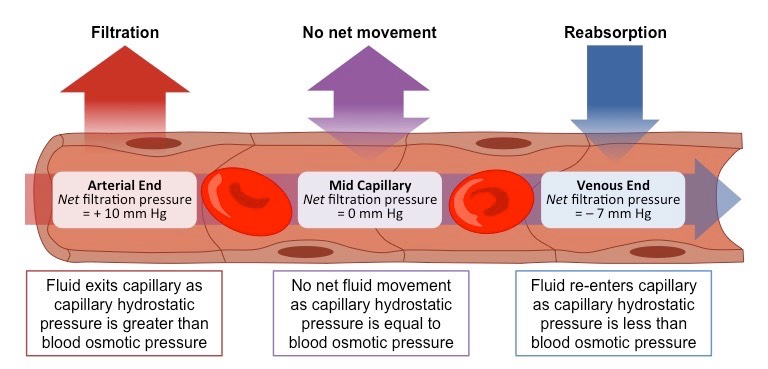
Hydrostatic Pressure → Pressure exerted by blood on capillary walls (or fluid on cell walls)
Higher within capillaries at arterial end (hence fluid moves out)
Hydrostatic Pressure > Osmotic at arterial end
Osmotic Pressure → Pressure to move water towards/into itself
E.g. Movement of water into cell/capillary
How to identify vessels
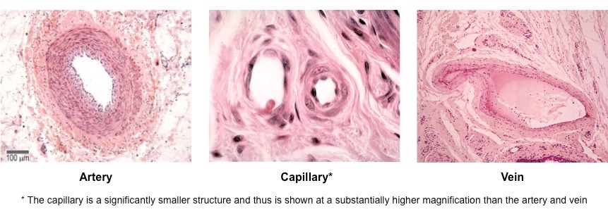
Arteries have thick walls composed of three distinct layers (tunica)
Veins have thin walls but typically have wider lumen (lumen size may vary depending on specific artery or vein)
Capillaries are very small and will not be easily detected under the same magnification as arteries and veins
What is the cause of Coronary Occlusion
Atherosclerosis is the hardening and narrowing of the arteries due to the deposition of cholesterol
Atheromas (fatty deposits) develop in the arteries and significantly reduce the diameter of the lumen (stenosis)
The restricted blood flow increases pressure in the artery, leading to damage to the arterial wall (from shear stress)
The damaged region is repaired with fibrous tissue which significantly reduces the elasticity of the vessel wall
As the smooth lining of the artery is progressively degraded, lesions form called atherosclerotic plaques
If the plaque ruptures, blood clotting is triggered, forming a thrombus that restricts blood flow
If the thrombus is dislodged it becomes an embolus and can cause a blockage in a smaller arteriole = Blockage of coronary arteries
Define Coronary Arteries
Coronary arteries are the blood vessels that surround the heart and nourish the cardiac tissue to keep the heart working
If coronary arteries become occluded, the region of heart tissue nourished by the blocked artery will die and cease to function
What are the risk factors of coronary heart disease [A Goddess]
Age – Blood vessels become less flexible with advancing age
Genetics – Having hypertension predispose individuals to developing CHD
Obesity – Being overweight places an additional strain on the heart
Diseases – Certain diseases increase the risk of CHD (e.g. diabetes)
Diet – Diets rich in saturated fats, salts and alcohol increases the risk
Exercise – Sedentary lifestyles increase the risk of developing CHD
Sex – Males are at a greater risk due to lower oestrogen levels
Smoking – Nicotine causes vasoconstriction, raising blood pressure
What are the consequences of Coronary Occlusion
Atherosclerosis can lead to blood clots which cause coronary heart disease when they occur in coronary arteries
Myocardial tissue requires the oxygen and nutrients transported via the coronary arteries in order to function
If a coronary artery becomes completely blocked, an acute myocardial infarction (heart attack) will result
Blockages of coronary arteries are typically treated by by-pass surgery or creating a stent (e.g. balloon angioplasty)
What does it mean when ‘Heart contraction is myogenic’
The signal for cardiac compression arises within the heart tissue itself
In other words, the signal for a heart beat is initiated by the heart muscle cells (cardiomyocytes) rather than from brain signals
Explain the Electrical Conduction of the heart
The sinoatrial node sends out an electrical impulse that stimulates contraction of the myocardium (heart muscle tissue)
This impulse directly causes the atria to contract and stimulates AV node at the junction between the atrium and ventricle
This second node – the atrioventricular node (AV node) – sends signals down the septum via a nerve bundle (Bundle of His)
The Bundle of His innervates nerve fibres (Purkinje fibres) in the ventricular wall, causing ventricular contraction
What are the Accelerator and Decelerator nerves [Brain’s role in heart rate regulation]
Nerves running from medulla (brain stem. Most primitive part of brain, regulating autonomic functions] to Sino-Atrial node
Stimulation of Accelerator - Increases heart rate
releases the neurotransmitter noradrenaline (a.k.a. norepinephrine)
Stimulation of Decelerator - Lowers heart rate
Release of neurotransmitter acetylcholine
What else may control the heart rate
Nerve signals from the brain can trigger rapid changes, while endocrine signals can trigger more sustained changes
Changes to blood pressure levels or CO concentrations (and thereby blood pH) will trigger changes in heart rate
How is heart rate affected by adrenaline
The hormone adrenaline (a.k.a. epinephrine) is released from the adrenal glands (located above the kidneys)
Adrenaline increases heart rate by activating the same chemical pathways as the neurotransmitter noradrenaline
Results in : Faster heart rate and glucose metabolism. Draws blood away from surface to core organs, resulting in hyper-awareness

What are the consequences of interference of heart’s pacemakers
The interference of the pacemakers will lead to the irregular and uncoordinated contraction of the heart muscle (fibrillation)
When fibrillation occurs, normal sinus rhythm may be re-established with a controlled electrical current (defibrillation)
What is blood composed of
Plasma
Consists mainly of water, which dissolves materials and functions as a transport medium
Contains electrolytes (minerals that carry a charge), which are important for maintaining fluid balance and blood pH
Proteins in the blood plasma maintain osmotic potential (albumin), transport lipids (globulin) and help clot (fibrinogen)
Blood plasma also functions to transport various materials needed by the body and wastes produced by body cells
Red Blood Cells
Red blood cells (erythrocytes) are responsible for transporting oxygen around the body
Oxygen is bound to haemoglobin at the lungs and released from the red blood cell at respiring body tissues
Buffy Coat
The buffy coat is the fraction of a blood sample that contains white blood cells and platelets
White blood cells (leukocytes) are involved in the body’s immune defence (eliminating infections)
Phagocytes - Engulf and destroy foreign particles
Lymphocytes - Synthesize antibodies
Platelets (thrombocytes) are involved in blood clotting (repairing damaged vessels to prevent blood loss)
Outline the composition of blood plasma
Nutrients – needed by cells to make chemical energy (e.g. glucose)
Antibodies – involved in pathogen identification and elimination
Carbon dioxide – waste produced as a by-product of cell respiration
Hormones – chemical messengers that move through the bloodstream
Oxygen – required by body tissues in order to respire aerobically
Urea – compound that is excreted to remove nitrogen from the body
Heat – not a molecule, but still an important component of blood plasma
Mnemonic: Nacho, Uh!