EXAM 4 objectives
1/12
There's no tags or description
Looks like no tags are added yet.
Name | Mastery | Learn | Test | Matching | Spaced | Call with Kai |
|---|
No analytics yet
Send a link to your students to track their progress
13 Terms
Describe the functions of the nervous system.
The nervous system is like the body's control center. It takes in information from the world, processes it, and tells the body how to respond. It helps with movement, thinking, emotions, and automatic functions like breathing. Super important for keeping everything running smoothly!
Explain the concept of integration and levels of integration
Integration, in the context of the nervous system, refers to the process of combining and interpreting sensory information to generate appropriate responses. It ensures that different signals from various parts of the body are coordinated, allowing for smooth and efficient functioning.
Spinal Cord Level: Reflexes and simple responses occur here without involving the brain. For example, pulling your hand away from a hot surface is a spinal cord-mediated reflex.
Brainstem Level: This level controls basic life functions, such as breathing, heart rate, and digestion, often operating without conscious thought.
Subcortical Level: The limbic system and basal ganglia contribute to emotions, movement coordination, and subconscious processing.
Cortical Level: The highest level of integration happens in the cerebral cortex, where complex thinking, problem-solving, learning, and voluntary movements take place.
Describe and draw the microscopic structure of a multipolar neuron
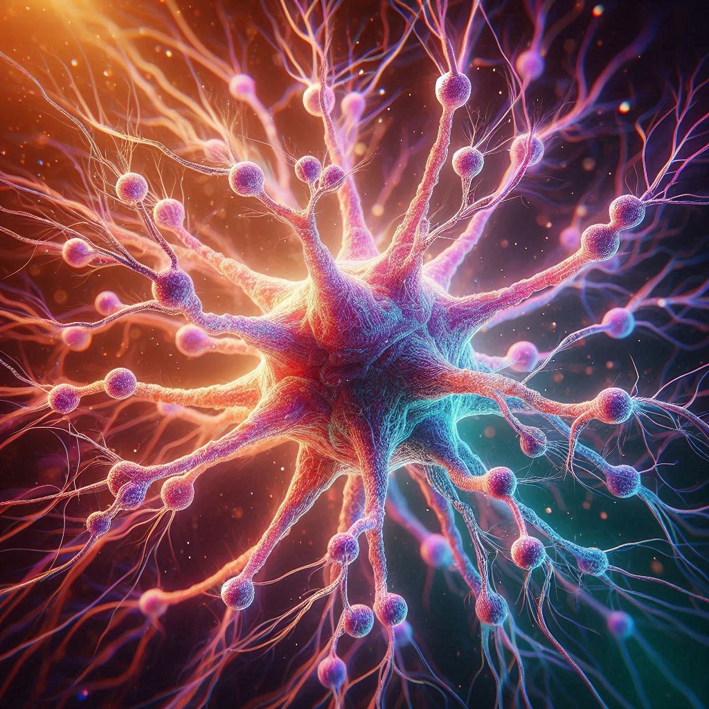
A multipolar neuron is a type of neuron that has multiple dendrites and a single axon. It's the most common type found in the central nervous system, especially in the brain and spinal cord.
Cell Body (Soma): Contains the nucleus and is responsible for maintaining the cell.
Dendrites: Branch-like structures that receive signals from other neurons.
Axon: A long projection that transmits impulses away from the cell body.
Axon Hillock: The base of the axon where action potentials begin.
Myelin Sheath: A protective covering (in myelinated neurons) that speeds up signal transmission.
Nodes of Ranvier: Gaps between myelin sheaths that help boost the signal.
Synaptic Terminals: Endings of the axon where neurotransmitters are released to communicate with other neurons.

Explain the origin of the resting membrane potential
The resting membrane potential originates from the unequal distribution of ions across the cell membrane, creating an electrical charge difference.
Explain the concept of “all or none”
The "all or none" principle refers to the way nerve impulses (action potentials) work. A neuron either fires completely or does not fire at all—there’s no in-between.
Explain how stimulation of a neuron produces an action potential and secretion of neurotransmitter
Stimulation & Depolarization
A stimulus (like a touch or chemical signal) causes sodium (Na⁺) channels in the neuron's membrane to open.
Na⁺ rushes into the cell, making the inside more positive.
If the change in voltage reaches the threshold, an action potential is triggered.
2. Propagation of Action Potential
The action potential moves down the axon, opening more Na⁺ channels along the way.
To reset, potassium (K⁺) exits the cell, restoring the negative resting potential.
This process continues until the signal reaches the axon terminals.
3. Neurotransmitter Release
When the action potential reaches the end of the axon, it triggers calcium (Ca²⁺) channels to open.
Ca²⁺ enters the neuron, causing synaptic vesicles to fuse with the membrane.
These vesicles release neurotransmitters into the synaptic cleft.
The neurotransmitters bind to receptors on the next neuron, passing the signal along.
Describe the structure and function of a synapse.
A synapse is the junction between two neurons or between a neuron and its target cell (like a muscle or gland). It serves as the site where neurons communicate by transmitting signals through chemical messengers called neurotransmitters.
Structure of a Synapse
Presynaptic Neuron – The neuron sending the signal.
Synaptic Terminal – The end of the presynaptic neuron’s axon, where neurotransmitters are stored in vesicles.
Synaptic Cleft – The small gap between two neurons where neurotransmitters are released.
Postsynaptic Neuron (or Target Cell) – The receiving cell that has specialized receptors for neurotransmitters.
Neurotransmitters – Chemical messengers (like dopamine or serotonin) that carry signals across the synaptic cleft.
Function of a Synapse
When an action potential reaches the synaptic terminal, calcium (Ca²⁺) channels open, triggering the release of neurotransmitters into the synaptic cleft.
Neurotransmitters bind to receptors on the postsynaptic neuron, causing an electrical or chemical response.
The signal is then passed on or inhibited, depending on the type of neurotransmitter.
Describe the development of the brain from the embryonic ectoderm
Neural Plate Formation (Week 3)
The ectoderm thickens to form the neural plate under the influence of signaling molecules.
This plate gives rise to the entire nervous system.
2. Neural Tube Formation (Week 4)
The neural plate folds, forming the neural groove, then closes to become the neural tube.
The neural tube later differentiates into the brain and spinal cord.
Improper closure can lead to defects like spina bifida.
3. Brain Vesicle Development
The rostral (head) end of the neural tube expands into three primary brain vesicles:
Forebrain (Prosencephalon) → Becomes the cerebrum and diencephalon.
Midbrain (Mesencephalon) → Forms the brainstem midbrain.
Hindbrain (Rhombencephalon) → Develops into the pons, medulla, and cerebellum.
4. Neuronal Migration & Cortical Development
Cells migrate to different brain regions, forming distinct layers and structures.
The cerebral cortex develops as neurons organize into functional circuits.
5. Synaptogenesis & Myelination (Later Fetal & Postnatal)
Neurons connect through synapses, refining brain communication.
Myelin begins to form, increasing the speed of neural transmission.
The brain continues developing well into childhood and adolescence.
list the major parts of the brain and give their functions
Cerebrum
Function: Controls thinking, memory, emotions, voluntary movements, and sensory processing.
Divisions: Divided into lobes—frontal, parietal, temporal, and occipital—that handle specific tasks.
2. Cerebellum
Function: Coordinates movement, balance, and posture.
Role: Helps with precision and smooth muscle movements.
3. Brainstem
Parts: Includes the midbrain, pons, and medulla oblongata.
Function: It regulates automatic functions like breathing, heart rate, digestion, and reflexes.
4. Diencephalon
Parts: Includes the thalamus, hypothalamus, and epithalamus.
Function: Controls sensory relay, hormone regulation, sleep-wake cycles, and homeostasis.
Draw and label mid-sagittal and frontal sections of the brain
List the major parts of the spinal cord and give their functions.
Cervical Region (C1–C8)
Controls movement and sensation in the neck, arms, and diaphragm.
C3–C5 are essential for breathing by controlling the diaphragm.
2. Thoracic Region (T1–T12)
Manages signals for the chest, upper back, and abdominal muscles.
Supports posture and stability.
3. Lumbar Region (L1–L5)
Governs movement and sensation in the hips, thighs, and lower legs.
Plays a key role in walking and balance.
4. Sacral Region (S1–S5)
Controls pelvic organs, including the bladder and reproductive functions.
Regulates movement in the feet and lower legs.
5. Coccygeal Region
The lowest part, linked to the tailbone.
Primarily involved in minor sensory functions.
Additional Structures:
Gray Matter: Processes incoming and outgoing signals.
White Matter: Transmits nerve impulses up and down the spinal cord.
Central Canal: Carries cerebrospinal fluid, which cushions and nourishes the spinal cord.
Identify the locations of the major myelinated tracts of the spinal cord and give their functions
The spinal cord has several major parts, each with a specific function:
Cervical region: Controls the neck, arms, and breathing muscles.
Thoracic region: Manages chest and abdominal muscles.
Lumbar region: Controls leg movement and balance.
Sacral region: Regulates bladder, reproductive organs, and foot movement.
Coccygeal region: Handles minor sensory functions near the tailbone.
Additionally:
White matter: Sends signals up and down the spinal cord.
Gray matter: Processes incoming and outgoing nerve signals.
Central canal: Holds cerebrospinal fluid for protection.
Describe the structure and function of the cerebellum.
The cerebellum helps with balance, coordination, and movement control.
Structure:
Location: At the back of the brain, under the cerebrum.
Hemispheres: Two sides connected by the vermis.
Arbor Vitae: Tree-like pattern of nerve fibers inside.
Cortex: Outer layer that processes movement signals.
Function:
Controls balance and posture.
Fine-tunes movement for precision.
Helps learn new motor skills.
Coordinates voluntary muscle movements.