Muscles of the Axial Skeleton on Head and Neck
1/87
There's no tags or description
Looks like no tags are added yet.
Name | Mastery | Learn | Test | Matching | Spaced |
|---|
No study sessions yet.
88 Terms
Depression of lower lip (pout)
Origin: Mandible
Insertion: muscles of lower lip
Innervation: Facial nerve (CN VII)
Depressor labii inferioris m.
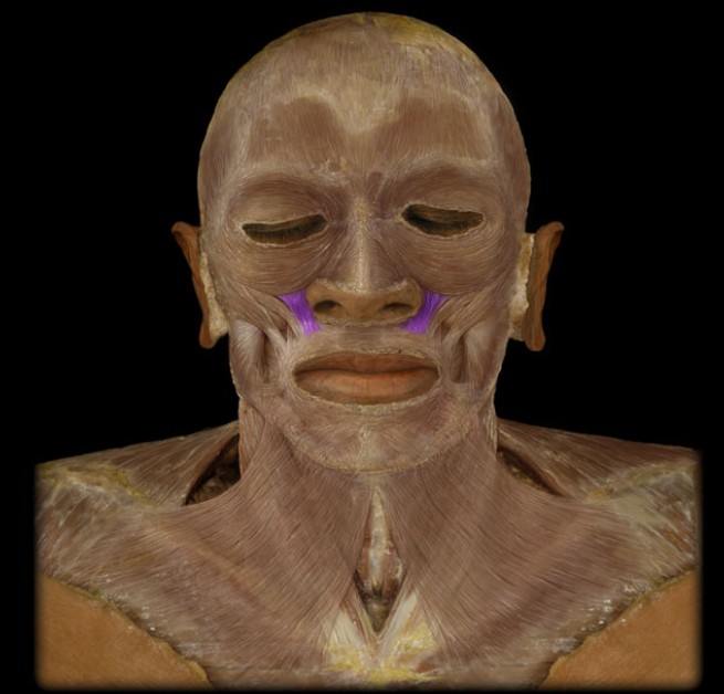
Action: Elevation of upper lip (sneer)
Origin: Maxilla, Zygomatic bone
Insertion: muscles of upper lip
Innervation: Facial nerve (CN VII)
Levator labii superioris m.
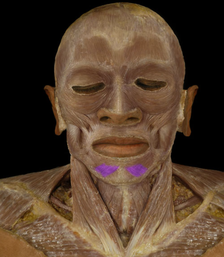
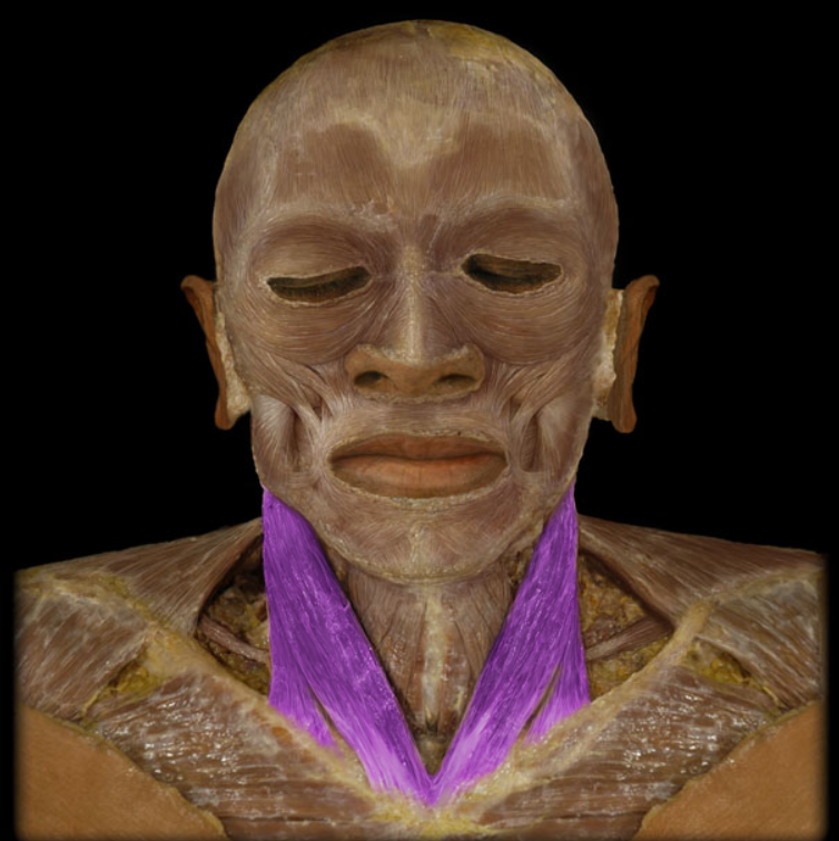
Action: Depression of hyoid bone
Origin: Scapula (superior border)
Insertion: Hyoid bone
Innervation: Ventral rami of C1-3 spinal nerves (ansa cervicalis)
Omohyoid m.
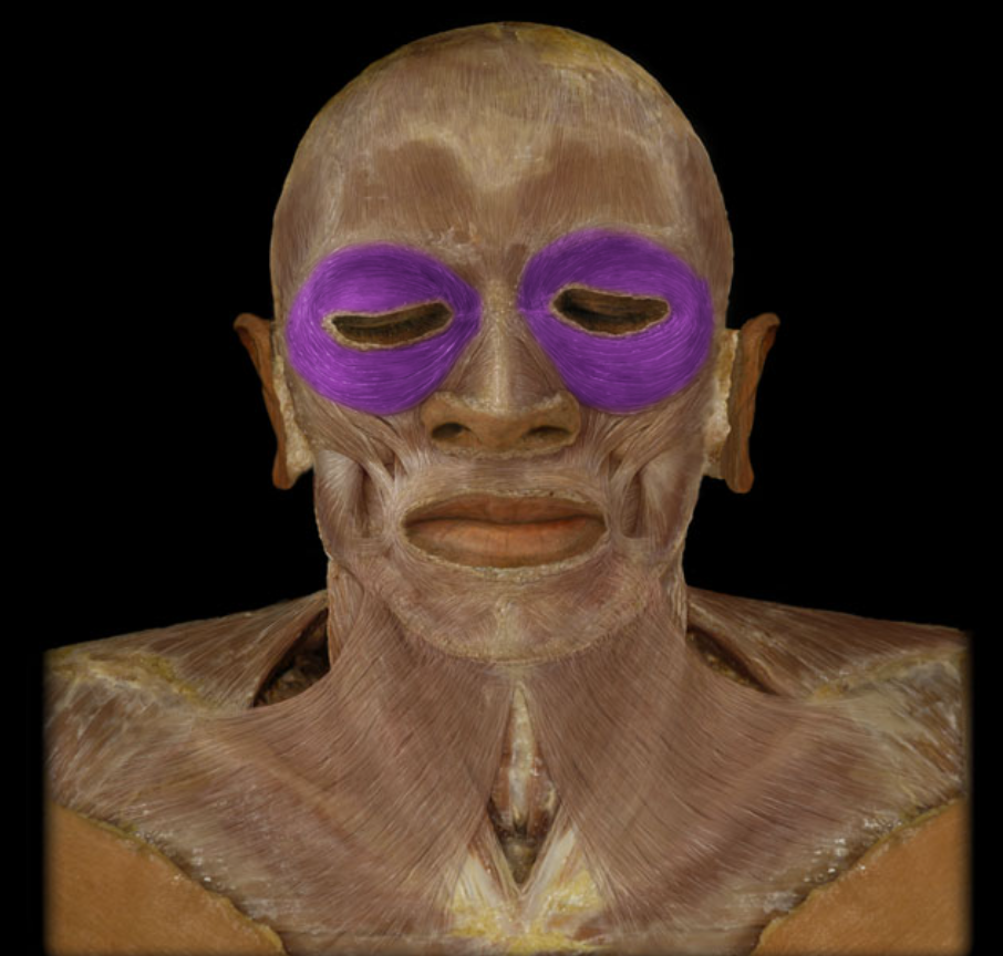
Action: Closes eyelids (blink/wink)
Origin: Frontal bone, Maxilla
Insertion: lateral side of orbit
Innervation: Facial nerve (CN VII)
Obicularis oculi m.
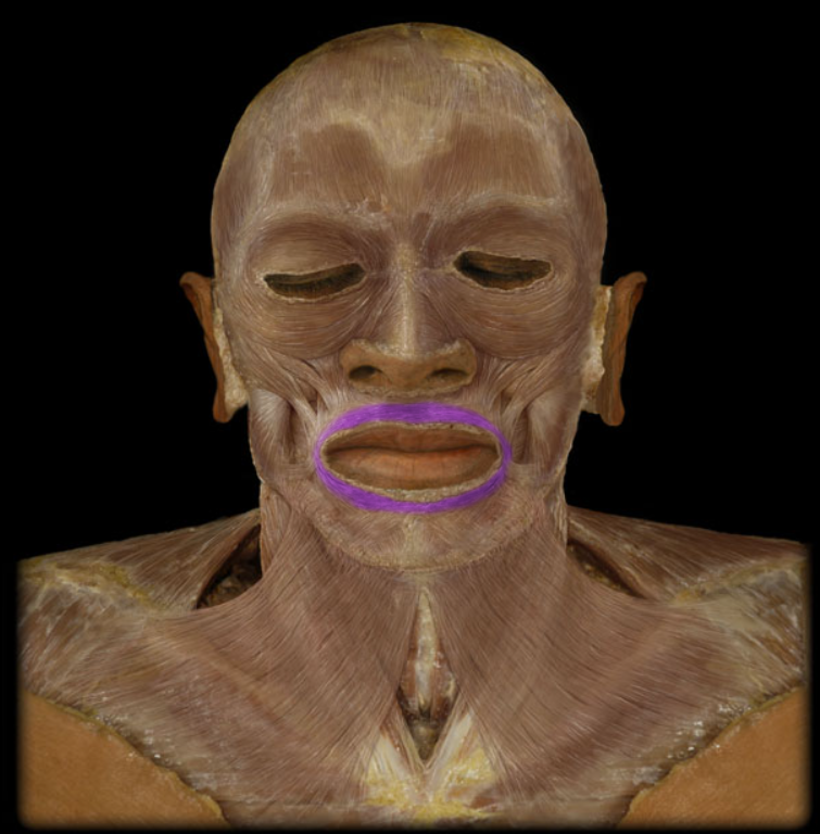
Action: Closes lips, Protrudes lips (pucker)
Origin: Skin around mouth, Mandible (midline), Maxilla (midline)
Insertion:, Lips, Interdigitation with fibers from opposite side
Innervation: Facial nerve (CN VII)
Obicularis oris m.
Action: Unilateral: rotation of head so face turns to opposite side, Bilateral: flexion of neck & extension of head
Origin: Clavicle (medial), Sternum (manubrium)
Insertion: Mastoid process of temporal bone, Lateral one-half of superior nuchal line of occipital bone
Innervation: Accessory nerve (CN XI)
Sternocleidomastoid m. (frontal view)
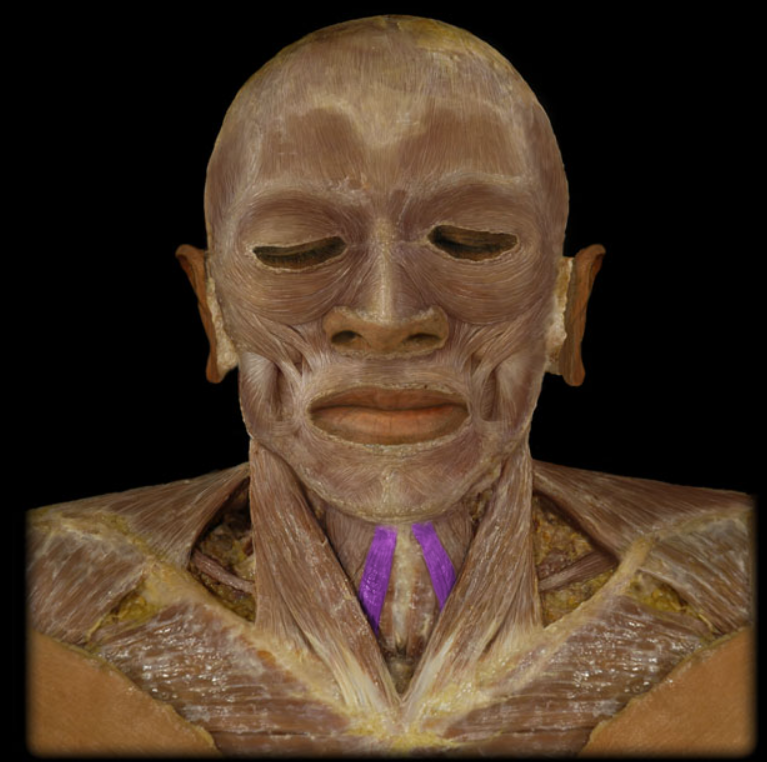
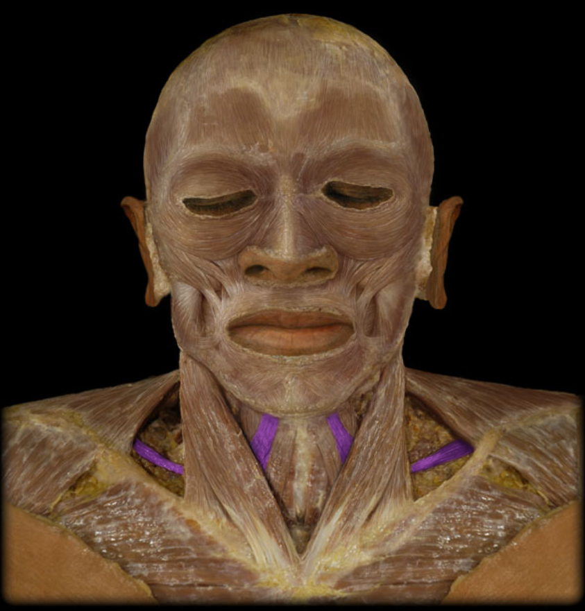
Action: Depression of hyoid bone
Origin: Sternum (manubrium), Clavicle (medial end)
Insertion: Hyoid bone
Innervation: Ventral rami of C1-3 spinal nerves (ansa cervicalis)
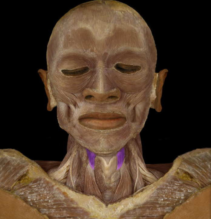
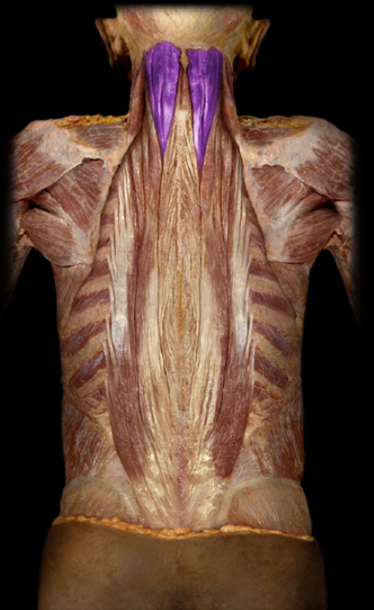
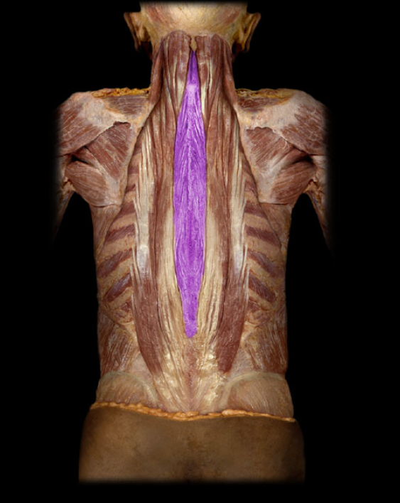
Sternphyoid m.
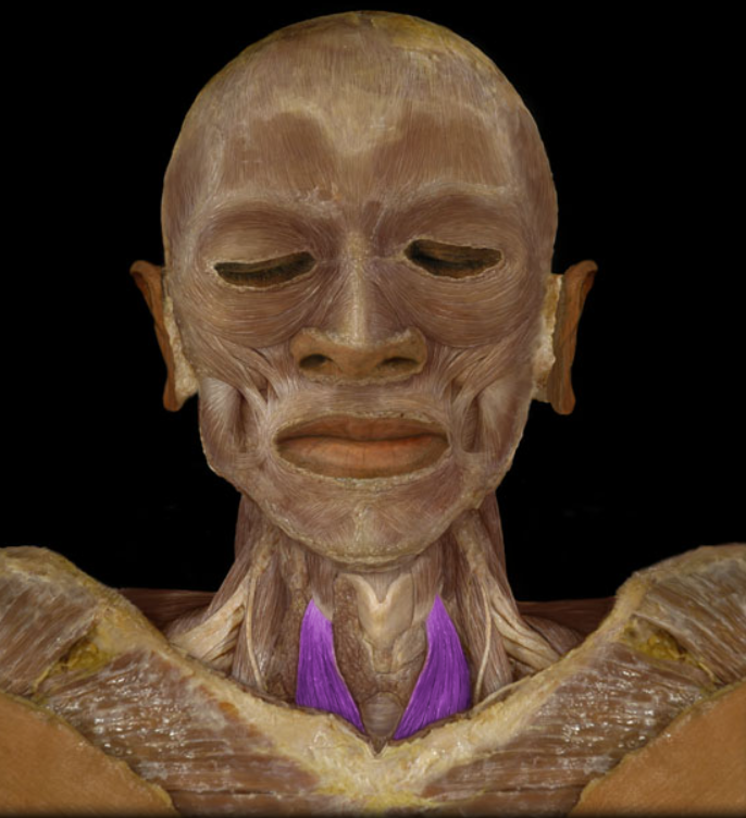
Action: Depression of larynx
Origin: Sternum (manubrium)
Insertion: Thyroid cartilage of larynx
Innervation: Ventral rami of C1-3 spinal nerves (ansa cervicalis)
Sternothyroid m.
Action: Elevation of larynx, Depression of hyoid bone
Origin: Thyroid cartilage of larynx
Insertion: Hyoid bone
Innervation: Ventral ramus of C1 spinal nerve
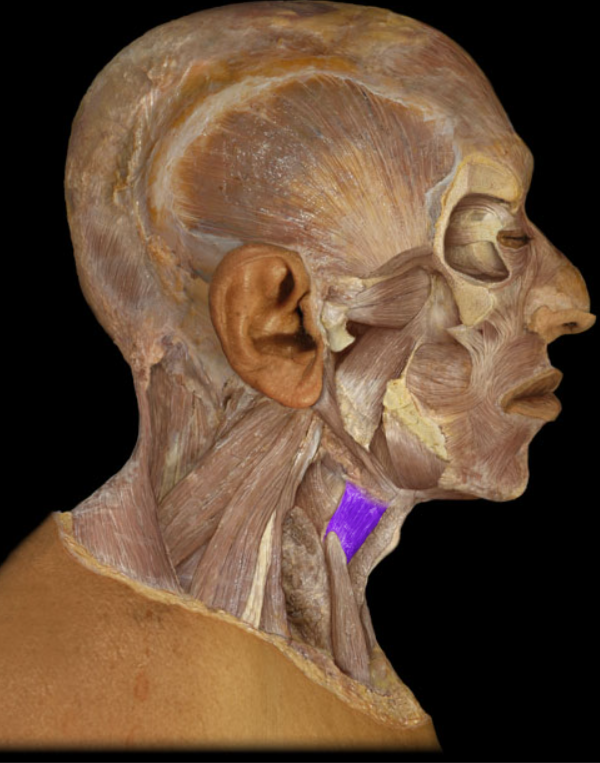
Thyrohyoid m. (frontal view)
Action: Elevation of corner of mouth (smile)
Origin: Zygomatic bone
Insertion: Blends with muscles at angle of mouth
Innervation: Facial nerve (CN VII)
Zygomaticus major m.
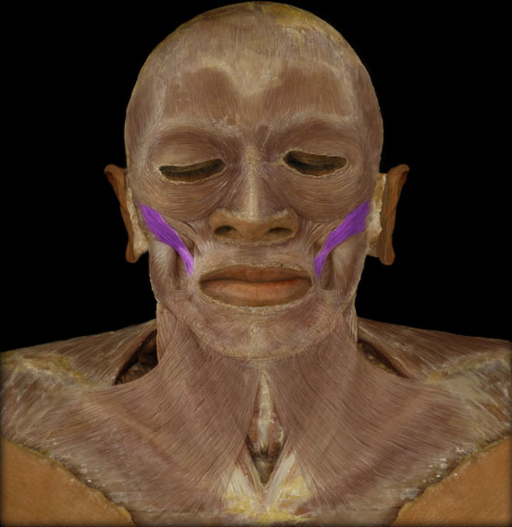
Action: Compression of cheek (e.g., inflating a balloon; playing a wind musical instrument)
Origin: Pterygomandibular raphe, Maxilla (lateral aspect), Mandible (lateral body)
Insertion: Blends with muscles at angle of mouth
Innervation: Facial nerve (CN VII)
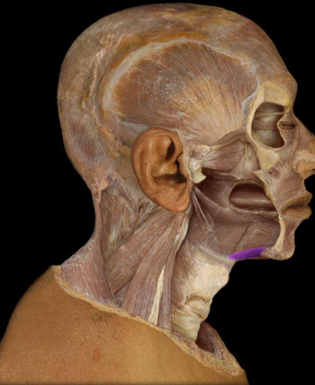
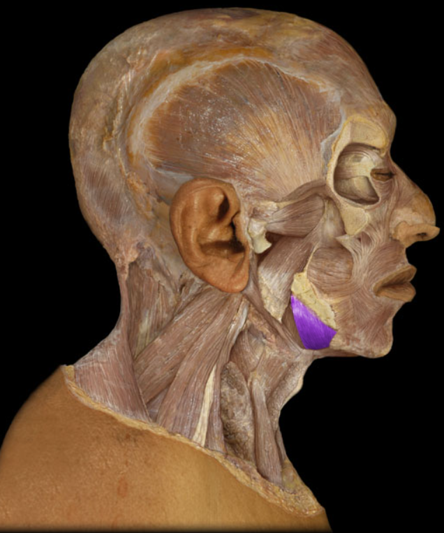
Buccinator m.
Action: Depression of mandible (opens mouth), Elevation of hyoid bone
Origin: Anterior belly: mandible (inner aspect of body near midline), Posterior belly: temporal bone (mastoid process)
Insertion: Hyoid bone
Innervation: Anterior belly: trigeminal nerve (mandibular division - CN V3), Posterior belly: facial nerve (CN VII)
Digastric m.
Geniohyoid m.
Action: Elevation of hyoid bone
Origin: Mandible (mental tubercle)
Insertion: Hyoid bone
Innervation: C1 spinal nerve (ventral ramus)
Action: Elevation of mandible (e.g., closing mouth), Protraction of mandible
Origin: Zygomatic arch
Insertion: Mandible (external surface of angle and ramus)
Innervation: Trigeminal nerve (mandibular division - CN V3)
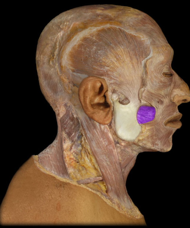
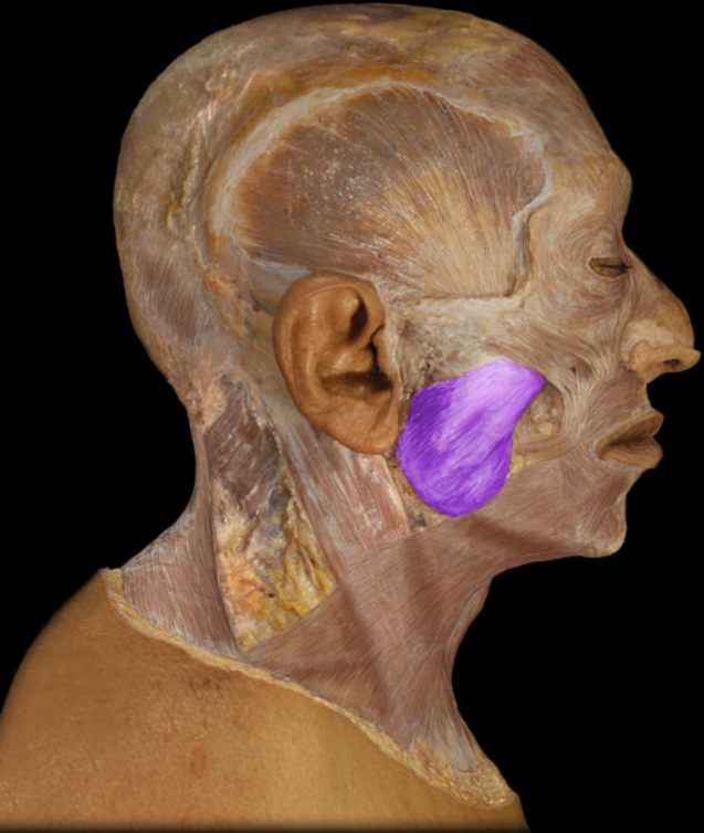
Masseter m.
Action: Elevation of floor of mouth
Origin: Mandible (inner aspect of body)
Insertion: Hyoid bone (posterior fibers), Midline fibrous raphe (anterior fibers)
Innervation: Trigeminal nerve (mandibular division - CN V3)
Mylohyoid m.
Action: Elevation of eyebrows, Creases skin of forehead, Moves scalp backward and forward
Origin: Occipitalis: occipital bone (superior nuchal line), Frontalis: skin of eyebrow and forehead (blends with orbicularis oculi muscle)
Insertion: Epicranial aponeurosis
Innervation: Facial nerve (CN VII)
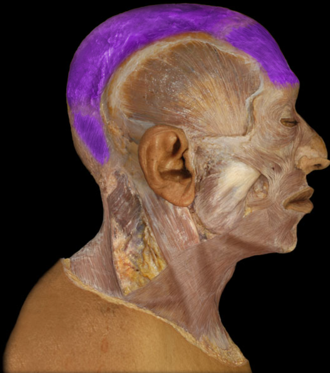
Occipitofrontalis m.
Action: Depression of hyoid bone
Origin: Scapula (superior border)
Insertion: Hyoid bone
Innervation: Ventral rami of C1-3 spinal nerves (ansa cervicalis)
Omohyoid m. (lateral view)
Action: Unilateral: rotation of head so face turns to opposite side, Bilateral: flexion of neck, extension of head
Origin: Clavicle (medial), Sternum (manubrium)
Insertion: Mastoid process of temporal bone, Lateral one-half of superior nuchal line of occipital bone
Innervation: Accessory nerve (CN XI)
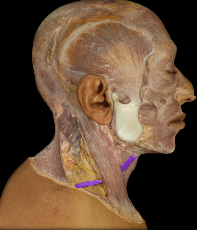
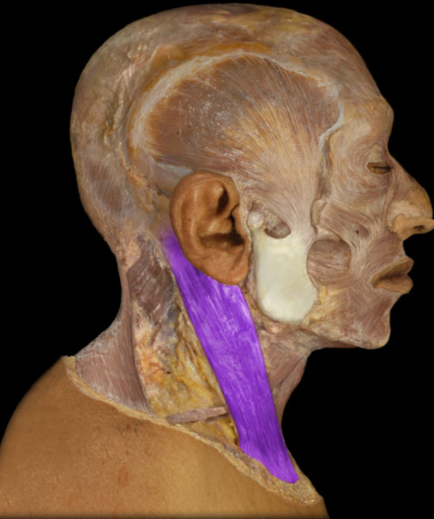
Sternocleidomastoid m.
Action: Depression of hyoid bone
Origin: Sternum (manubrium), Clavicle (medial end)
Insertion: Hyoid bone
Innervation: Ventral rami of C1-3 spinal nerves (ansa cervicalis)
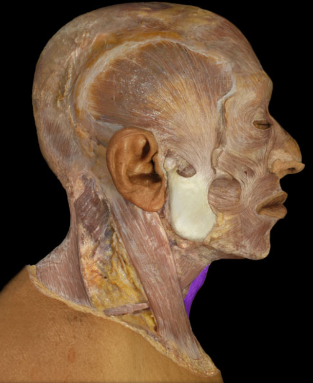
Sternohyoid m. (lateral view)
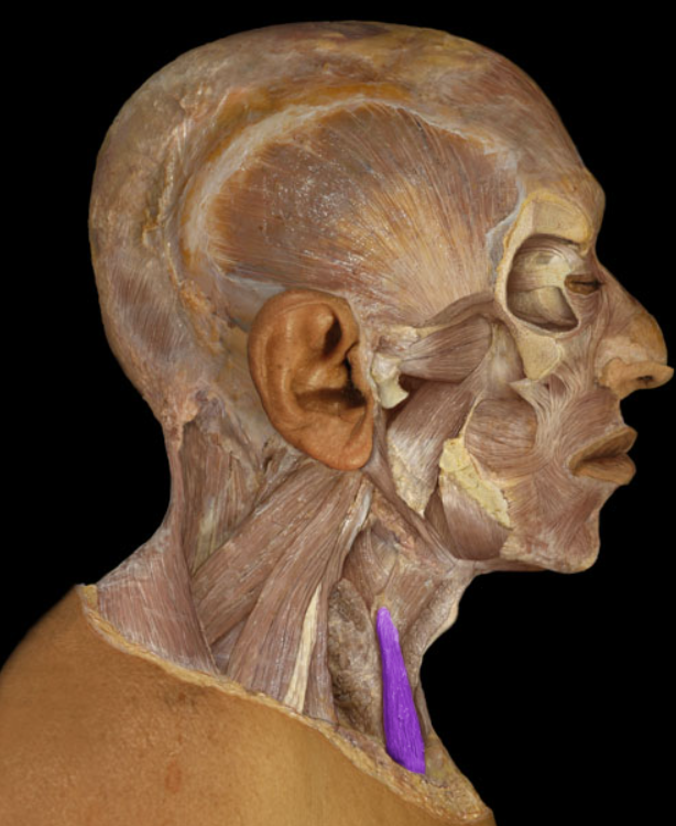
Action: Depression of larynx
Origin: Sternum (manubrium)
Insertion: Thyroid cartilage of larynx
Innervation: Ventral rami of C1-3 spinal nerves (ansa cervicalis)
Sternothyroid m.
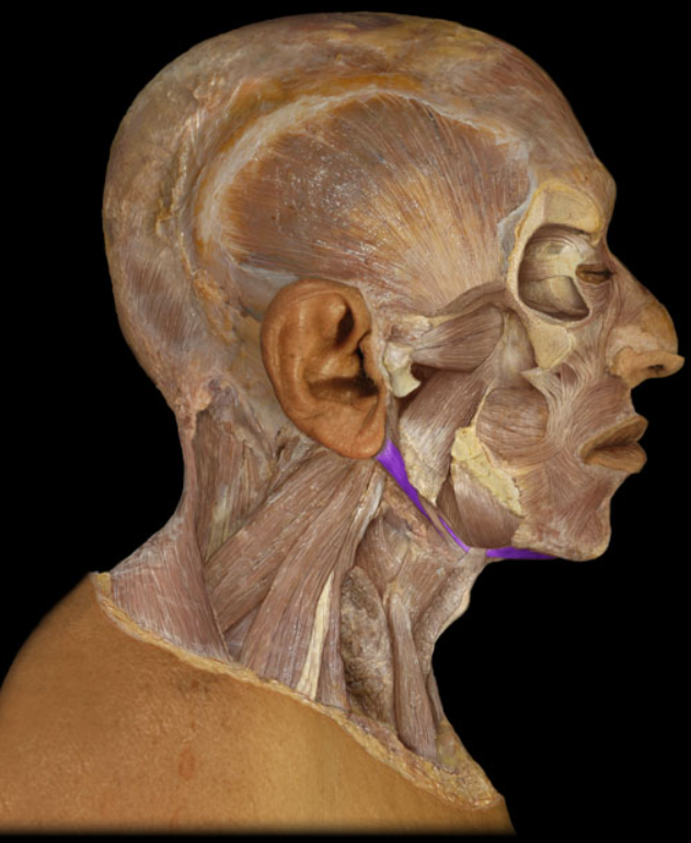
Action: Elevation of hyoid bone
Origin: Styloid process of temporal bone
Insertion: Hyoid bone
Innervation: Facial nerve (CN VII)
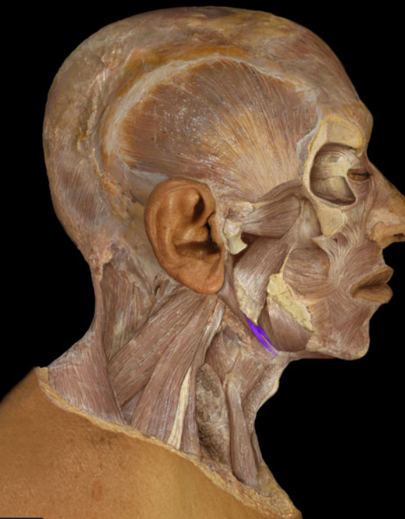
Stylohyoid m.
Action: Elevation of mandible (e.g., closing mouth), Retraction of mandible
Origin: Temporal fossa (formed by parts of frontal, parietal, sphenoid (greater wing), and temporal bones)
Insertion: Mandible (coronoid process and ramus)
Innervation: Trigeminal nerve (mandibular division - CN V3)
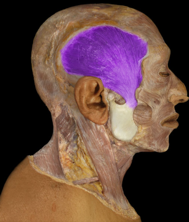
Temporalis m.
Action: Elevation of larynx, Depression of hyoid bone
Origin: Thyroid cartilage of larynx
Insertion: Hyoid bone
Innervation: Ventral ramus of C1 spinal nerve
Thyrohyoid m.
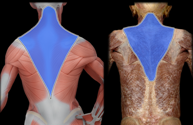
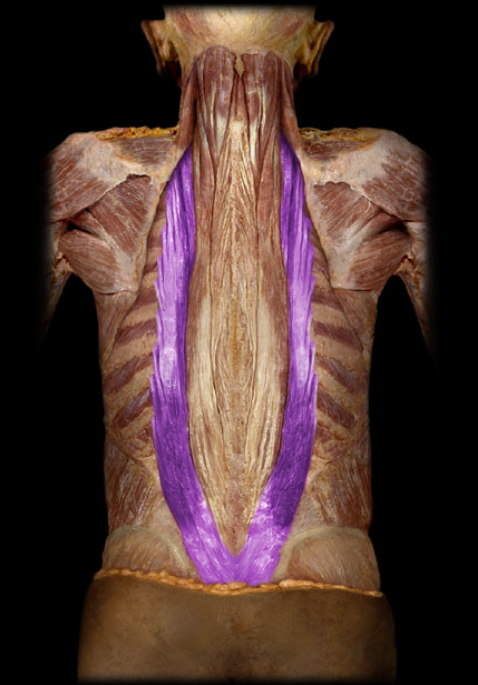
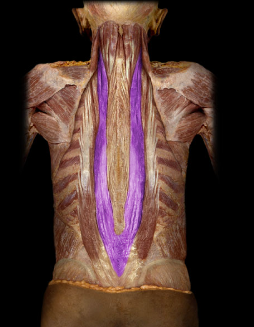
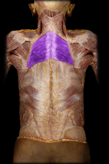
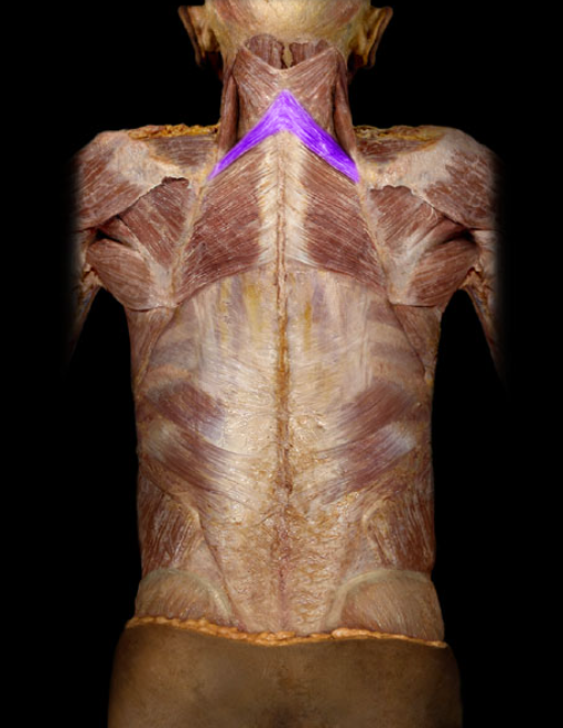
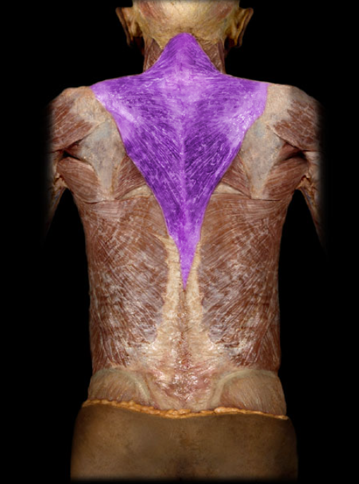
Action: Superior fibers: elevation of scapula; superior rotation of scapula (glenoid cavity directed superiorly), Middle fibers: retraction (adduction) of scapula, Inferior fibers: depression of scapula
Origin: Occipital bone (superior nuchal line), Nuchal ligament, Vertebra prominens (spinous process of C7 vertebra), Spinous processes of T1-12 vertebrae
Insertion: Clavicle (lateral), Scapula (spine and acromion)
Innervation: Accessory nerve (CN XI)
Trapezius m.
Action: Protraction (abduction) of scapula, Superior rotation of scapula (directs glenoid cavity superiorly), Stabilizes scapula (holds it against thoracic wall)
Origin: Ribs 1-9
Insertion: Scapula (medial border)
Innervation: Long thoracic nerve
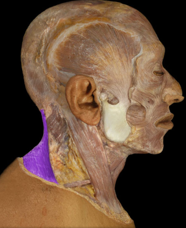
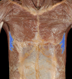
Serratus anterior m.
Action: Dome of diaphragm flattens during inspiration, Contraction increases vertical dimension of thoracic cavity
Origin: Sternal part (not always present): xiphoid process, Costal part: ribs 5-10 and their costal cartilage, Lumbar part: arcuate ligaments and L1-3 vertebral bodies
Insertion: Central tendon
Innervation: Phrenic nerve
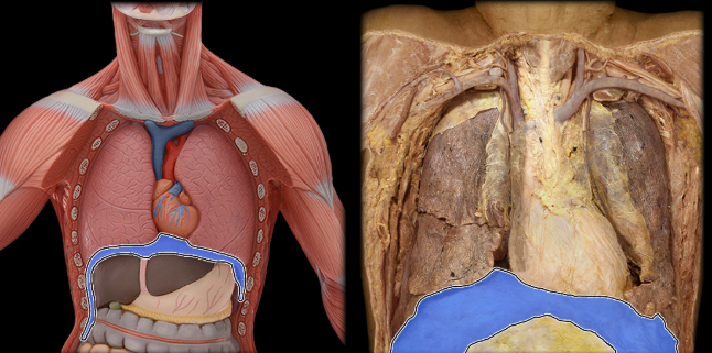
Diaphragm
Action: Muscles of respiration
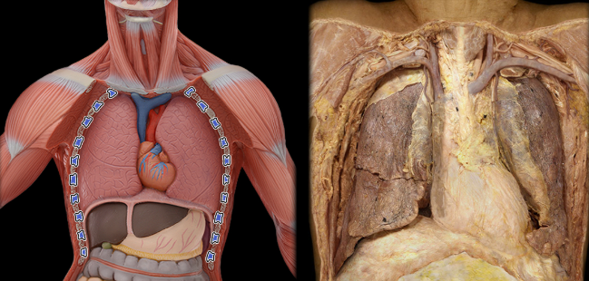
Location: Found between ribs 1-11
Innervation: Intercostal nerves (ventral rami of T1-11 spinal nerves)
Intercostal mm.
Action: Superior fibers: elevation of scapula; superior rotation of scapula (glenoid cavity directed superiorly), Middle fibers: retraction (adduction) of scapula, Inferior fibers: depression of scapula
Origin: Occipital bone (superior nuchal line), Nuchal ligament, Vertebra prominens (spinous process of C7 vertebra), Spinous processes of T1-12 vertebrae
Insertion: Clavicle (lateral), Scapula (spine and acromion)
Innervation: Accessory nerve (CN XI)
Trapezius m.
Action: Unilateral: lateral flexion of vertebral column, Bilateral: extension of vertebral column, Depression of ribs
Origin: Common origin and ribs
Insertion: Ribs, Transverse processes (cervical)
Innervation: Dorsal rami of spinal nerves
Iliocostalis part of erector spinae m.
Action: Unilateral: lateral flexion of vertebral column and rotation of head to same side, Bilateral: extension of vertebral column and head, Depresses ribs
Origin: Iliac crest, Dorsal sacrum, Sacral and lumbar spinous processes, Supraspinous ligament
Insertion: Transverse processes (cervical and thoracic), ribs, and mastoid process
Innervation: Dorsal rami of spinal nerves
Longissimus part of erector spinae m.
Action: Retraction (adduction) of scapula, Elevation of scapula
Origin: Spinous processes of T2-5 vertebrae
Insertion: Scapula (medial border)
Innervation: Dorsal scapular nerve
Rhomboid major m.
Action: Retraction (adduction) of scapula, Elevation of scapula
Origin: Spinous processes of C7-T1 vertebrae
Insertion: Scapula (medial border)
Innervation: Dorsal scapular nerve
Rhomboid minor m.
Action: Unilateral: rotation of head to opposite side, Bilateral: extension of head and neck
Origin: Transverse processes of C7-T6 vertebrae
Insertion: Occipital bone
Innervation: Cervical spinal nerves (dorsal rami)
Semispinalis capitis m.
Action: Unilateral: lateral flexion of vertebral column, Bilateral: extension of vertebral column
Origin: Spinous processes and nuchal ligament
Insertion: Spinous processes (cervical and thoracic) and occipital bone
Innervation: Dorsal rami of spinal nerves
Spinalis part of erector spinae m.
Action: Superior fibers: elevation of scapula; superior rotation of scapula (glenoid cavity directed superiorly), Middle fibers: retraction (adduction) of scapula, Inferior fibers: depression of scapula
Origin: Occipital bone (superior nuchal line), Nuchal ligament, Vertebra prominens (spinous process of C7 vertebra), Spinous processes of T1-12 vertebrae
Insertion: Clavicle (lateral), Scapula (spine and acromion)
Innervation: Accessory nerve (CN XI)
Trapezius m.
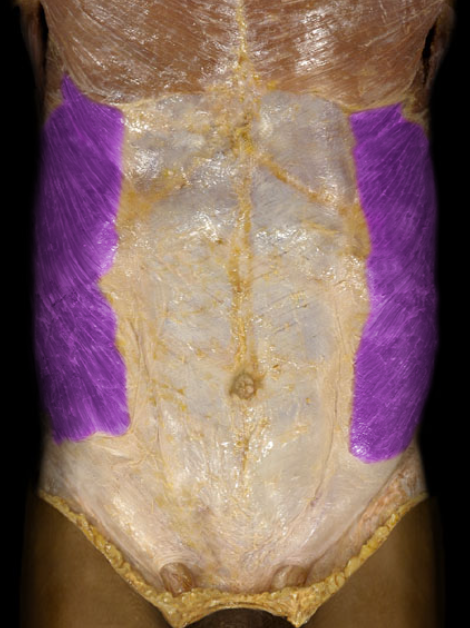
Action: Unilateral: rotation of trunk, Bilateral: flexion of trunk (sit-ups), Compression of anterior abdominal wall and viscera (increases intra-abdominal pressure)
Origin: Ribs and costal cartilages 5-12
Insertion: Linea alba, Ilium (crest)
Innervation: Ventral rami of T8-L1 spinal nerves
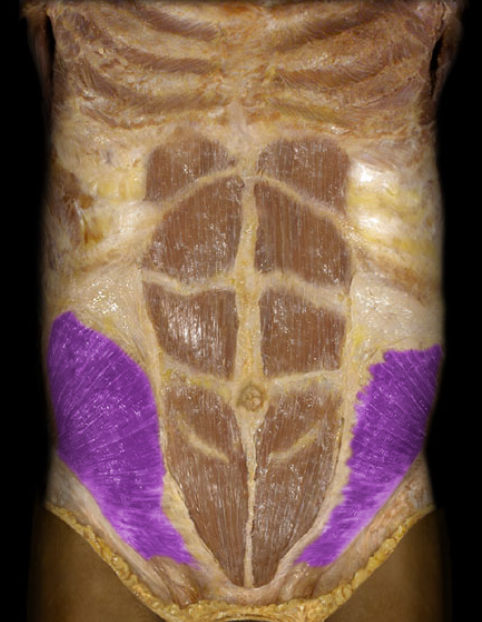
External abdominal oblique m.
Action: Unilateral: rotation of trunk, Bilateral: flexion of trunk (sit-ups), Compression of anterior abdominal wall and viscera (increases intra-abdominal pressure)
Origin: Inguinal ligament, Ilium (crest), Thoracolumbar fascia
Insertion: Linea alba, Costal cartilages 7-10
Innervation: Ventral rami of T8-L1 spinal nerves
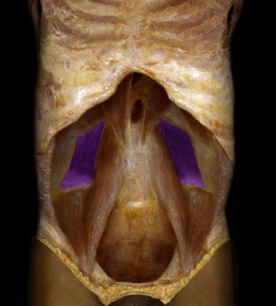
Internal abdominal oblique
Action: Unilateral: lateral flexion of trunk, Bilateral: extension of trunk, Fix/depress rib 12
Origin: Ilium (crest)
Insertion: Rib 12, L1-4 vertebrae (transverse processes)
Innervation: Ventral rami of T12-L4 spinal nerves
Quadratus lumborum m.
Action: Flexion of trunk (sit-ups), Compression of anterior abdominal wall
Origin: Pubis (crest and symphysis)
Insertion: Sternum (xiphoid process), Costal cartilages 5-7
Innervation: Ventral rami of T7-12 spinal nerves
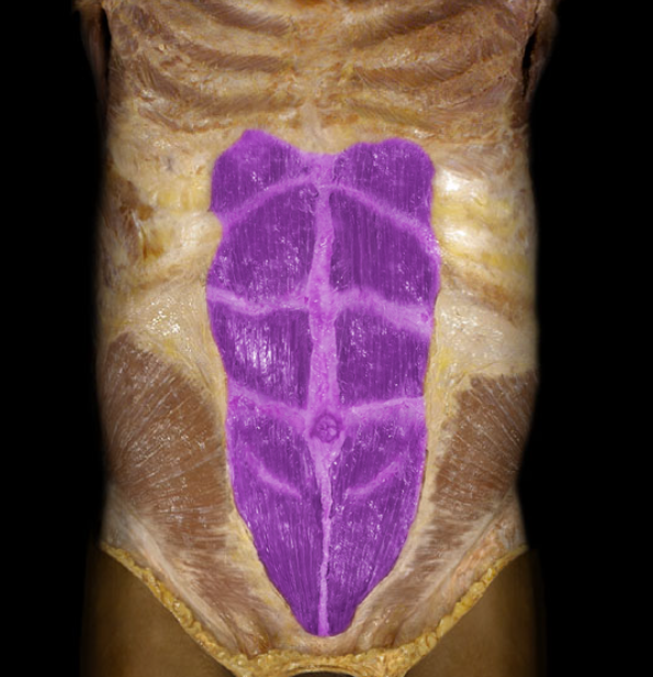
Rectus abdominis m.
Action: Compression of anterior abdominal wall and viscera (increases intra-abdominal pressure)
Origin: Ilium (crest), Inguinal ligament, Thoracolumbar fascia, Costal cartilages 7-12
Insertion: Pubis (body), Linea alba, Sternum (xiphoid process)
Innervation: Ventral rami of T8-L1 spinal nerves
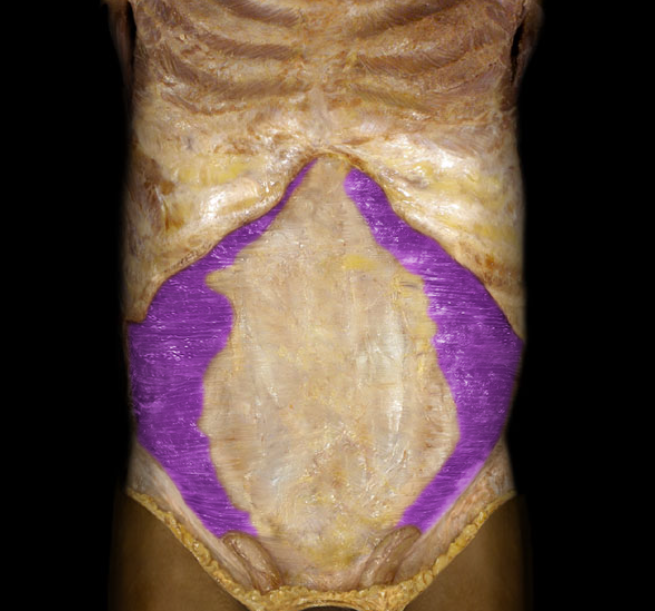
Transversus abdominis m.
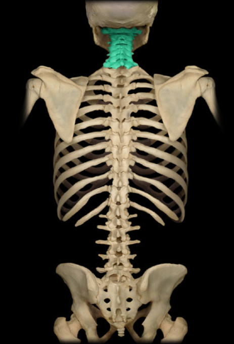
Location: Neck, Between occipital bone and T1 vertebra
Description: Seven individual vertebrae, Characteristic features include transverse foramen and bifid (split) spinous process on C3-C6
Cervical vertebra (holistic view)
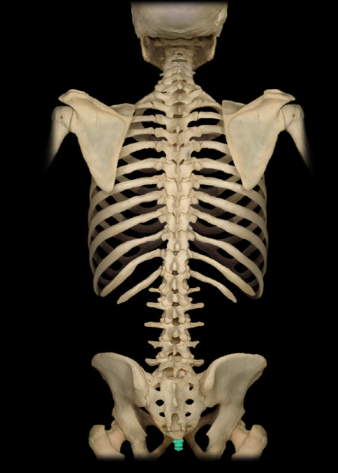
Location: Posterior pelvic wall, Lower back, inferior to S5 vertebra
Description: Small, triangular bone, Consists of three to five, variably fused, poorly developed vertebrae
Also known as: "Tailbone"
Coccyx
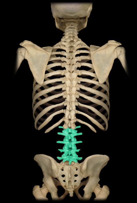
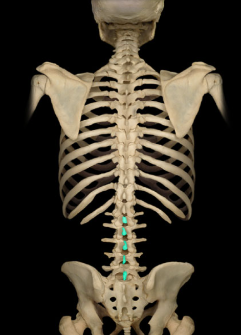
Location: Lower back, Between T12 and S1 vertebrae
Description: Five individual vertebrae, Characteristic features include large size, kidney bean-shaped body, and a thick, blunt spinous process
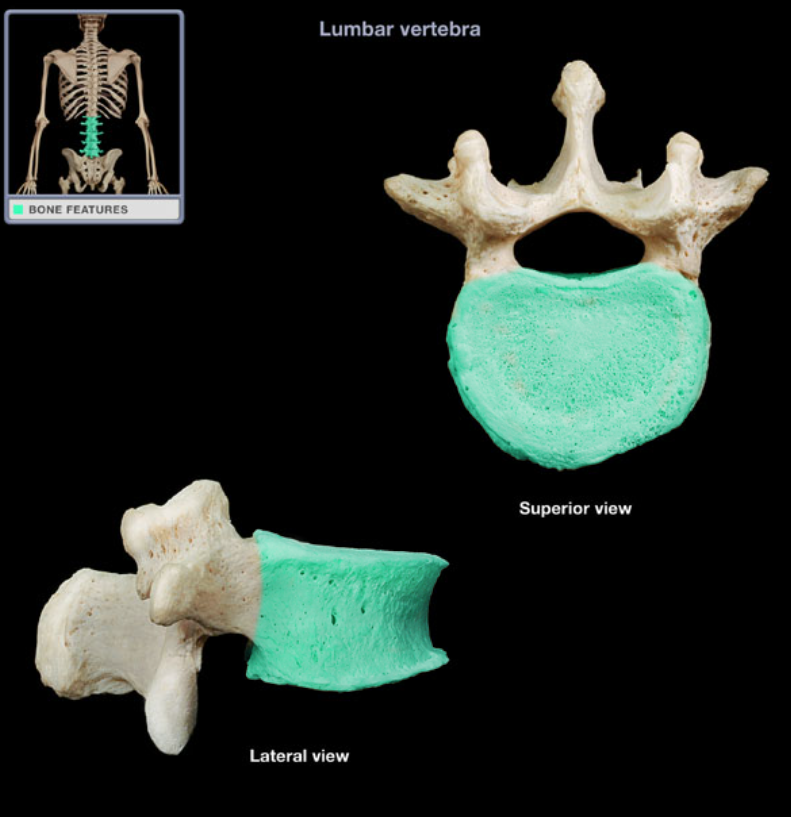
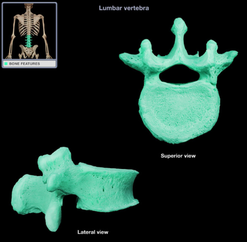
Lumbar vertebra
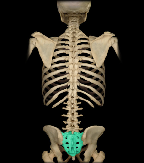
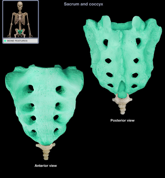
Location: Lower back, Between L5 and Co1 vertebrae, Posterior wall of pelvis
Description: Five fused vertebrae, Triangular bone wedged between hip bones
Sacrum
Location: Vertebrae (posterior aspect)
Description: Unpaired, posterior projection from midline of vertebral arch, Has characteristic thick, blunt form
Spinous process of lumbar vertebra (holistic view)
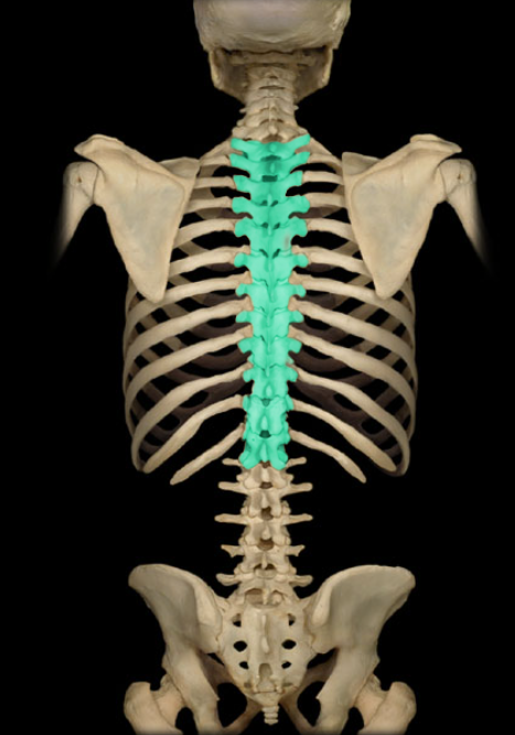
Location: Trunk, Between C7 and L1 vertebrae
Description: 12 individual vertebrae, Characteristic features include costal demifacets (or facets) for articulation with head of rib, spinous process slopes inferiorly, and heart-shaped vertebral body
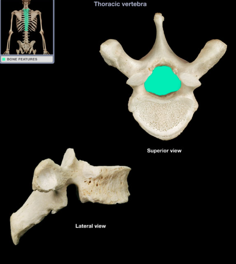
Thoracic vertebra
Location: Lateral aspect of cervical vertebra
Description: Prominent, paired, laterally-directed process
Comment: Provides attachment for intrinsic and extrinsic back muscles
Transverse process of cervical vertebra
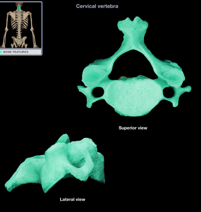
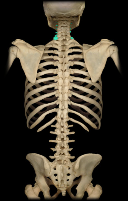
Location: Neck, Between occipital bone and T1 vertebra
Description: Seven individual vertebrae, Characteristic features include transverse foramen and bifid (split) spinous process on C3-C6
Cervical vertebra
Location: Vertebrae (posterior aspect)
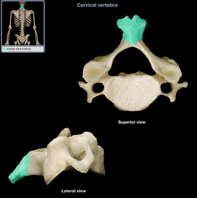
Description: Unpaired, posterior projection from midline of vertebral arch, Usually bifid
Spinous process of cervical vertebra
Location: Cervical vertebra, Superior articular process
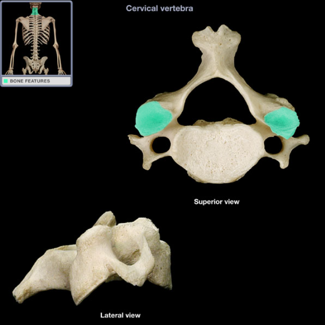
Description: Smooth area
Comment: Provides articulation between adjacent vertebrae
Superior articular facet of cervical vertebra
Location: Cervical vertebra (superior aspect)
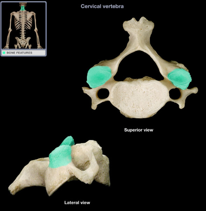
Description: Paired process at junction of pedicle and lamina, Has superiorly-directed articular facet (smooth area)
Superior articular process of cervical vertebra
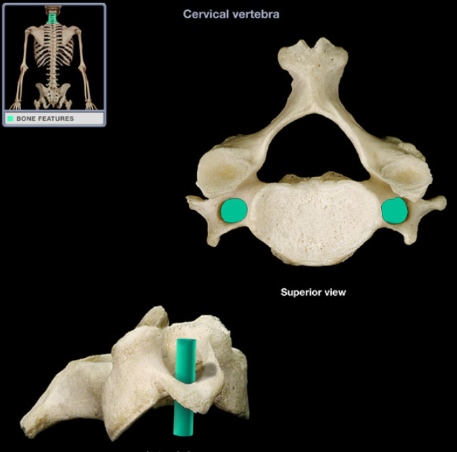
Location: Cervical vertebra, Transverse process
Description: Foramen in each transverse process
Comment: Vertebral artery passes through transverse foramina of C1-6 vertebrae (not through C7 transverse foramen)
Transverse foramen of cervical vertebra
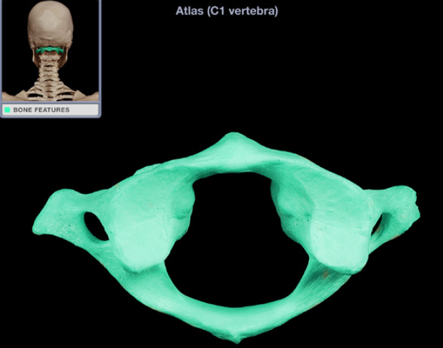
Location: Neck, Between occipital bone and axis (C2 vertebra)
Description: Ring-shaped vertebra, Characteristic features include transverse foramen, Lacks vertebral body, spinous process, or lamina
Also known as: C1 vertebra
Atlas
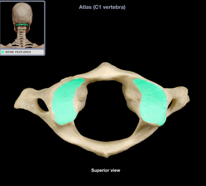
Location: Atlas (C1 vertebra), Superior aspect of lateral mass
Description: Superior-facing, smooth concavity, Receives corresponding occipital condyle of occipital bone
Function: Part of atlanto-occipital joint
Comment: Adapted for "nodding" movements of head
Superior articular facet of atlas
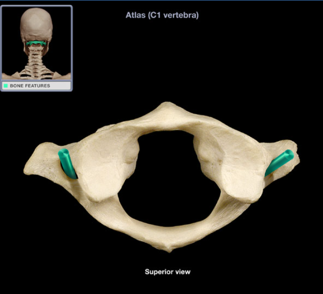
Location: Atlas, Transverse process
Description: Foramen in each transverse process of atlas
Comment: Vertebral artery passes through transverse foramina of C1-6 vertebrae (not through C7 transverse foramen), Atlas also known as C1 vertebra
Transverse foramen of atlas
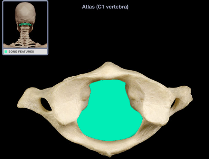
Location: Atlas
Description: Large foramen formed by anterior and posterior arches of atlas
Comment: Vertebral canal formed by combined vertebral foramina, Contains spinal cord, meninges, spinal nerve roots, blood vessels, and fat, Atlas also known as C1 vertebra
Vertebral foramen of atlas
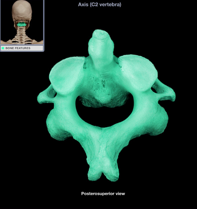
Location: Neck, Between atlas (C1 vertebra) and C3 vertebra
Description: Characteristic features include transverse foramen, bifid (split) spinous process, and dens (prominent superior projection from body)
Also known as: C2 vertebra
Axis (C2 vertebra)
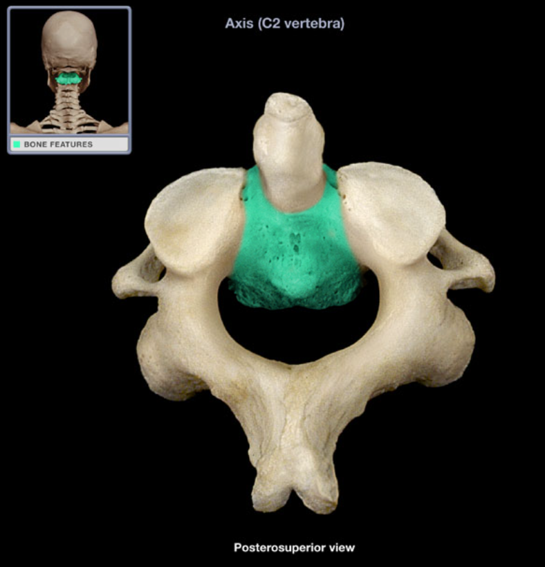
Location: Axis (anterior aspect)
Description: Has superior projection (dens), Attached to pedicle on either side, Separated from body of C3 by intervertebral disc
Body of axis
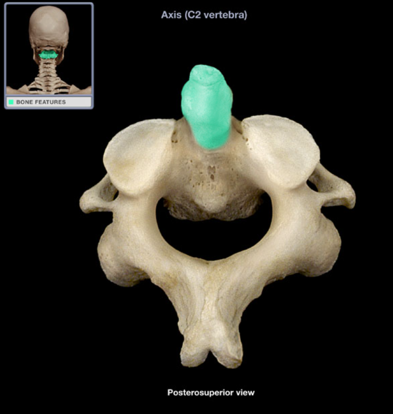
Location: Axis (C2 vertebra)
Description: Prominent, finger-like projection from superior aspect of body of axis (C2 vertebra)
Also known as: Odontoid process
Comment: Represents body of atlas (C1 vertebra), No other vertebra has a dens
Dens
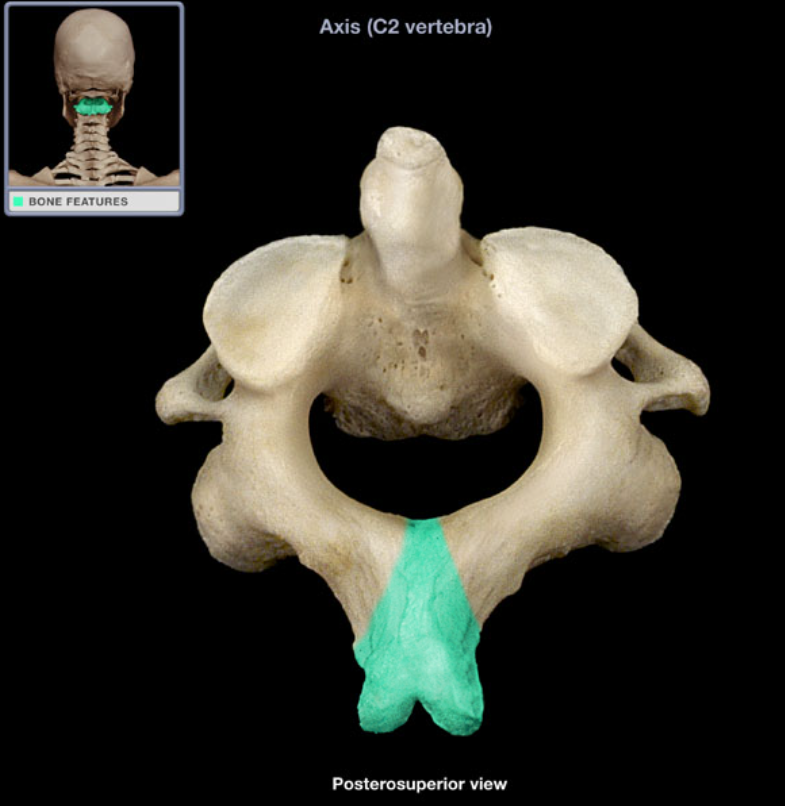
Location: Axis (posterior)
Description: Unpaired posterior midline projection from vertebral arch, Process has characteristic bifid (split) form
Spinous process of axis
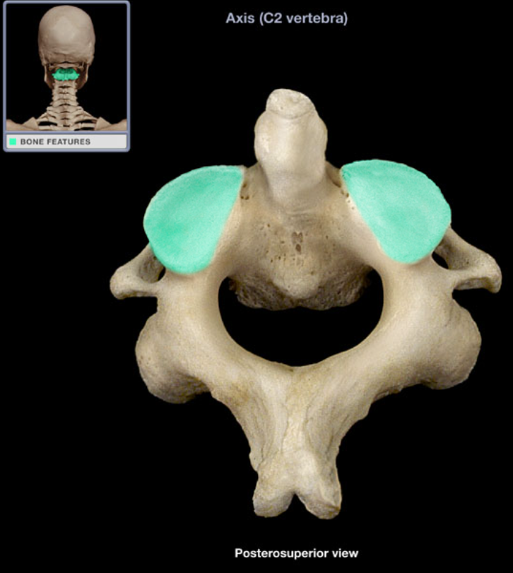
Location: Axis, Superior articular process
Description: Smooth area
Superior articular facet of axis
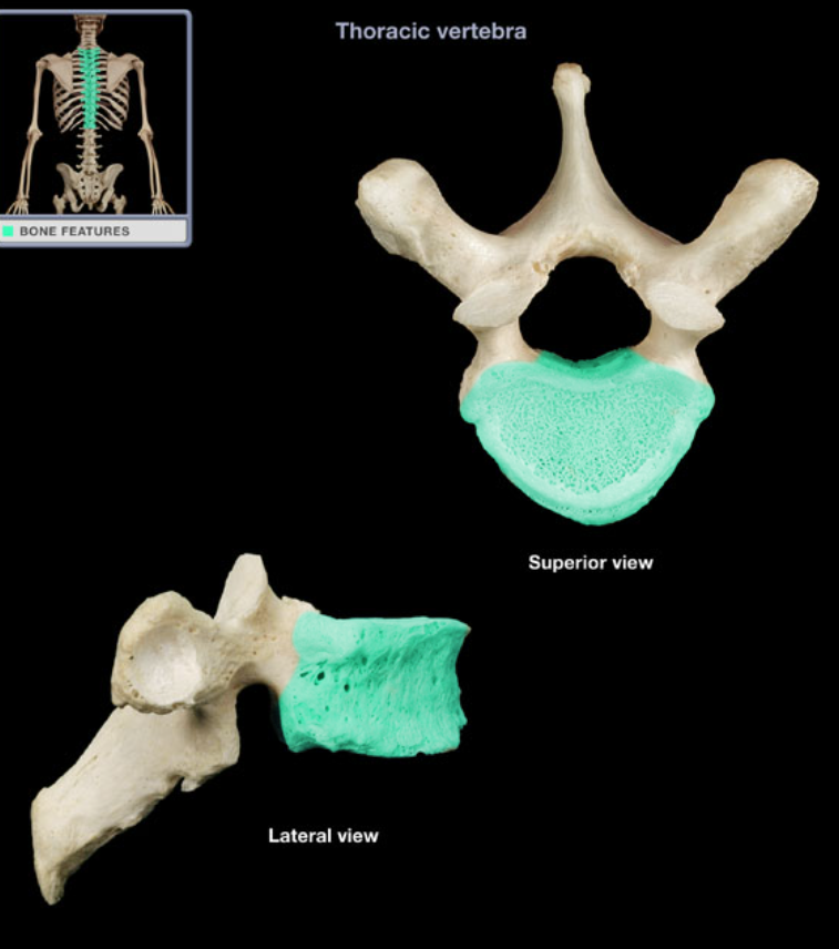
Location: Thoracic vertebra (anterior aspect)
Description: Progressive increase in size from superior to inferior, Contains paired costal demifacets on each side, Attached to pedicle on either side
Comment: Supports body weight
Body of thoracic vertebra
Location: T1-T12 vertebrae
Description: Paired plates that form dorsal wall of vertebral canal, Connects pedicle to spinous process
Lamina of thoracic vertebra
Location: T1-T12 vertebrae
Description: Short, thick pillar, Connects vertebral body to lamina on each side
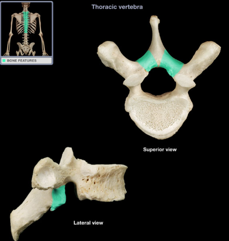
Pedicle of thoracic vertebra
Location: Thoracic vertebra
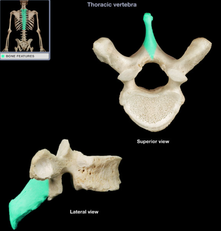
Description: Unpaired posterior midline projection from vertebral arch, Has characteristic long, slender form, Processes of inferior thoracic vertebrae directed inferiorly
Spinous process of thoracic vertebra
Location: Thoracic vertebra (superior aspect)
Description: Paired process at junction of pedicle and lamina, Has posteriorly-directed articular facet (smooth area
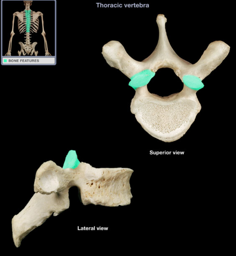
Superior articular process of thoracic vertebra
Location: Thoracic vertebral body (lateral aspect), At junction with pedicle
Description: Smooth area
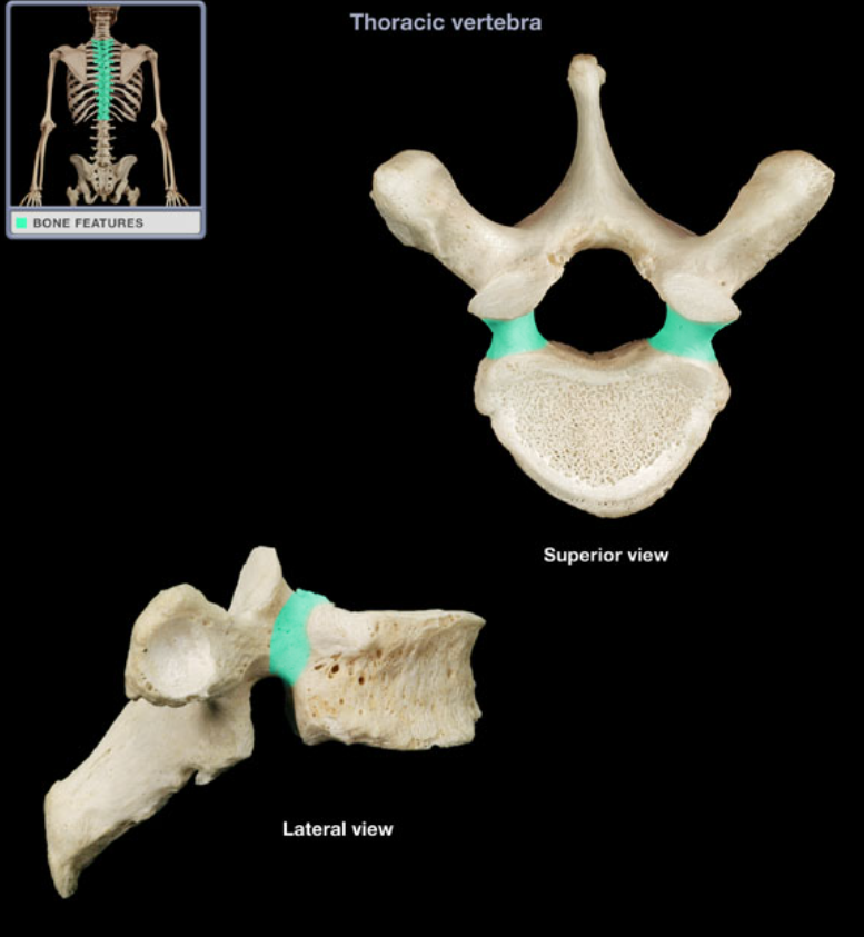
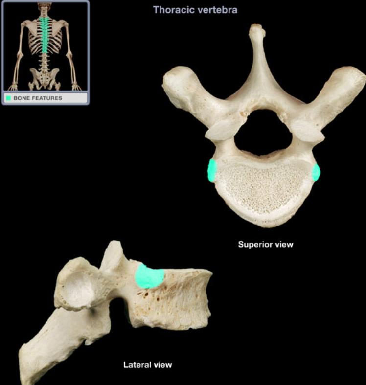
Superior costal facet of thoracic vertebra
Location: Thoracic vertebra 1-10, Transverse process
Description: Smooth area
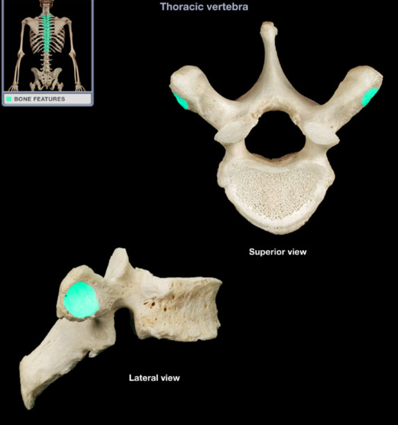
Transverse costal facet of thoracic vertebra
Location: Thoracic vertebra
Description: Large foramen formed by vertebral arch and posterior aspect of vertebral body
Vertebral foramen of thoracic vertebra
Location: Vertebra (anterior aspect)
Description: Oval ventral portion, Forms ventral wall of vertebral canal
Comment: Supports body weight
Body of lumbar vertebra
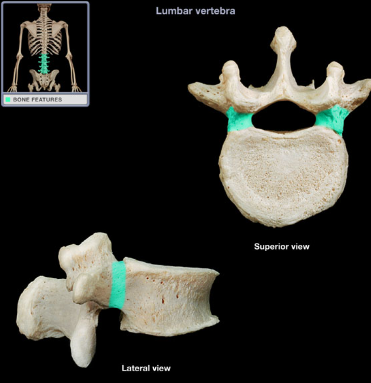
Location: Lumbar vertebra, At junction of pedicles and laminae
Description: Paired processes, Has laterally-directed articular facet (smooth area)
Inferior articular process of lumbar vertebra
Location: L1-L5 vertebrae
Description: Paired plate that forms dorsal wall of vertebral canal, Connects pedicle to spinous process
Lamina of lumbar vertebra
Location: Lower back, Between T12 and S1 vertebrae
Description: Five individual vertebrae, Characteristic features include large size, kidney bean-shaped body, and a thick, blunt spinous process
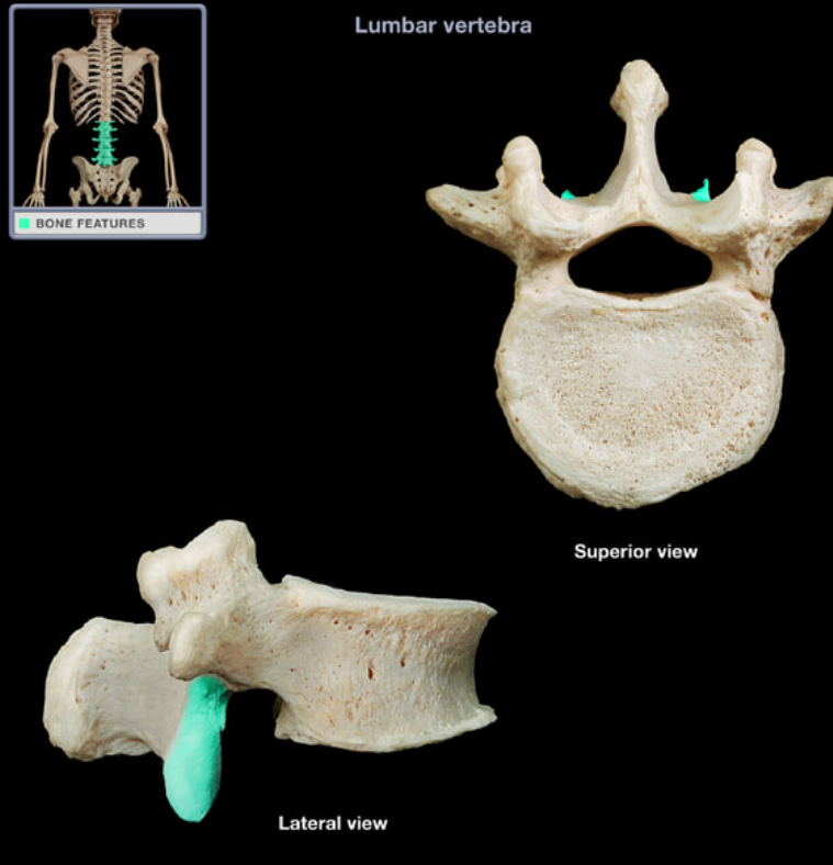
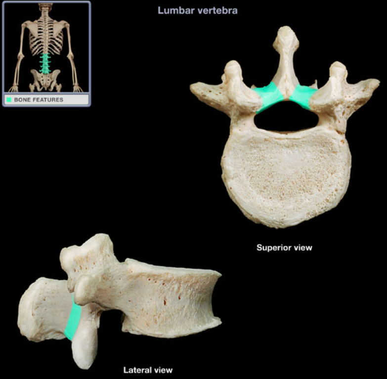
Lumbar vertebra
Location: Vertebrae L1-5 (lateral aspect)
Description: Short, thick pillar, Connects vertebral body to lamina on each side
Comment: Adjacent pedicles contribute to each intervertebral foramen
Pedicle of lumbar vertebra
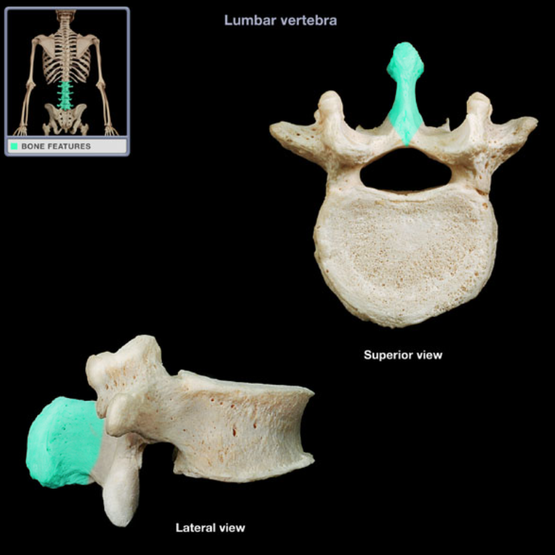
Location: Vertebrae (posterior aspect)
Description: Unpaired, posterior projection from midline of vertebral arch, Has characteristic thick, blunt form
Spinous process of lumbar vertebra
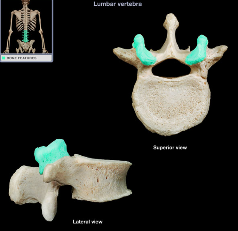
Location: Lumbar vertebra (superior aspect)
Description: Paired process at junction of pedicle and lamina, Has medially-directed articular facet (smooth area)
Superior articular process of lumbar vertebra
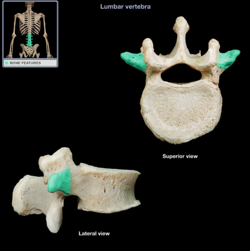
Location: Lateral aspect of lumbar vertebra
Description: Prominent, paired, laterally-directed process
Comment: Provides attachment for intrinsic back muscles and ligaments
Transverse process of lumbar vertebra
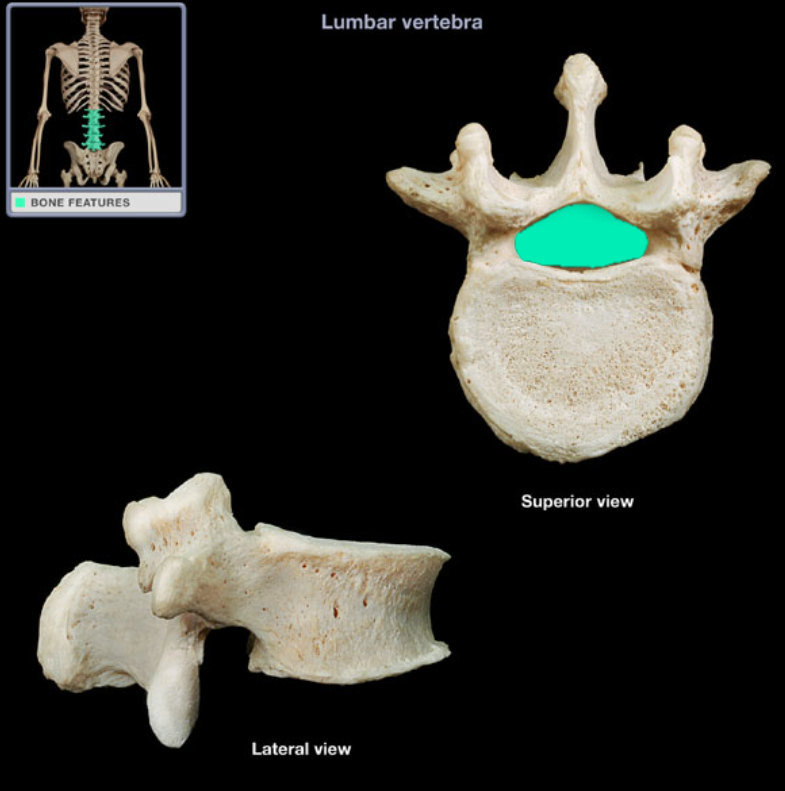
Location: Lumbar vertebra
Description: Large foramen formed by vertebral arch and posterior aspect of vertebral body
Vertebral foramen of lumbar vertebra
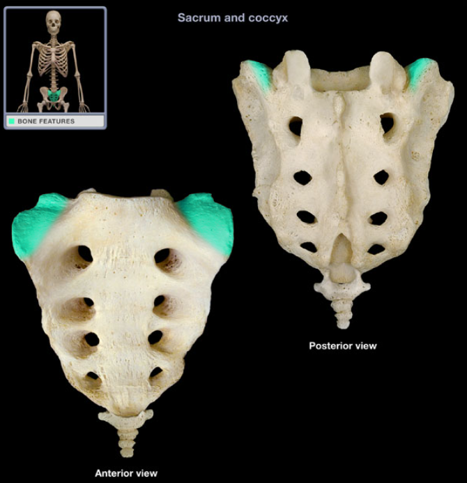
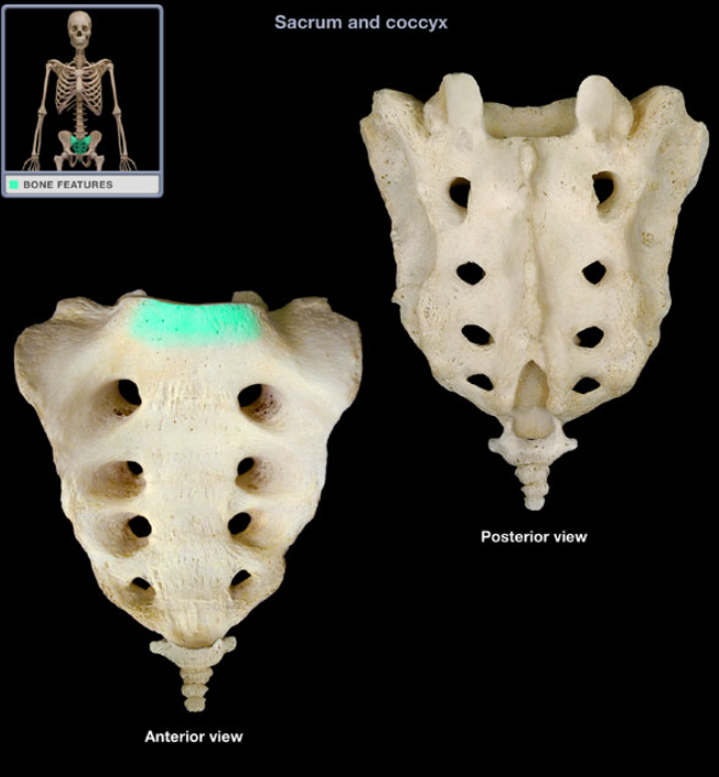
Location: Sacrum (base)
Description: Paired, wing-like, laterally projecting masses, Modification of S1 vertebral transverse process
Comment: Base of sacrum is superior surface of S1 vertebra, Latin: ala = wing
Ala of sacrum
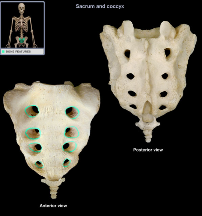
Location: Sacrum (anterior surface)
Description: Four pairs of openings, Communicate with sacral canal
Function: Transmit ventral rami of S1-4 spinal nerves
Anterior sacral foramen
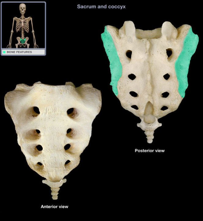
Location: Sacrum (lateral surface)
Description: Paired, elongated, ear-shaped, lateral surface of sacrum
Function: Articulation with ilium (synovial sacroiliac joint)
Comment: Latin: auris = ear
Auricular surface of sacrum
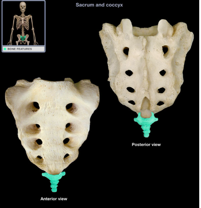
Location: Posterior pelvic wall, Lower back, inferior to S5 vertebra
Description: Small, triangular bone, Consists of three to five, variably fused, poorly developed vertebrae
Also known as: "Tailbone"
Comment: Rudiment of the tail in other vertebrates
Coccyx (Tailbone)
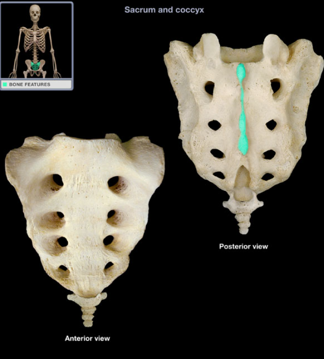
Location: Sacrum, Along dorsal midline
Description: Prominent, interrupted longitudinal ridges, Formed by three or four spinous tubercles, Represent fused spinous processes of S1-3(4) vertebrae
Function: Attachment for aponeurosis of erector spinae muscles
Median sacral crest
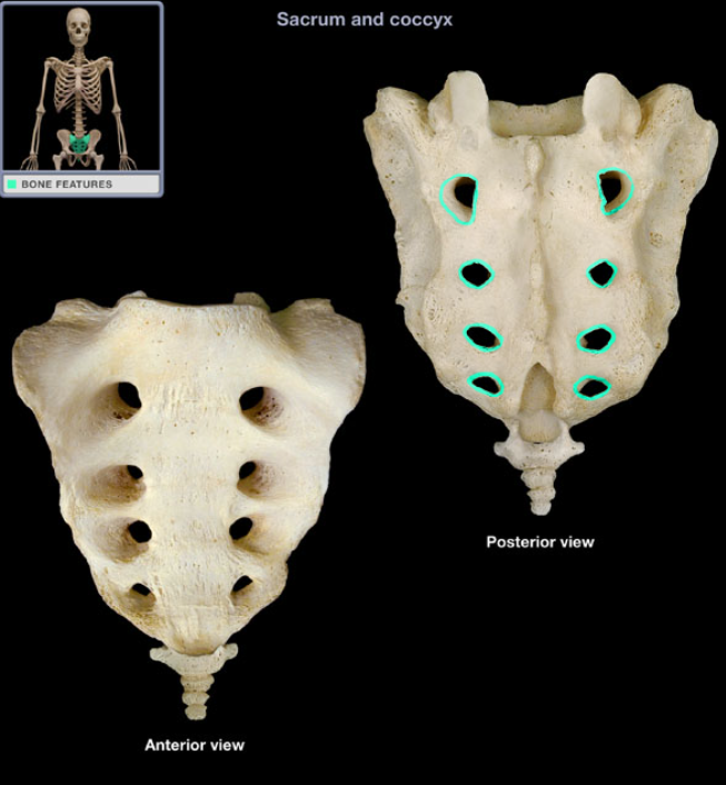
Location: Sacrum (posterior surface)
Description: Four pairs of openings, Communicate with sacral canal
Function: Transmit dorsal rami of S1-4 spinal nerves
Posterior sacral foramen
Location: Sacrum (anterior)
Description: Prominent, projecting edge of base of sacrum formed by superior border of S1 vertebral body. Forms posterior part of pelvic brim at midline
Comment: Sacral promontory is landmark for establishing female pelvic dimensions
Promontory of sacrum
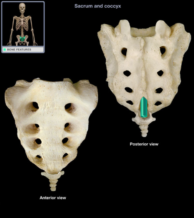
Location: Sacrum (posterior surface)
Description: Arched opening, Communicates with sacral canal
Function: Transmits filum terminale
Sacral hiatus
Location: Lower back, Between L5 and Co1 vertebrae, Posterior wall of pelvis
Description: Five fused vertebrae, Triangular bone wedged between hip bones
Sacrum (posterior view)
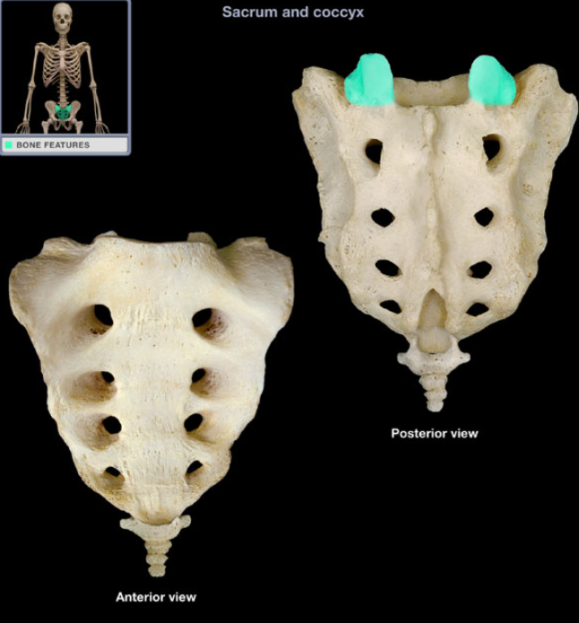
Location: Sacrum (base)
Description: Paired, superiorly-directed process, Has articular facet
Function: Articulation with inferior articular process of L5 vertebra
Comment: Base of sacrum is superior surface of S1 vertebra
Superior articular process of sacrum