The Heart
0.0(0)
0.0(0)
Card Sorting
1/157
There's no tags or description
Looks like no tags are added yet.
Study Analytics
Name | Mastery | Learn | Test | Matching | Spaced |
|---|
No study sessions yet.
158 Terms
1
New cards
How many chambers does the heart have?
Four
2
New cards
Where is the heart situated?
Middle Mediastinum
3
New cards
What angle does the heart sit at?
45
4
New cards
Two Collecting Chambers
Atria
5
New cards
Two Pumping Chambers
Ventricles
6
New cards
Sac surrounding heart and roots of great vessels
Pericardium
7
New cards
Tough external layer of pericardium
Fibrous Pericardium
8
New cards
What 3 things make up the Serous Pericardium
Parietal Pericardium, Pericardial Cavity, Epicardium
9
New cards
Lines internal surface of fibrous pericardium
Parietal Pericardium
10
New cards
Contains lubricating serous fluid
Pericardial Cavity
11
New cards
Outer layer of heart
Epicardium
12
New cards
What are the three layers of the heart
Epicardium, Myocardium, Endocardium
13
New cards
Thin, smooth, outer layer
Epicardium
14
New cards
Thick muscular layer
Myocardium
15
New cards
Thin endothelial lining
Endocardium
16
New cards
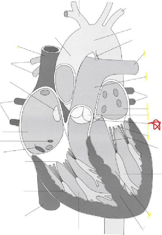
Aortic Valve
17
New cards
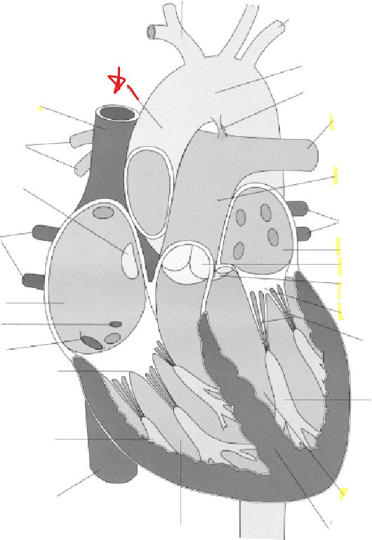
Ascending Aorta
18
New cards
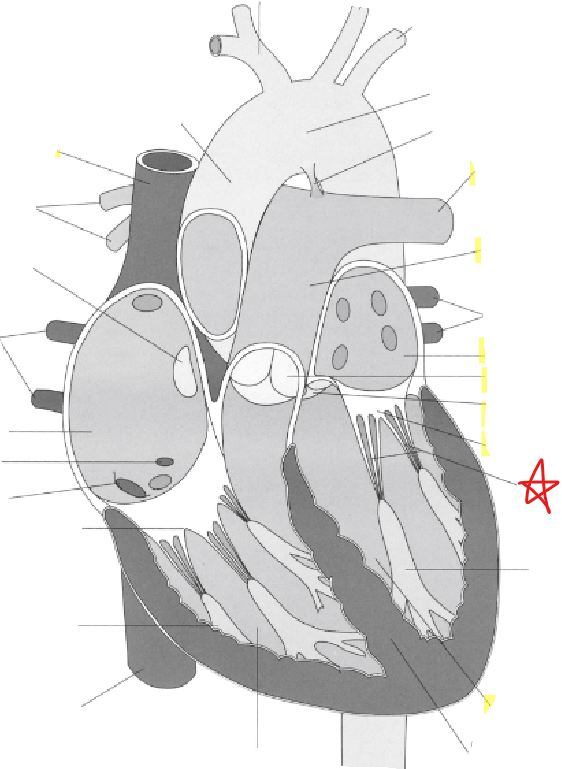
Chordae Tendineae
19
New cards
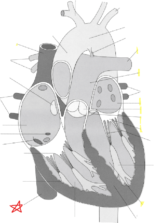
Inferior Vena Cava
20
New cards
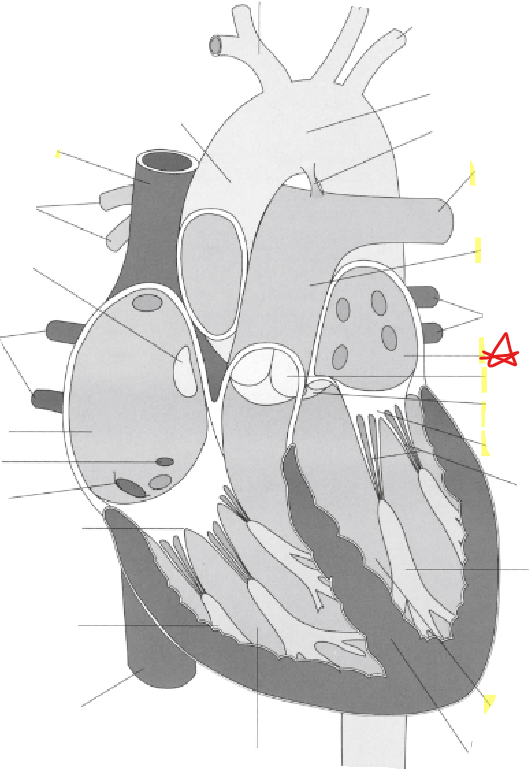
Left Atrium
21
New cards
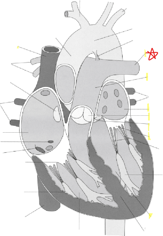
Left Pulmonary Artery
22
New cards
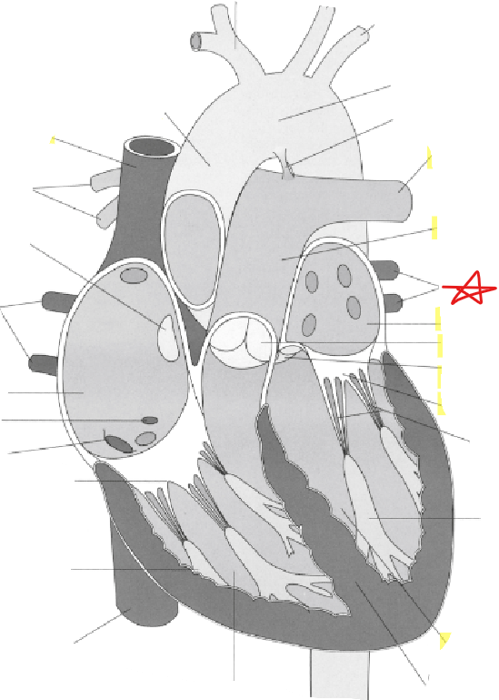
Left Pulmonary Veins
23
New cards
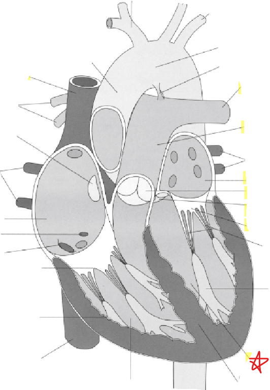
Left Ventricle
24
New cards
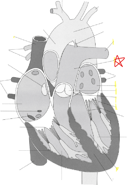
Main Pulmonary Artery
25
New cards
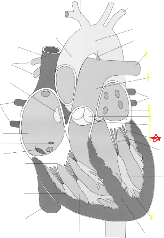
Mitral Valve
26
New cards
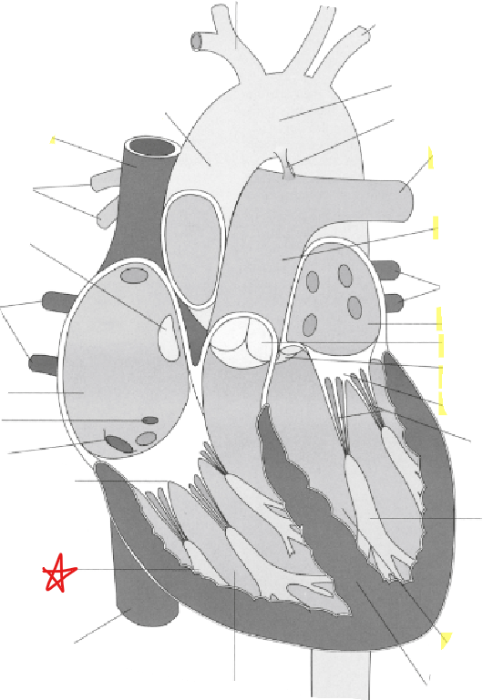
Papillary Muscle
27
New cards
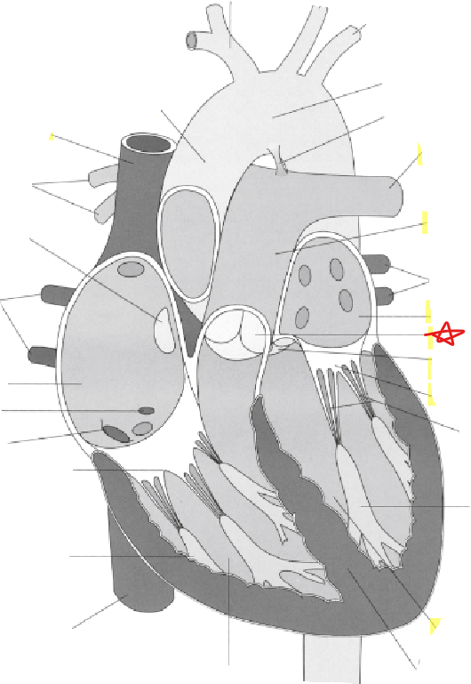
Pulmonary Valve
28
New cards
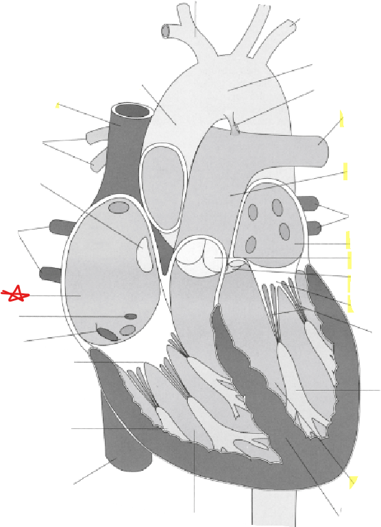
Right Atrium
29
New cards
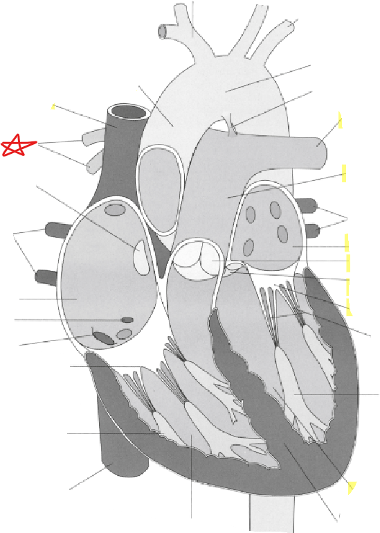
Right Pulmonary Arteries
30
New cards
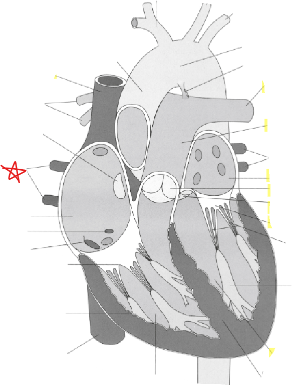
Right Pulmonary Veins
31
New cards
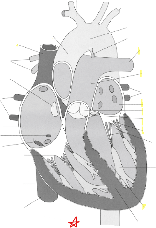
Right Ventricle
32
New cards
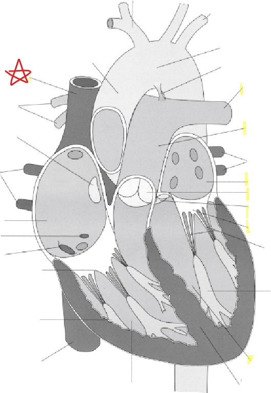
Superior Vena Cava
33
New cards
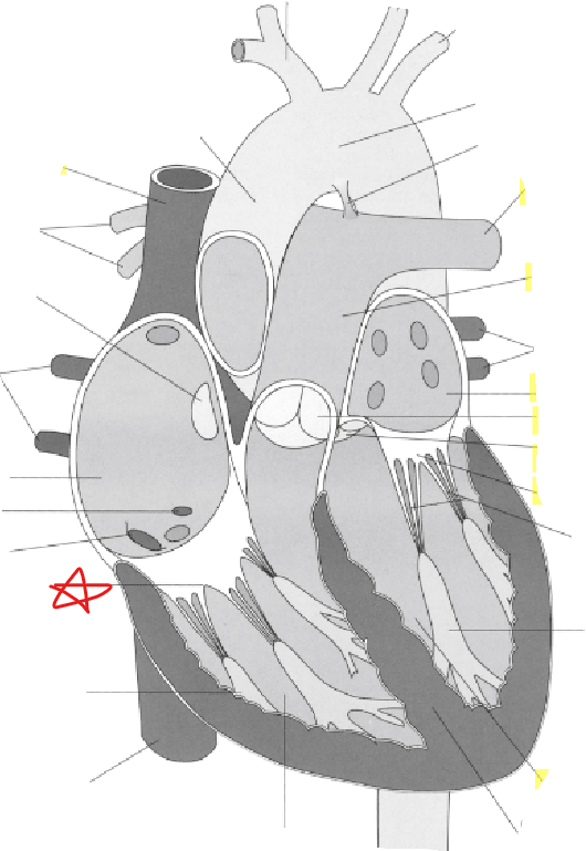
\
Tricuspid Valve
34
New cards
(BF) Inferior Vena Cava & Superior Vena Cava into -
Right Atrium
35
New cards
(BF) Right Atrium through Tricuspid Valve into -
Right Ventricle
36
New cards
(BF) Right Ventricle through Pulmonary Valve to -
Lungs
37
New cards
(BF) From Lungs through Pulmonary veins into -
Left Atrium
38
New cards
(BF) Left Atrium through Mitral Valve into -
Left Ventricle
39
New cards
(BF) Left Ventricle through Aortic Valve into -
Aorta
40
New cards
Receives deoxygenated blood from the entire body via the IVC and SVC
Right Atrium
41
New cards
Valve comprised of three leaflets allows blood to flow only from the right atrium to the right ventricle
Tricuspid Valve
42
New cards
Strong fibrous connections between the valve leaflets and the papillary muscles - prevent leaflets from moving backwards into atrium
Chordae Tendineae
43
New cards
Small muscles arising from walls of left and right ventricles - contract simultaneously with ventricles and prevent prolapse of valves into atria
Papillary Muscles
44
New cards
Pumps deoxygenated blood to lungs
Right Ventricle
45
New cards
Valve comprised of 3 cusps and separates the right ventricle from the pulmonary trunk
Pulmonary Valve
46
New cards
Transports deoxygenated blood from the right ventricle to the lungs
Main Pulmonary Artery
47
New cards
Carry oxygen rich blood from the lungs to the left atrium
Pulmonary Veins
48
New cards
Receives oxygen rich blood from the pulmonary veins
Left Atrium
49
New cards
Comprised of two leaflets - allows bloodflow from left atrium to left ventricle
Mitral Valve
50
New cards
Pumps oxygenated blood to the body via the aorta
Left Ventricle
51
New cards
Comprised of three leaflets - allows blood flow from the left ventricle into the ascending aorta
Aortic Valve
52
New cards
Supply blood to the cardiac muscle
Coronary Arteries
53
New cards
What is the origin of the coronary arteries
Aortic Root
54
New cards
What does the left coronary artery bifurcate into
Left Anterior Descending Artery (LAD)
Left Circumplex Artery (Cx)
Left Circumplex Artery (Cx)
55
New cards
What are the 4 views of cardiac ultrasound
Parasternal Long Axis (PLAX)
Parasternal Short Axis (PSAX)
Apical 4 Chamber
Subxiphoid 4 chamber
Parasternal Short Axis (PSAX)
Apical 4 Chamber
Subxiphoid 4 chamber
56
New cards
Where does the indicator point in PLAX view
Towards patients right shoulder at 10
57
New cards
Where is the apex of the heart in a PLAX view
To the Left
58
New cards
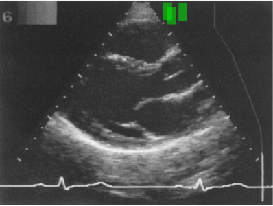
Name that Image View
PLAX
59
New cards
What does PLAX stand for?
Parasternal Long Axis
60
New cards
What does PSAX stand for?
Parasternal Short Axis
61
New cards
Where is the indicator on a PSAX view
Towards Patient’s Left Shoulder at 2
62
New cards
Where is transducer for PSAX
To left of sternum in 3rd or 4th space
63
New cards
PSAX: Images taken at level of Aortic Valve →
Mercedes Benz
64
New cards
PSAX: Images taken at level of Mitral Valve →
Fish Mouth
65
New cards
Where is the transducer for an Apical Four Chamber view
In 4th or 5th rib space
66
New cards
Where is the indicator for an Apical Four Chamber view
Towards Left shoulder to 2-3
67
New cards
Where is the sound beam angled to in the Apical Four Chamber view
Towards the patient’s head
68
New cards
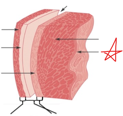
Endocardium
69
New cards
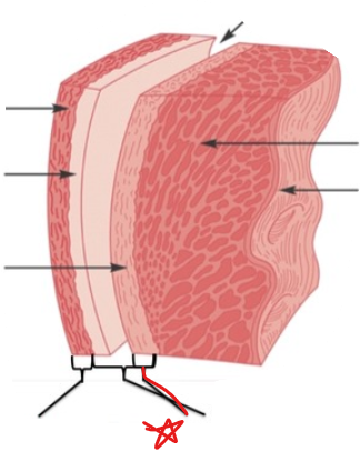
Epicardium
70
New cards
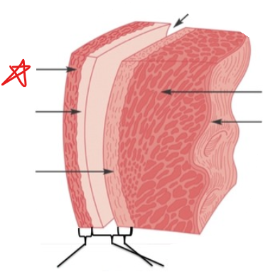
Fibrous Layer
71
New cards
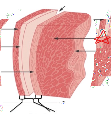
Myocardium
72
New cards
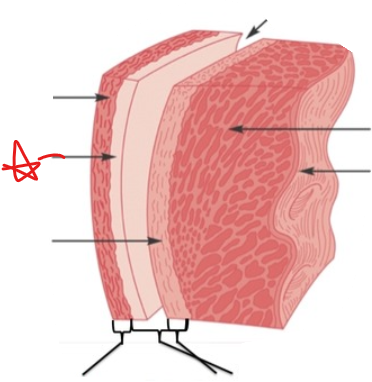
Parietal Pericardium
73
New cards
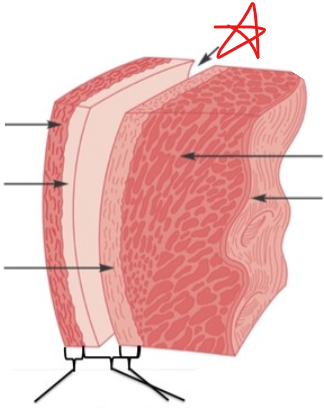
Pericardial Cavity
74
New cards
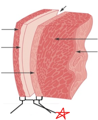
Serous Pericardium
75
New cards
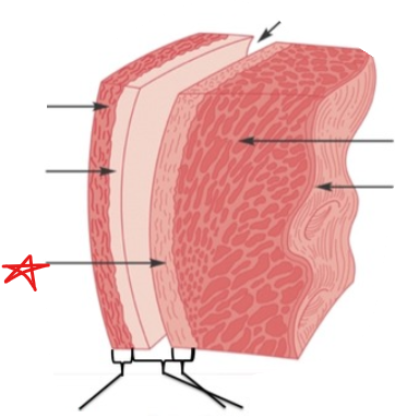
Visceral Pericardium
76
New cards
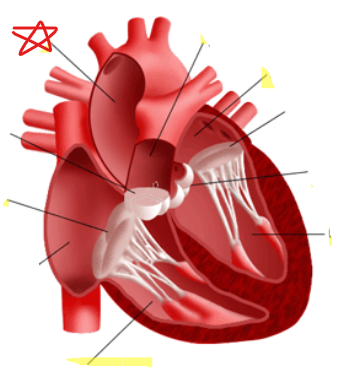
Aorta
77
New cards
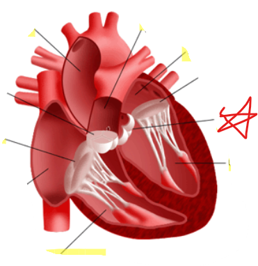
Aortic Valve
78
New cards
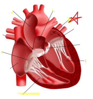
Left Atrium
79
New cards
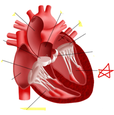
Left Ventricle
80
New cards
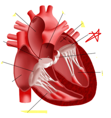
Mitral Valve
81
New cards
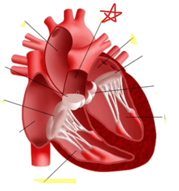
Pulmonary Trunk
82
New cards
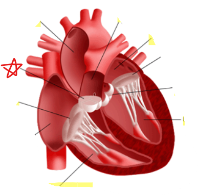
Pulmonary Valve
83
New cards
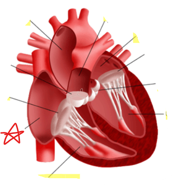
Right Atrium
84
New cards
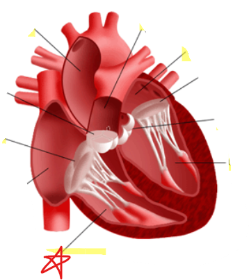
Right Ventricle
85
New cards
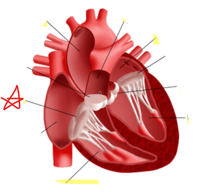
Tricuspid Valve
86
New cards
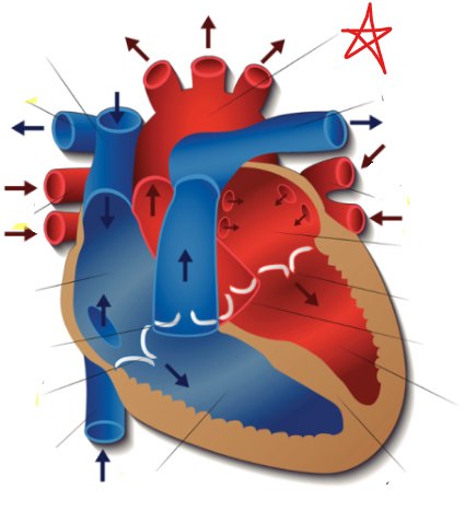
Aorta
87
New cards
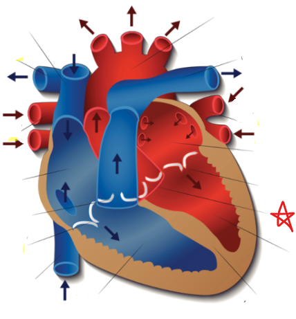
Aortic Valve
88
New cards
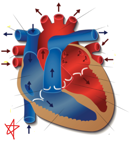
Inferior Vena Cava
89
New cards
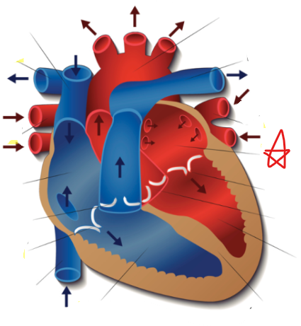
Left Atrium
90
New cards
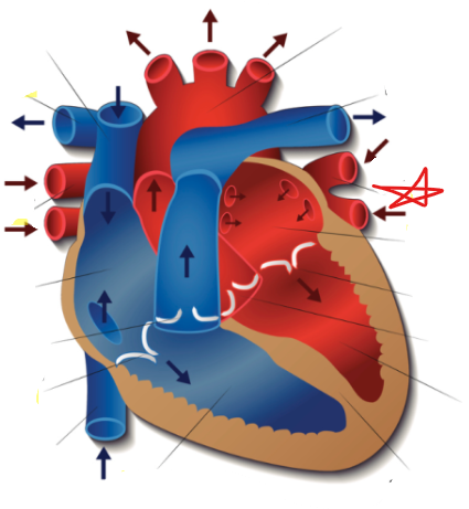
Left Pulmonary Veins
91
New cards
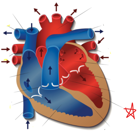
Left Ventricle
92
New cards
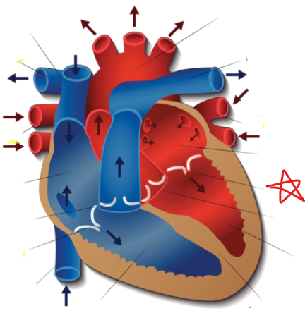
Mitral Valve
93
New cards
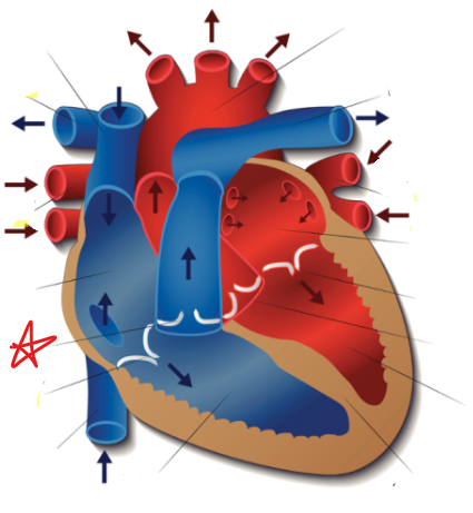
Pulmonary Valve
94
New cards
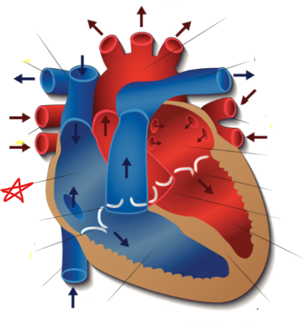
Right Atrium
95
New cards
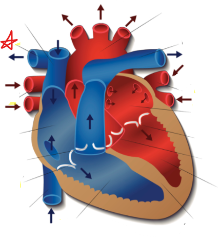
Right Pulmonary Arteries
96
New cards
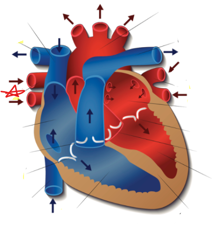
Right Pulmonary Veins
97
New cards
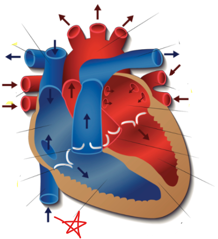
Right Ventricle
98
New cards
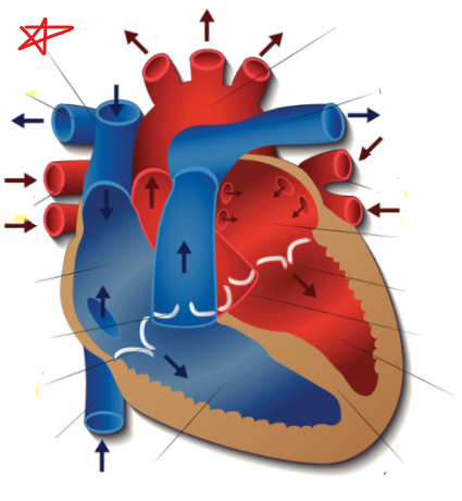
Superior Vena Cava
99
New cards
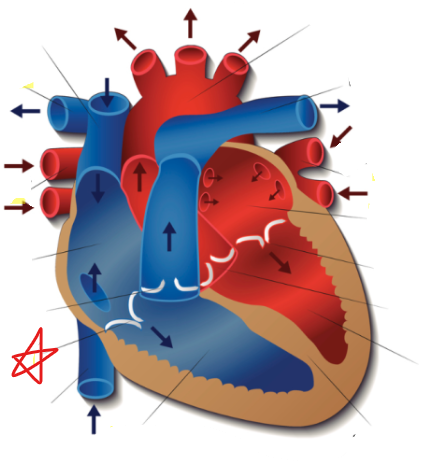
Tricuspid Valve
100
New cards
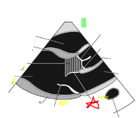
Anterior Mitral Valve Leaflet