Head and Neck Surgery
 Call Kai
 Learn
Learn Practice Test
 Spaced Repetition
 Match
Match Flashcards
 Knowt Play
1/39

There's no tags or description
Looks like no tags are added yet.
Name | Mastery | Learn | Test | Matching | Spaced | Call with Kai | Chat |
|---|
No analytics yet
Send a link to your students to track their progress
40 Terms
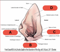
Antitragus
what is structure A?
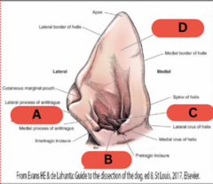
Tragus
what is structure B?
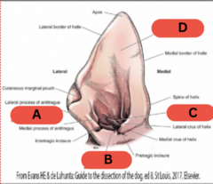
Anthelix
what is structure C?
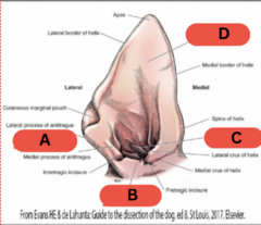
Scapha
what is structure D?
structures of the external ear?
vertical canal
horizontal canal
cartilages
glands (e.g. seruminous)
hair
the middle ear is also known as the
tympanic cavity
structure of the middle ear that connects to the nasopharynx
eustachian tube
species with a middle ear that is divided into two compartments by a septum
cat
structures of the inner ear?
labyrinth
semicircular canals
pinna laceration tx options?
Manage as wound
Primary wound closure
Pinnectomy
when can pinna lacerations be treated with primary wound closure?
If adequate and healthy tissue is present
Injury occurred within 6 hours
Suture patterns to close skin of pinnae lacerations?
Simple interrupted
Ford interlocking
Suture patterns to close skin and cartilage of pinna laceration together?
Vertical mattress
Figure of eight
criteria for partial or complete pinnectomy?
malignant tumor
ulcerated tumor
painful tumor
a ? thickness partial pinnectomy is indicated for malignant tumors
full
painful hemorrhage within layers of the pinna causing the layers of cartilage to separate due to trauma or an autoimmune response
aural hematoma
aural hematoma tx?
Address underlying cause
Treat quickly
Continuous drainage
Steroids/PRP possibly
TF needle aspiration of aural hematoma is an effective treatment
F
Methods for continuous drainage of aural hematoma?
teat cannula
penrose drain
closed suction drain
S shaped incision
circular fenestration
goals of aural hematoma tx?
remove clot
remove fibrin
close dead space
provide cosmesis and function
prevent recurrence
Indications for external ear surgery?
Stenosis
Poor/no response to medical management
Ear masses
Dystrophic mineralization
Trauma
TF most external ear surgeries are salvage procedures
T
Best diagnostic imaging method for evaluation of ear canal, extent of disease, and secondary changes from otitis externa/media?
CT
three most common surgical procedures employed for otitis externa?
Lateral wall resection
Vertical canal ablation
Total ear canal ablation and lateral bulla ostectomy (TECALBO)
rarely indicated procedure through which the lateral wall of the vertical canal is opened
lateral wall resection
indications for lateral wall resection?
Small tumor of lateral wall
Mild otitis externa affecting vertical canal
Congenital stenosis
rarely indicated procedure through which the vertical canal is removed and an opening is created for the horizontal canal
vertical canal ablation
vertical canal ablation indications?
Small tumor of vertical canal
Mild otitis externa affecting vertical canal
Congenital stenosis
TF lateral wall resection and vertical canal ablations are useful and effective procedures to treat otitis
F
commonly fails if treating otitis and becomes stenotic
procedure defined by complete removal of the vertical and horizontal canal as well as the lateral wall of the bulla that requires special equipment
total ear canal ablation and lateral bulla ostectomy (TECALBO)
indications for TECALBO?
severe chronic otitis
neoplasia
Steps of TECALBO?
Make incision around ear canal opening
Incision along vertical canal and make a flap for erect ears
Elevate auricular muscles from canal
Elevate canal from opening of bulla
Remove lateral wall of bulla
Remove epithelial lining from bulla cavity
Lavage
Culture and submit for histopath
Complications of TECALBO?
Facial nerve injury (temporary vs. permanent)
Deep infection (draining tract/abscess)
Hemorrhage
Damage to inner ear
Pinna necrosis
Airway swelling
Incisional complications
Loss of hearing
more common and temporary facial nerve injury that results from stretching of the nerve after ear surgery - resolves in 6-8 weeks
neuropraxia
failure to ? in a TECALBO can and often will result in infection/abscesses and draining tract formation within weeks to a year of surgery
remove all of the canal cartilage and epithelial lining of bulla
TF facial nerve injury is a very serious and life-threatening complication following ear surgery
F
mainly cosmetic, need to lubricate eyes with artificial tears to prevent corneal ulcers
important vessels to avoid when performing ear surgery?
retroarticular vein
external carotid
performing a bilateral TECABO raises the risk of what complication?
airway swelling
TF inner ear clinical signs that appear AFTER ear surgery are typically permanent
F
typically resolve in time
TECABO post op care?
Pain management
Abx and change based on culture
Incision care